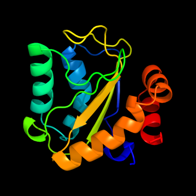
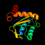
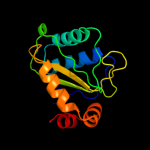
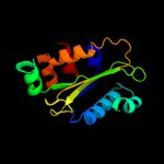
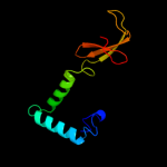
Region: 3 - 146 Aligned: 144 Modelled: 144 Confidence: 100.0% Identity: 38% PDB header:transferase Chain: B: PDB Molecule:phosphotransferase enzyme ii, a component; PDBTitle: structure of phosphotransferase enzyme ii, a component from yersinia2 pestis co92 at 1.2 a resolution
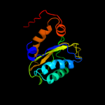
Region: 3 - 146 Aligned: 144 Modelled: 144 Confidence: 100.0% Identity: 38% PDB header:transferase Chain: A: PDB Molecule:phosphotransferase enzyme ii, a component; PDBTitle: structure of phosphotransferase enzyme ii, a component from yersinia2 pestis co92 at 1.2 a resolution
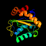
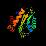
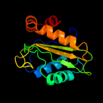
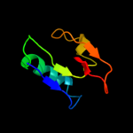
Region: 1 - 146 Aligned: 144 Modelled: 146 Confidence: 100.0% Identity: 19% Fold: Phoshotransferase/anion transport protein Superfamily: Phoshotransferase/anion transport protein Family: IIA domain of mannitol-specific and ntr phosphotransferase EII
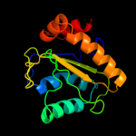
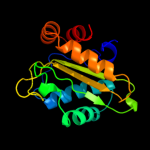
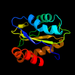
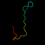
Region: 7 - 146 Aligned: 137 Modelled: 140 Confidence: 100.0% Identity: 28% Fold: Phoshotransferase/anion transport protein Superfamily: Phoshotransferase/anion transport protein Family: IIA domain of mannitol-specific and ntr phosphotransferase EII
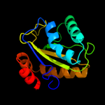
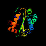
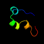
Region: 3 - 146 Aligned: 143 Modelled: 144 Confidence: 100.0% Identity: 15% Fold: Phoshotransferase/anion transport protein Superfamily: Phoshotransferase/anion transport protein Family: IIA domain of mannitol-specific and ntr phosphotransferase EII
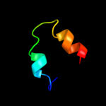
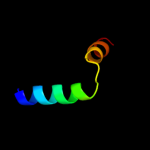
Region: 3 - 99 Aligned: 94 Modelled: 97 Confidence: 8.4% Identity: 14% PDB header:motor protein/protein transport Chain: A: PDB Molecule:myosin viia isoform 1; PDBTitle: structure of myosin viia myth4-ferm-sh3 in complex with the cen1 of2 sans