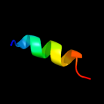
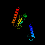
1 c2f9jP_
35.2
50
PDB header: rna binding proteinChain: P: PDB Molecule: splicing factor 3b subunit 1;PDBTitle: 3.0 angstrom resolution structure of a y22m mutant of the spliceosomal2 protein p14 bound to a region of sf3b155
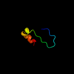
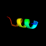
2 c3kztB_
25.0
17
PDB header: structural genomics, unknown functionChain: B: PDB Molecule: uncharacterized protein;PDBTitle: crystal structure of protein of unknown function (np_812423.1) from2 bacteroides thetaiotaomicron vpi-5482 at 2.10 a resolution
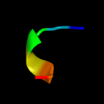
3 d1xl7a2
24.8
17
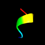
Fold: CoA-dependent acyltransferasesSuperfamily: CoA-dependent acyltransferasesFamily: Choline/Carnitine O-acyltransferase4 d1w6ga2
16.0
17
Fold: Cystatin-likeSuperfamily: Amine oxidase N-terminal regionFamily: Amine oxidase N-terminal region5 c3hp7A_
12.9
9
PDB header: structural genomics, unknown functionChain: A: PDB Molecule: hemolysin, putative;PDBTitle: putative hemolysin from streptococcus thermophilus.
6 d2qi2a1
12.1
25
Fold: Sm-like foldSuperfamily: Dom34/Pelota N-terminal domain-likeFamily: Dom34/Pelota N-terminal domain-like7 c3lg8B_
10.9
25
PDB header: hydrolaseChain: B: PDB Molecule: a-type atp synthase subunit e;PDBTitle: crystal structure of the c-terminal part of subunit e (e101-206) from2 methanocaldococcus jannaschii of a1ao atp synthase
8 c3lydA_
10.4
28
PDB header: structural genomics, unknown functionChain: A: PDB Molecule: uncharacterized protein;PDBTitle: crystal structure of putative uncharacterized protein from jonesia2 denitrificans
9 c1xl8B_
9.6
26
PDB header: transferaseChain: B: PDB Molecule: peroxisomal carnitine o-octanoyltransferase;PDBTitle: crystal structure of mouse carnitine octanoyltransferase in2 complex with octanoylcarnitine
10 d2vgna1
9.6
33
Fold: Sm-like foldSuperfamily: Dom34/Pelota N-terminal domain-likeFamily: Dom34/Pelota N-terminal domain-like11 d1whza_
9.0
19
Fold: dsRBD-likeSuperfamily: YcfA/nrd intein domainFamily: YcfA-like12 c2qi2A_
8.7
21
PDB header: cell cycleChain: A: PDB Molecule: cell division protein pelota related protein;PDBTitle: crystal structure of the thermoplasma acidophilum pelota2 protein
13 d1em8b_
8.6
19
Fold: DNA polymerase III psi subunitSuperfamily: DNA polymerase III psi subunitFamily: DNA polymerase III psi subunit14 c1e2vB_
8.3
67
PDB header: electron transport proteinsChain: B: PDB Molecule: cytochrome f;PDBTitle: n153q mutant of cytochrome f from chlamydomonas reinhardtii
15 d1vf5c1
8.1
67
Fold: Common fold of diphtheria toxin/transcription factors/cytochrome fSuperfamily: Cytochrome f, large domainFamily: Cytochrome f, large domain16 d1ci3m1
8.0
67
Fold: Common fold of diphtheria toxin/transcription factors/cytochrome fSuperfamily: Cytochrome f, large domainFamily: Cytochrome f, large domain17 d1e2wa1
7.9
67
Fold: Common fold of diphtheria toxin/transcription factors/cytochrome fSuperfamily: Cytochrome f, large domainFamily: Cytochrome f, large domain18 c3obyB_
7.8
29
PDB header: hydrolaseChain: B: PDB Molecule: protein pelota homolog;PDBTitle: crystal structure of archaeoglobus fulgidus pelota reveals inter-2 domain structural plasticity
19 c1q6xA_
7.7
19
PDB header: transferaseChain: A: PDB Molecule: choline o-acetyltransferase;PDBTitle: crystal structure of rat choline acetyltransferase
20 c3agjD_
7.7
21
PDB header: translation/hydrolaseChain: D: PDB Molecule: protein pelota homolog;PDBTitle: crystal structure of archaeal pelota and gtp-bound ef1 alpha complex
21 c3agjB_
not modelled
7.7
21
PDB header: translation/hydrolaseChain: B: PDB Molecule: protein pelota homolog;PDBTitle: crystal structure of archaeal pelota and gtp-bound ef1 alpha complex
22 d1hcza1
not modelled
7.5
67
Fold: Common fold of diphtheria toxin/transcription factors/cytochrome fSuperfamily: Cytochrome f, large domainFamily: Cytochrome f, large domain23 c3m9vA_
not modelled
7.0
23
PDB header: oxidoreductaseChain: A: PDB Molecule: fad-dependent oxidoreductase;PDBTitle: x-ray structure of a kijd3 in complex with dtdp
24 d1t1ua2
not modelled
7.0
17
Fold: CoA-dependent acyltransferasesSuperfamily: CoA-dependent acyltransferasesFamily: Choline/Carnitine O-acyltransferase25 d1vbka2
not modelled
6.8
16
Fold: THUMP domainSuperfamily: THUMP domain-likeFamily: THUMP domain26 c3obwA_
not modelled
6.7
21
PDB header: hydrolaseChain: A: PDB Molecule: protein pelota homolog;PDBTitle: crystal structure of two archaeal pelotas reveal inter-domain2 structural plasticity
27 d1y1pa1
not modelled
6.4
8
Fold: NAD(P)-binding Rossmann-fold domainsSuperfamily: NAD(P)-binding Rossmann-fold domainsFamily: Tyrosine-dependent oxidoreductases28 d1gsoa2
not modelled
6.3
17
Fold: PreATP-grasp domainSuperfamily: PreATP-grasp domainFamily: BC N-terminal domain-like29 c2h4tB_
not modelled
6.2
20
PDB header: transferaseChain: B: PDB Molecule: carnitine o-palmitoyltransferase ii,PDBTitle: crystal structure of rat carnitine palmitoyltransferase ii
30 c2e75C_
not modelled
6.1
67
PDB header: photosynthesisChain: C: PDB Molecule: apocytochrome f;PDBTitle: crystal structure of the cytochrome b6f complex with 2-nonyl-4-2 hydroxyquinoline n-oxide (nqno) from m.laminosus
31 c3b54A_
not modelled
6.0
14
PDB header: transferaseChain: A: PDB Molecule: nucleoside diphosphate kinase;PDBTitle: saccharomyces cerevisiae nucleoside diphosphate kinase
32 c1q90A_
not modelled
5.8
67
PDB header: photosynthesisChain: A: PDB Molecule: apocytochrome f;PDBTitle: structure of the cytochrome b6f (plastohydroquinone : plastocyanin2 oxidoreductase) from chlamydomonas reinhardtii
33 c3fa4D_
not modelled
5.7
22
PDB header: lyaseChain: D: PDB Molecule: 2,3-dimethylmalate lyase;PDBTitle: crystal structure of 2,3-dimethylmalate lyase, a pep mutase/isocitrate2 lyase superfamily member, triclinic crystal form
34 d1tu2b1
not modelled
5.6
67
Fold: Common fold of diphtheria toxin/transcription factors/cytochrome fSuperfamily: Cytochrome f, large domainFamily: Cytochrome f, large domain35 c2jxmB_
not modelled
5.5
50
PDB header: electron transportChain: B: PDB Molecule: cytochrome f;PDBTitle: ensemble of twenty structures of the prochlorothrix2 hollandica plastocyanin- cytochrome f complex
36 d1ndba2
not modelled
5.5
16
Fold: CoA-dependent acyltransferasesSuperfamily: CoA-dependent acyltransferasesFamily: Choline/Carnitine O-acyltransferase37 c1ctmA_
not modelled
5.4
67
PDB header: electron transport(cytochrome)Chain: A: PDB Molecule: cytochrome f;PDBTitle: crystal structure of chloroplast cytochrome f reveals a2 novel cytochrome fold and unexpected heme ligation
38 d1ukua_
not modelled
5.4
35
Fold: Ferredoxin-likeSuperfamily: GlnB-likeFamily: Divalent ion tolerance proteins CutA (CutA1)39 c3jywW_
not modelled
5.3
44
PDB header: ribosomeChain: W: PDB Molecule: 60s ribosomal protein l31(a);PDBTitle: structure of the 60s proteins for eukaryotic ribosome based on cryo-em2 map of thermomyces lanuginosus ribosome at 8.9a resolution
40 c2vu5A_
not modelled
5.3
22
PDB header: transferaseChain: A: PDB Molecule: nucleoside diphosphate kinase;PDBTitle: crystal structure of pndk from bacillus anthracis
41 d1xqia1
not modelled
5.2
3
Fold: Ferredoxin-likeSuperfamily: Nucleoside diphosphate kinase, NDKFamily: Nucleoside diphosphate kinase, NDK42 d1zs6a1
not modelled
5.2
17
Fold: Ferredoxin-likeSuperfamily: Nucleoside diphosphate kinase, NDKFamily: Nucleoside diphosphate kinase, NDK43 d1kx5b_
not modelled
5.1
19
Fold: Histone-foldSuperfamily: Histone-foldFamily: Nucleosome core histones44 c2fipA_
not modelled
5.1
30
PDB header: transcriptionChain: A: PDB Molecule: late genes activator;PDBTitle: phage phi29 transcription regulator p4
45 d1vhfa_
not modelled
5.0
24
Fold: Ferredoxin-likeSuperfamily: GlnB-likeFamily: Divalent ion tolerance proteins CutA (CutA1)