Phyre Home Retrieve Phyre Job Id
Emaill.a.kelley@imperial.ac.uk
DescriptionP64476
DateThu Jan 5 12:08:44 GMT 2012
Unique Job IDc0739c190bdb1230

Summary 

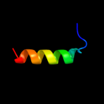
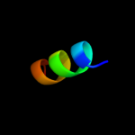
Top model
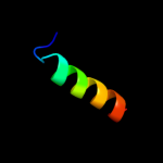
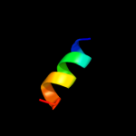
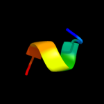
Image coloured by rainbow N → C terminus
Model (left) based on template c2qx0A_
Top template information
PDB header:transferase
Chain: A: PDB Molecule:7,8-dihydro-6-hydroxymethylpterin-
PDBTitle: crystal structure of yersinia pestis hppk (ternary complex)
Confidence and coverage
Confidence: 28.5% Coverage: 32%
20 residues ( 32% of your sequence) have been modelled with 28.5% confidence by the single highest scoring template.
You may wish to submit your sequence to Phyrealarm. This will automatically scan your sequence every week for new potential templates as they appear in the Phyre2 library.
Please note: You must be registered and logged in to use Phyrealarm.
3D viewing
Interactive 3D view in Jmol

Sequence analysis 

Secondary structure and disorder prediction 

   1........10.........20.........30.........40.........50.........60
Sequence  MSTQLDPTQLAIEFLRRDQSNLSPAQYLKRLKQLELEFADLLTLSSAELKEEIYFAWRLG
Secondary structure 


SS confidence 


Disorder  ??????


?
?
???


?


?


???
Disorder confidence 


   ..
Sequence  VH
Secondary structure 

SS confidence 

Disorder  ??
Disorder confidence 


Confidence Key
High(9)                    Low (0)
?Disordered
Alpha helix
Beta strand

Domain analysis 

Hover over an aligned region to see model and summary info

Please note, only up to the top 20 hits are modelled to reduce computer load

RankAligned region

PDB 2qx0 chain A

3D model

Region: 19 - 38
Aligned: 20
Modelled: 20
Confidence: 28.5%
Identity: 25%
PDB header:transferase
Chain: A: PDB Molecule:7,8-dihydro-6-hydroxymethylpterin-
PDBTitle: crystal structure of yersinia pestis hppk (ternary complex)

Phyre2

PDB 1f9y chain A

3D model

Region: 19 - 38
Aligned: 20
Modelled: 20
Confidence: 28.1%
Identity: 25%
Fold: Ferredoxin-like
Superfamily: 6-hydroxymethyl-7,8-dihydropterin pyrophosphokinase, HPPK
Family: 6-hydroxymethyl-7,8-dihydropterin pyrophosphokinase, HPPK

Phyre2

PDB 1cbk chain A

3D model

Region: 19 - 38
Aligned: 20
Modelled: 20
Confidence: 24.3%
Identity: 35%
Fold: Ferredoxin-like
Superfamily: 6-hydroxymethyl-7,8-dihydropterin pyrophosphokinase, HPPK
Family: 6-hydroxymethyl-7,8-dihydropterin pyrophosphokinase, HPPK

Phyre2

PDB 2qdm chain A

3D model

Region: 7 - 41
Aligned: 35
Modelled: 35
Confidence: 18.7%
Identity: 29%
PDB header:hydrolase
Chain: A: PDB Molecule:tyrosine-protein phosphatase non-receptor type 7;
PDBTitle: crystal structure of the heptp catalytic domain c270s/d236a/q314a2 mutant

Phyre2

PDB 3mcn chain A

3D model

Region: 19 - 38
Aligned: 20
Modelled: 20
Confidence: 17.0%
Identity: 40%
PDB header:transferase
Chain: A: PDB Molecule:2-amino-4-hydroxy-6-hydroxymethyldihydropteridine
PDBTitle: crystal structure of the 6-hyroxymethyl-7,8-dihydropterin2 pyrophosphokinase dihydropteroate synthase bifunctional enzyme from3 francisella tularensis

Phyre2

PDB 1onv chain B

3D model

Region: 32 - 42
Aligned: 11
Modelled: 11
Confidence: 13.9%
Identity: 55%
PDB header:transcription
Chain: B: PDB Molecule:serine phosphatase fcp1a;
PDBTitle: nmr structure of a complex containing the tfiif subunit2 rap74 and the rnap ii ctd phosphatase fcp1

Phyre2

PDB 2cg8 chain B

3D model

Region: 19 - 38
Aligned: 20
Modelled: 20
Confidence: 12.1%
Identity: 25%
PDB header:lyase/transferase
Chain: B: PDB Molecule:dihydroneopterin aldolase 6-hydroxymethyl-7,8-
PDBTitle: the bifunctional dihydroneopterin aldolase 6-hydroxymethyl-2 7,8-dihydropterin synthase from streptococcus pneumoniae

Phyre2

PDB 1kdd chain C

3D model

Region: 30 - 41
Aligned: 12
Modelled: 12
Confidence: 8.9%
Identity: 50%
PDB header:de novo protein
Chain: C: PDB Molecule:gcn4 acid base heterodimer acid-d12la16i;
PDBTitle: x-ray structure of the coiled coil gcn4 acid base2 heterodimer acid-d12la16i base-d12la16l

Phyre2

PDB 1kdd chain F

3D model

Region: 30 - 41
Aligned: 12
Modelled: 12
Confidence: 8.7%
Identity: 50%
PDB header:de novo protein
Chain: F: PDB Molecule:gcn4 acid base heterodimer acid-d12la16i;
PDBTitle: x-ray structure of the coiled coil gcn4 acid base2 heterodimer acid-d12la16i base-d12la16l

Phyre2

PDB 1kdd chain A

3D model

Region: 30 - 41
Aligned: 12
Modelled: 12
Confidence: 8.7%
Identity: 50%
PDB header:de novo protein
Chain: A: PDB Molecule:gcn4 acid base heterodimer acid-d12la16i;
PDBTitle: x-ray structure of the coiled coil gcn4 acid base2 heterodimer acid-d12la16i base-d12la16l

Phyre2

PDB 3igm chain A

3D model

Region: 7 - 17
Aligned: 11
Modelled: 11
Confidence: 8.6%
Identity: 27%
PDB header:transcription/dna
Chain: A: PDB Molecule:pf14_0633 protein;
PDBTitle: a 2.2a crystal structure of the ap2 domain of pf14_0633 from p.2 falciparum, bound as a domain-swapped dimer to its cognate dna

Phyre2

PDB 1kd9 chain F

3D model

Region: 30 - 41
Aligned: 12
Modelled: 12
Confidence: 8.5%
Identity: 50%
PDB header:de novo protein
Chain: F: PDB Molecule:gcn4 acid base heterodimer acid-d12la16l;
PDBTitle: x-ray structure of the coiled coil gcn4 acid base2 heterodimer acid-d12la16l base-d12la16l

Phyre2

PDB 1kd9 chain A

3D model

Region: 30 - 41
Aligned: 12
Modelled: 12
Confidence: 8.5%
Identity: 50%
PDB header:de novo protein
Chain: A: PDB Molecule:gcn4 acid base heterodimer acid-d12la16l;
PDBTitle: x-ray structure of the coiled coil gcn4 acid base2 heterodimer acid-d12la16l base-d12la16l

Phyre2

PDB 1kd9 chain C

3D model

Region: 30 - 41
Aligned: 12
Modelled: 12
Confidence: 8.5%
Identity: 50%
PDB header:de novo protein
Chain: C: PDB Molecule:gcn4 acid base heterodimer acid-d12la16l;
PDBTitle: x-ray structure of the coiled coil gcn4 acid base2 heterodimer acid-d12la16l base-d12la16l

Phyre2

PDB 3kf8 chain D

3D model

Region: 5 - 22
Aligned: 18
Modelled: 18
Confidence: 8.2%
Identity: 33%
PDB header:structural protein
Chain: D: PDB Molecule:protein ten1;
PDBTitle: crystal structure of c. tropicalis stn1-ten1 complex

Phyre2

PDB 2bmb chain A

3D model

Region: 19 - 38
Aligned: 20
Modelled: 20
Confidence: 7.7%
Identity: 35%
PDB header:transferase
Chain: A: PDB Molecule:folic acid synthesis protein fol1;
PDBTitle: x-ray structure of the bifunctional 6-hydroxymethyl-7,8-2 dihydroxypterin pyrophosphokinase dihydropteroate synthase3 from saccharomyces cerevisiae

Phyre2

PDB 2v4x chain A

3D model

Region: 22 - 29
Aligned: 8
Modelled: 8
Confidence: 6.8%
Identity: 50%
PDB header:viral protein
Chain: A: PDB Molecule:capsid protein p27;
PDBTitle: crystal structure of jaagsiekte sheep retrovirus capsid n-2 terminal domain

Phyre2

PDB 2pa2 chain A domain 1

3D model

Region: 1 - 26
Aligned: 26
Modelled: 26
Confidence: 6.7%
Identity: 31%
Fold: alpha/beta-Hammerhead
Superfamily: Ribosomal protein L16p/L10e
Family: Ribosomal protein L10e

Phyre2

PDB 1b8i chain A

3D model

Region: 14 - 57
Aligned: 40
Modelled: 44
Confidence: 6.6%
Identity: 25%
Fold: DNA/RNA-binding 3-helical bundle
Superfamily: Homeodomain-like
Family: Homeodomain

Phyre2

PDB 3pn1 chain A

3D model

Region: 8 - 46
Aligned: 39
Modelled: 39
Confidence: 5.9%
Identity: 33%
PDB header:ligase/ligase inhibitor
Chain: A: PDB Molecule:dna ligase;
PDBTitle: novel bacterial nad+-dependent dna ligase inhibitors with broad2 spectrum potency and antibacterial efficacy in vivo

Phyre2
1

c2qx0A_
2

d1f9ya_
3

d1cbka_
4

c2qdmA_
5

c3mcnA_
6

c1onvB_
7

c2cg8B_
8

c1kddC_
9

c1kddF_
10

c1kddA_
11

c3igmA_
12

c1kd9F_
13

c1kd9A_
14

c1kd9C_
15

c3kf8D_
16

c2bmbA_
17

c2v4xA_
18

d2pa2a1
19

d1b8ia_
20

c3pn1A_
21


22


23


24


25


26


27


28


29


30


Detailed template information 

#
Template Alignment Coverage3D Model Confidence
% i.d. Template Information
1c2qx0A_


28.5 25 PDB header:transferase
Chain: A: PDB Molecule:7,8-dihydro-6-hydroxymethylpterin-
PDBTitle: crystal structure of yersinia pestis hppk (ternary complex)
2d1f9ya_


28.1 25 Fold:Ferredoxin-like
Superfamily:6-hydroxymethyl-7,8-dihydropterin pyrophosphokinase, HPPK
Family:6-hydroxymethyl-7,8-dihydropterin pyrophosphokinase, HPPK
3d1cbka_


24.3 35 Fold:Ferredoxin-like
Superfamily:6-hydroxymethyl-7,8-dihydropterin pyrophosphokinase, HPPK
Family:6-hydroxymethyl-7,8-dihydropterin pyrophosphokinase, HPPK
4c2qdmA_


18.7 29 PDB header:hydrolase
Chain: A: PDB Molecule:tyrosine-protein phosphatase non-receptor type 7;
PDBTitle: crystal structure of the heptp catalytic domain c270s/d236a/q314a2 mutant
5c3mcnA_


17.0 40 PDB header:transferase
Chain: A: PDB Molecule:2-amino-4-hydroxy-6-hydroxymethyldihydropteridine
PDBTitle: crystal structure of the 6-hyroxymethyl-7,8-dihydropterin2 pyrophosphokinase dihydropteroate synthase bifunctional enzyme from3 francisella tularensis
6c1onvB_


13.9 55 PDB header:transcription
Chain: B: PDB Molecule:serine phosphatase fcp1a;
PDBTitle: nmr structure of a complex containing the tfiif subunit2 rap74 and the rnap ii ctd phosphatase fcp1
7c2cg8B_


12.1 25 PDB header:lyase/transferase
Chain: B: PDB Molecule:dihydroneopterin aldolase 6-hydroxymethyl-7,8-
PDBTitle: the bifunctional dihydroneopterin aldolase 6-hydroxymethyl-2 7,8-dihydropterin synthase from streptococcus pneumoniae
8c1kddC_


8.9 50 PDB header:de novo protein
Chain: C: PDB Molecule:gcn4 acid base heterodimer acid-d12la16i;
PDBTitle: x-ray structure of the coiled coil gcn4 acid base2 heterodimer acid-d12la16i base-d12la16l
9c1kddF_


8.7 50 PDB header:de novo protein
Chain: F: PDB Molecule:gcn4 acid base heterodimer acid-d12la16i;
PDBTitle: x-ray structure of the coiled coil gcn4 acid base2 heterodimer acid-d12la16i base-d12la16l
10c1kddA_


8.7 50 PDB header:de novo protein
Chain: A: PDB Molecule:gcn4 acid base heterodimer acid-d12la16i;
PDBTitle: x-ray structure of the coiled coil gcn4 acid base2 heterodimer acid-d12la16i base-d12la16l
11c3igmA_


8.6 27 PDB header:transcription/dna
Chain: A: PDB Molecule:pf14_0633 protein;
PDBTitle: a 2.2a crystal structure of the ap2 domain of pf14_0633 from p.2 falciparum, bound as a domain-swapped dimer to its cognate dna
12c1kd9F_


8.5 50 PDB header:de novo protein
Chain: F: PDB Molecule:gcn4 acid base heterodimer acid-d12la16l;
PDBTitle: x-ray structure of the coiled coil gcn4 acid base2 heterodimer acid-d12la16l base-d12la16l
13c1kd9A_


8.5 50 PDB header:de novo protein
Chain: A: PDB Molecule:gcn4 acid base heterodimer acid-d12la16l;
PDBTitle: x-ray structure of the coiled coil gcn4 acid base2 heterodimer acid-d12la16l base-d12la16l
14c1kd9C_


8.5 50 PDB header:de novo protein
Chain: C: PDB Molecule:gcn4 acid base heterodimer acid-d12la16l;
PDBTitle: x-ray structure of the coiled coil gcn4 acid base2 heterodimer acid-d12la16l base-d12la16l
15c3kf8D_


8.2 33 PDB header:structural protein
Chain: D: PDB Molecule:protein ten1;
PDBTitle: crystal structure of c. tropicalis stn1-ten1 complex
16c2bmbA_


7.7 35 PDB header:transferase
Chain: A: PDB Molecule:folic acid synthesis protein fol1;
PDBTitle: x-ray structure of the bifunctional 6-hydroxymethyl-7,8-2 dihydroxypterin pyrophosphokinase dihydropteroate synthase3 from saccharomyces cerevisiae
17c2v4xA_


6.8 50 PDB header:viral protein
Chain: A: PDB Molecule:capsid protein p27;
PDBTitle: crystal structure of jaagsiekte sheep retrovirus capsid n-2 terminal domain
18d2pa2a1


6.7 31 Fold:alpha/beta-Hammerhead
Superfamily:Ribosomal protein L16p/L10e
Family:Ribosomal protein L10e
19d1b8ia_


6.6 25 Fold:DNA/RNA-binding 3-helical bundle
Superfamily:Homeodomain-like
Family:Homeodomain
20c3pn1A_


5.9 33 PDB header:ligase/ligase inhibitor
Chain: A: PDB Molecule:dna ligase;
PDBTitle: novel bacterial nad+-dependent dna ligase inhibitors with broad2 spectrum potency and antibacterial efficacy in vivo
21c2kgfA_


not modelled 5.7 38 PDB header:viral protein
Chain: A: PDB Molecule:capsid protein p27;
PDBTitle: n-terminal domain of capsid protein from the mason-pfizer2 monkey virus
22d1d1da2


not modelled 5.5 50 Fold:Retrovirus capsid protein, N-terminal core domain
Superfamily:Retrovirus capsid protein, N-terminal core domain
Family:Retrovirus capsid protein, N-terminal core domain
23c1zuoA_


not modelled 5.5 17 PDB header:ligase
Chain: A: PDB Molecule:hypothetical protein loc92912;
PDBTitle: structure of human ubiquitin-conjugating enzyme (ubci)2 involved in embryo attachment and implantation
24d1zuoa1


not modelled 5.5 17 Fold:UBC-like
Superfamily:UBC-like
Family:UBC-related
25c2q97T_


not modelled 5.3 53 PDB header:structural protein/cell invasion
Chain: T: PDB Molecule:toxofilin;
PDBTitle: complex of mammalian actin with toxofilin from toxoplasma gondii
26d1em9a_


not modelled 5.2 50 Fold:Retrovirus capsid protein, N-terminal core domain
Superfamily:Retrovirus capsid protein, N-terminal core domain
Family:Retrovirus capsid protein, N-terminal core domain
27d1b04a_


not modelled 5.2 22 Fold:ATP-grasp
Superfamily:DNA ligase/mRNA capping enzyme, catalytic domain
Family:Adenylation domain of NAD+-dependent DNA ligase
28c1xdtT_


not modelled 5.2 33 PDB header:complex (toxin/growth factor)
Chain: T: PDB Molecule:diphtheria toxin;
PDBTitle: complex of diphtheria toxin and heparin-binding epidermal growth2 factor
29d1ta8a_


not modelled 5.1 36 Fold:ATP-grasp
Superfamily:DNA ligase/mRNA capping enzyme, catalytic domain
Family:Adenylation domain of NAD+-dependent DNA ligase
30c1kd8F_


not modelled 5.0 42 PDB header:de novo protein
Chain: F: PDB Molecule:gcn4 acid base heterodimer acid-d12ia16v;
PDBTitle: x-ray structure of the coiled coil gcn4 acid base2 heterodimer acid-d12ia16v base-d12la16l

Binding site prediction 

Due to computational demand, binding site predictions are not run for batch jobs

If you want to predict binding sites, please manually submit your model of choice to 3DLigandSite


Phyre is for academic use only

Please cite: Protein structure prediction on the web: a case study using the Phyre server
Kelley LA and Sternberg MJE. Nature Protocols 4, 363 - 371 (2009) [pdf] [Import into BibTeX]
 
If you use the binding site predictions from 3DLigandSite, please also cite:
3DLigandSite: predicting ligand-binding sites using similar structures.
Wass MN, Kelley LA and Sternberg MJ Nucleic Acids Research 38, W469-73 (2010) [PubMed]
 
© Structural Bioinformatics Group
Imperial College London
Lawrence Kelley, Benjamin Jefferys
Disclaimer
Terms and Conditions
Component software
Template detection: HHpred 1.51
Secondary structure prediction: Psi-pred 2.5
Disorder prediction: Disopred 2.4
Transmembrane prediction: Memsat_SVM
Multi-template modelling and ab initio: Poing 1.0