Phyre Home Retrieve Phyre Job Id
Emaill.a.kelley@imperial.ac.uk
DescriptionP0A9K7
DateThu Jan 5 11:10:34 GMT 2012
Unique Job IDb651e3ec4422ac7c

Summary 

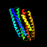
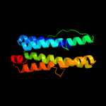
Top model
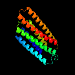
Image coloured by rainbow N → C terminus
Model (left) based on template d1sumb_
Top template information
Fold:Spectrin repeat-like
Superfamily:PhoU-like
Family:PhoU-like
Confidence and coverage
Confidence:100.0% Coverage: 90%
217 residues ( 90% of your sequence) have been modelled with 100.0% confidence by the single highest scoring template.
3D viewing
Interactive 3D view in Jmol

Sequence analysis 

Secondary structure and disorder prediction 

   1........10.........20.........30.........40.........50.........60
Sequence  MDSLNLNKHISGQFNAELESIRTQVMTMGGMVEQQLSDAITAMHNQDSDLAKRVIEGDKN
Secondary structure 


SS confidence 


Disorder  ???????


Disorder confidence 


   .........70.........80.........90.........100.........110.........120
Sequence  VNMMEVAIDEACVRIIAKRQPTASDLRLVMVISKTIAELERIGDVADKICRTALEKFSQQ
Secondary structure 


SS confidence 


Disorder 


??


Disorder confidence 


   .........130.........140.........150.........160.........170.........180
Sequence  HQPLLVSLESLGRHTIQMLHDVLDAFARMDIDEAVRIYREDKKVDQEYEGIVRQLMTYMM
Secondary structure 


SS confidence 


Disorder 
?


Disorder confidence 


   .........190.........200.........210.........220.........230.........240
Sequence  EDSRTIPSVLTALFCARSIERIGDRCQNICEFIFYYVKGQDFRHVGGDELDKLLAGKDSD
Secondary structure 


SS confidence 


Disorder 


?


???????
Disorder confidence 


   .
Sequence  K
Secondary structure 
SS confidence 
Disorder  ?
Disorder confidence 
 

Confidence Key
High(9)                    Low (0)
?Disordered
Alpha helix
Beta strand

Domain analysis 

Hover over an aligned region to see model and summary info

Please note, only up to the top 20 hits are modelled to reduce computer load

RankAligned region

PDB 1sum chain B

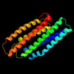
3D model

Region: 12 - 228
Aligned: 217
Modelled: 217
Confidence: 100.0%
Identity: 26%
Fold: Spectrin repeat-like
Superfamily: PhoU-like
Family: PhoU-like

Phyre2

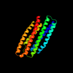
PDB 1xwm chain A

3D model

Region: 14 - 222
Aligned: 209
Modelled: 209
Confidence: 100.0%
Identity: 30%
Fold: Spectrin repeat-like
Superfamily: PhoU-like
Family: PhoU-like

Phyre2

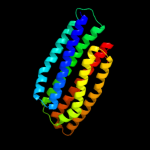
PDB 1t72 chain A

3D model

Region: 11 - 220
Aligned: 210
Modelled: 209
Confidence: 100.0%
Identity: 31%
Fold: Spectrin repeat-like
Superfamily: PhoU-like
Family: PhoU-like

Phyre2

PDB 2i0m chain A

3D model

Region: 13 - 223
Aligned: 206
Modelled: 211
Confidence: 100.0%
Identity: 31%
PDB header:structural genomics, unknown function
Chain: A: PDB Molecule:phosphate transport system protein phou;
PDBTitle: crystal structure of the phosphate transport system regulatory protein2 phou from streptococcus pneumoniae

Phyre2

PDB 3l39 chain A

3D model

Region: 23 - 217
Aligned: 188
Modelled: 195
Confidence: 99.9%
Identity: 8%
PDB header:phosphate-binding protein
Chain: A: PDB Molecule:putative phou-like phosphate regulatory protein;
PDBTitle: crystal structure of putative phou-like phosphate regulatory2 protein (bt4638) from bacteroides thetaiotaomicron vpi-54823 at 1.93 a resolution

Phyre2

PDB 2olt chain B

3D model

Region: 23 - 217
Aligned: 186
Modelled: 186
Confidence: 99.8%
Identity: 13%
PDB header:unknown function
Chain: B: PDB Molecule:hypothetical protein;
PDBTitle: crystal structure of a phou-like protein (so_3770) from shewanella2 oneidensis mr-1 at 2.00 a resolution

Phyre2

PDB 1vct chain A domain 1

3D model

Region: 121 - 214
Aligned: 93
Modelled: 94
Confidence: 99.7%
Identity: 12%
Fold: Spectrin repeat-like
Superfamily: PhoU-like
Family: PhoU-like

Phyre2

PDB 2bkn chain A

3D model

Region: 21 - 121
Aligned: 97
Modelled: 101
Confidence: 99.3%
Identity: 15%
PDB header:membrane protein
Chain: A: PDB Molecule:hypothetical protein ph0236;
PDBTitle: structure analysis of unknown function protein

Phyre2

PDB 1col chain A

3D model

Region: 200 - 240
Aligned: 39
Modelled: 41
Confidence: 51.2%
Identity: 26%
Fold: Toxins' membrane translocation domains
Superfamily: Colicin
Family: Colicin

Phyre2

PDB 1rh1 chain A domain 2

3D model

Region: 200 - 240
Aligned: 39
Modelled: 41
Confidence: 36.0%
Identity: 18%
Fold: Toxins' membrane translocation domains
Superfamily: Colicin
Family: Colicin

Phyre2

PDB 2qrx chain A

3D model

Region: 6 - 169
Aligned: 164
Modelled: 164
Confidence: 27.9%
Identity: 9%
PDB header:dna binding protein
Chain: A: PDB Molecule:gm27569p;
PDBTitle: crystal structure of drosophila melanogaster translin2 protein

Phyre2

PDB 1zke chain A domain 1

3D model

Region: 3 - 31
Aligned: 29
Modelled: 29
Confidence: 26.6%
Identity: 14%
Fold: ROP-like
Superfamily: HP1531-like
Family: HP1531-like

Phyre2

PDB 3qd8 chain M

3D model

Region: 62 - 213
Aligned: 143
Modelled: 152
Confidence: 25.9%
Identity: 17%
PDB header:metal binding protein
Chain: M: PDB Molecule:probable bacterioferritin bfrb;
PDBTitle: crystal structure of mycobacterium tuberculosis bfrb

Phyre2

PDB 1pq1 chain B

3D model

Region: 93 - 104
Aligned: 12
Modelled: 12
Confidence: 22.0%
Identity: 50%
PDB header:apoptosis
Chain: B: PDB Molecule:bcl2-like protein 11;
PDBTitle: crystal structure of bcl-xl/bim

Phyre2

PDB 1eq1 chain A

3D model

Region: 10 - 177
Aligned: 150
Modelled: 154
Confidence: 19.3%
Identity: 7%
Fold: Apolipophorin-III
Superfamily: Apolipophorin-III
Family: Apolipophorin-III

Phyre2

PDB 3izc chain W

3D model

Region: 66 - 109
Aligned: 44
Modelled: 44
Confidence: 17.6%
Identity: 14%
PDB header:ribosome
Chain: W: PDB Molecule:60s ribosomal protein rpl22 (l22e);
PDBTitle: localization of the large subunit ribosomal proteins into a 6.1 a2 cryo-em map of saccharomyces cerevisiae translating 80s ribosome

Phyre2

PDB 1jqs chain B

3D model

Region: 226 - 238
Aligned: 13
Modelled: 13
Confidence: 16.9%
Identity: 38%
PDB header:ribosome
Chain: B: PDB Molecule:elongation factor g;
PDBTitle: fitting of l11 protein and elongation factor g (domain g'2 and v) in the cryo-em map of e. coli 70s ribosome bound3 with ef-g and gmppcp, a nonhydrolysable gtp analog

Phyre2

PDB 3ci9 chain B

3D model

Region: 128 - 171
Aligned: 44
Modelled: 44
Confidence: 16.4%
Identity: 18%
PDB header:transcription
Chain: B: PDB Molecule:heat shock factor-binding protein 1;
PDBTitle: crystal structure of the human hsbp1

Phyre2

PDB 1set chain A domain 1

3D model

Region: 11 - 82
Aligned: 72
Modelled: 72
Confidence: 15.9%
Identity: 10%
Fold: Long alpha-hairpin
Superfamily: tRNA-binding arm
Family: Seryl-tRNA synthetase (SerRS)

Phyre2

PDB 2k7w chain B

3D model

Region: 95 - 104
Aligned: 10
Modelled: 10
Confidence: 13.6%
Identity: 60%
PDB header:apoptosis
Chain: B: PDB Molecule:bcl-2-like protein 11;
PDBTitle: bax activation is initiated at a novel interaction site

Phyre2
1

d1sumb_
2

d1xwma_
3

d1t72a_
4

c2i0mA_
5

c3l39A_
6

c2oltB_
7

d1vcta1
8

c2bknA_
9

d1cola_
10

d1rh1a2
11

c2qrxA_
12

d1zkea1
13

c3qd8M_
14

c1pq1B_
15

d1eq1a_
16

c3izcw_
17

c1jqsB_
18

c3ci9B_
19

d1seta1
20

c2k7wB_
21


22


23


24


25


26


27


28


29


30


31


32


33


34


35


36


37


38


39


40


41


42


43


44


45


46


47


48


49


50


51


Detailed template information 

#
Template Alignment Coverage3D Model Confidence
% i.d. Template Information
1d1sumb_


100.0 26 Fold:Spectrin repeat-like
Superfamily:PhoU-like
Family:PhoU-like
2d1xwma_


100.0 30 Fold:Spectrin repeat-like
Superfamily:PhoU-like
Family:PhoU-like
3d1t72a_


100.0 31 Fold:Spectrin repeat-like
Superfamily:PhoU-like
Family:PhoU-like
4c2i0mA_


100.0 31 PDB header:structural genomics, unknown function
Chain: A: PDB Molecule:phosphate transport system protein phou;
PDBTitle: crystal structure of the phosphate transport system regulatory protein2 phou from streptococcus pneumoniae
5c3l39A_


99.9 8 PDB header:phosphate-binding protein
Chain: A: PDB Molecule:putative phou-like phosphate regulatory protein;
PDBTitle: crystal structure of putative phou-like phosphate regulatory2 protein (bt4638) from bacteroides thetaiotaomicron vpi-54823 at 1.93 a resolution
6c2oltB_


99.8 13 PDB header:unknown function
Chain: B: PDB Molecule:hypothetical protein;
PDBTitle: crystal structure of a phou-like protein (so_3770) from shewanella2 oneidensis mr-1 at 2.00 a resolution
7d1vcta1


99.7 12 Fold:Spectrin repeat-like
Superfamily:PhoU-like
Family:PhoU-like
8c2bknA_


99.3 15 PDB header:membrane protein
Chain: A: PDB Molecule:hypothetical protein ph0236;
PDBTitle: structure analysis of unknown function protein
9d1cola_


51.2 26 Fold:Toxins' membrane translocation domains
Superfamily:Colicin
Family:Colicin
10d1rh1a2


36.0 18 Fold:Toxins' membrane translocation domains
Superfamily:Colicin
Family:Colicin
11c2qrxA_


27.9 9 PDB header:dna binding protein
Chain: A: PDB Molecule:gm27569p;
PDBTitle: crystal structure of drosophila melanogaster translin2 protein
12d1zkea1


26.6 14 Fold:ROP-like
Superfamily:HP1531-like
Family:HP1531-like
13c3qd8M_


25.9 17 PDB header:metal binding protein
Chain: M: PDB Molecule:probable bacterioferritin bfrb;
PDBTitle: crystal structure of mycobacterium tuberculosis bfrb
14c1pq1B_


22.0 50 PDB header:apoptosis
Chain: B: PDB Molecule:bcl2-like protein 11;
PDBTitle: crystal structure of bcl-xl/bim
15d1eq1a_


19.3 7 Fold:Apolipophorin-III
Superfamily:Apolipophorin-III
Family:Apolipophorin-III
16c3izcw_


17.6 14 PDB header:ribosome
Chain: W: PDB Molecule:60s ribosomal protein rpl22 (l22e);
PDBTitle: localization of the large subunit ribosomal proteins into a 6.1 a2 cryo-em map of saccharomyces cerevisiae translating 80s ribosome
17c1jqsB_


16.9 38 PDB header:ribosome
Chain: B: PDB Molecule:elongation factor g;
PDBTitle: fitting of l11 protein and elongation factor g (domain g'2 and v) in the cryo-em map of e. coli 70s ribosome bound3 with ef-g and gmppcp, a nonhydrolysable gtp analog
18c3ci9B_


16.4 18 PDB header:transcription
Chain: B: PDB Molecule:heat shock factor-binding protein 1;
PDBTitle: crystal structure of the human hsbp1
19d1seta1


15.9 10 Fold:Long alpha-hairpin
Superfamily:tRNA-binding arm
Family:Seryl-tRNA synthetase (SerRS)
20c2k7wB_


13.6 60 PDB header:apoptosis
Chain: B: PDB Molecule:bcl-2-like protein 11;
PDBTitle: bax activation is initiated at a novel interaction site
21d1krqa_


not modelled 13.6 12 Fold:Ferritin-like
Superfamily:Ferritin-like
Family:Ferritin
22c2nl9B_


not modelled 11.7 50 PDB header:apoptosis
Chain: B: PDB Molecule:bcl-2-like protein 11;
PDBTitle: crystal structure of the mcl-1:bim bh3 complex
23c3kj2B_


not modelled 11.3 50 PDB header:apoptosis
Chain: B: PDB Molecule:bcl-2-like protein 11;
PDBTitle: mcl-1 in complex with bim bh3 mutant f4ae
24c1kmiZ_


not modelled 11.2 7 PDB header:signaling protein
Chain: Z: PDB Molecule:chemotaxis protein chez;
PDBTitle: crystal structure of an e.coli chemotaxis protein, chez
25c1rimA_


not modelled 10.9 46 PDB header:de novo protein
Chain: A: PDB Molecule:e6apc2 peptide;
PDBTitle: e6-binding zinc finger (e6apc2)
26c2kdtA_


not modelled 10.8 15 PDB header:protein transport
Chain: A: PDB Molecule:neuroendocrine convertase 1;
PDBTitle: pc1/3 dcsg sorting domain structure in dpc
27c3d1dC_


not modelled 9.3 17 PDB header:nuclear protein
Chain: C: PDB Molecule:rna-induced transcriptional silencing complex
PDBTitle: hexagonal crystal structure of tas3 c-terminal alpha motif
28d1vlga_


not modelled 9.0 9 Fold:Ferritin-like
Superfamily:Ferritin-like
Family:Ferritin
29c3c98B_


not modelled 8.8 14 PDB header:endocytosis/exocytosis
Chain: B: PDB Molecule:syntaxin-1a;
PDBTitle: revised structure of the munc18a-syntaxin1 complex
30d1jgca_


not modelled 8.8 17 Fold:Ferritin-like
Superfamily:Ferritin-like
Family:Ferritin
31c3kz0C_


not modelled 7.9 42 PDB header:apoptosis
Chain: C: PDB Molecule:mcl-1 specific peptide mb7;
PDBTitle: mcl-1 complex with mcl-1-specific selected peptide
32c3kz0D_


not modelled 7.9 42 PDB header:apoptosis
Chain: D: PDB Molecule:mcl-1 specific peptide mb7;
PDBTitle: mcl-1 complex with mcl-1-specific selected peptide
33c3d7vB_


not modelled 7.8 43 PDB header:apoptosis
Chain: B: PDB Molecule:bcl-2-like protein 11;
PDBTitle: crystal structure of mcl-1 in complex with an mcl-12 selective bh3 ligand
34c3iz5w_


not modelled 6.1 14 PDB header:ribosome
Chain: W: PDB Molecule:60s ribosomal protein l22 (l22e);
PDBTitle: localization of the large subunit ribosomal proteins into a 5.5 a2 cryo-em map of triticum aestivum translating 80s ribosome
35d2dmda1


not modelled 6.0 43 Fold:beta-beta-alpha zinc fingers
Superfamily:beta-beta-alpha zinc fingers
Family:Classic zinc finger, C2H2
36d1a87a_


not modelled 6.0 20 Fold:Toxins' membrane translocation domains
Superfamily:Colicin
Family:Colicin
37c1a87A_


not modelled 6.0 20 PDB header:bacteriocin
Chain: A: PDB Molecule:colicin n;
PDBTitle: colicin n
38d1j0ta_


not modelled 6.0 27 Fold:Crustacean CHH/MIH/GIH neurohormone
Superfamily:Crustacean CHH/MIH/GIH neurohormone
Family:Crustacean CHH/MIH/GIH neurohormone
39d1uptb_


not modelled 6.0 14 Fold:GRIP domain
Superfamily:GRIP domain
Family:GRIP domain
40c3cd0B_


not modelled 5.7 32 PDB header:oxidoreductase
Chain: B: PDB Molecule:3-hydroxy-3-methylglutaryl-coenzyme a reductase;
PDBTitle: thermodynamic and structure guided design of statin hmg-coa2 reductase inhibitors
41c1vh6A_


not modelled 5.5 8 PDB header:structural genomics, unknown function
Chain: A: PDB Molecule:flagellar protein flis;
PDBTitle: crystal structure of a flagellar protein
42d1vh6a_


not modelled 5.5 8 Fold:Four-helical up-and-down bundle
Superfamily:Flagellar export chaperone FliS
Family:Flagellar export chaperone FliS
43c3znfA_


not modelled 5.4 44 PDB header:zinc finger dna binding domain
Chain: A: PDB Molecule:zinc finger;
PDBTitle: high-resolution three-dimensional structure of a single2 zinc finger from a human enhancer binding protein in3 solution
44d4znfa_


not modelled 5.4 44 Fold:beta-beta-alpha zinc fingers
Superfamily:beta-beta-alpha zinc fingers
Family:Classic zinc finger, C2H2
45c4znfA_


not modelled 5.4 44 PDB header:zinc finger dna binding domain
Chain: A: PDB Molecule:zinc finger;
PDBTitle: high-resolution three-dimensional structure of a single2 zinc finger from a human enhancer binding protein in3 solution
46d256ba_


not modelled 5.3 14 Fold:Four-helical up-and-down bundle
Superfamily:Cytochromes
Family:Cytochrome b562
47d1t6ua_


not modelled 5.3 11 Fold:Four-helical up-and-down bundle
Superfamily:Nickel-containing superoxide dismutase, NiSOD
Family:Nickel-containing superoxide dismutase, NiSOD
48c1hwjB_


not modelled 5.2 32 PDB header:oxidoreductase
Chain: B: PDB Molecule:hmg-coa reductase;
PDBTitle: complex of the catalytic portion of human hmg-coa reductase2 with cerivastatin
49d1hw8c2


not modelled 5.2 36 Fold:Substrate-binding domain of HMG-CoA reductase
Superfamily:Substrate-binding domain of HMG-CoA reductase
Family:Substrate-binding domain of HMG-CoA reductase
50c3qauA_


not modelled 5.1 27 PDB header:oxidoreductase
Chain: A: PDB Molecule:3-hydroxy-3-methylglutaryl-coenzyme a reductase;
PDBTitle: 3-hydroxy-3-methylglutaryl-coenzyme a reductase from streptococcus2 pneumoniae
51d1o9da_


not modelled 5.1 15 Fold:alpha-alpha superhelix
Superfamily:14-3-3 protein
Family:14-3-3 protein

Binding site prediction 

Due to computational demand, binding site predictions are not run for batch jobs

If you want to predict binding sites, please manually submit your model of choice to 3DLigandSite


Phyre is for academic use only

Please cite: Protein structure prediction on the web: a case study using the Phyre server
Kelley LA and Sternberg MJE. Nature Protocols 4, 363 - 371 (2009) [pdf] [Import into BibTeX]
 
If you use the binding site predictions from 3DLigandSite, please also cite:
3DLigandSite: predicting ligand-binding sites using similar structures.
Wass MN, Kelley LA and Sternberg MJ Nucleic Acids Research 38, W469-73 (2010) [PubMed]
 
© Structural Bioinformatics Group
Imperial College London
Lawrence Kelley, Benjamin Jefferys
Disclaimer
Terms and Conditions
Component software
Template detection: HHpred 1.51
Secondary structure prediction: Psi-pred 2.5
Disorder prediction: Disopred 2.4
Transmembrane prediction: Memsat_SVM
Multi-template modelling and ab initio: Poing 1.0