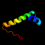
1 d2bsqe1
63.6
9
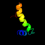
Fold: Ribbon-helix-helixSuperfamily: Ribbon-helix-helixFamily: Trafficking protein A-like2 d1mula_
46.0
21
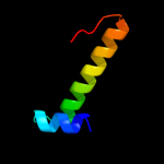
Fold: IHF-like DNA-binding proteinsSuperfamily: IHF-like DNA-binding proteinsFamily: Prokaryotic DNA-bending protein3 d1huua_
36.5
16
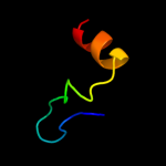
Fold: IHF-like DNA-binding proteinsSuperfamily: IHF-like DNA-binding proteinsFamily: Prokaryotic DNA-bending protein4 c3c4iA_
34.9
12
PDB header: dna binding proteinChain: A: PDB Molecule: dna-binding protein hu homolog;PDBTitle: crystal structure analysis of n terminal region containing the2 dimerization domain and dna binding domain of hu protein(histone like3 protein-dna binding) from mycobacterium tuberculosis [h37rv]
5 d1p71a_
34.3
19
Fold: IHF-like DNA-binding proteinsSuperfamily: IHF-like DNA-binding proteinsFamily: Prokaryotic DNA-bending protein6 d1owfa_
33.9
14
Fold: IHF-like DNA-binding proteinsSuperfamily: IHF-like DNA-binding proteinsFamily: Prokaryotic DNA-bending protein7 c2iifA_
33.2
16
PDB header: recombination/dnaChain: A: PDB Molecule: integration host factor;PDBTitle: single chain integration host factor mutant protein (scihf2-2 k45ae) in complex with dna
8 d1exea_
28.9
9
Fold: IHF-like DNA-binding proteinsSuperfamily: IHF-like DNA-binding proteinsFamily: Prokaryotic DNA-bending protein9 d1b8za_
28.3
18
Fold: IHF-like DNA-binding proteinsSuperfamily: IHF-like DNA-binding proteinsFamily: Prokaryotic DNA-bending protein10 c2k42A_
19.4
29
PDB header: signaling proteinChain: A: PDB Molecule: wiskott-aldrich syndrome protein;PDBTitle: solution structure of the gtpase binding domain of wasp in2 complex with espfu, an ehec effector
11 c2kl8A_
19.0
19
PDB header: de novo proteinChain: A: PDB Molecule: or15;PDBTitle: solution nmr structure of de novo designed ferredoxin-like2 fold protein, northeast structural genomics consortium3 target or15
12 c3gr5A_
18.5
7
PDB header: membrane proteinChain: A: PDB Molecule: escc;PDBTitle: periplasmic domain of the outer membrane secretin escc from2 enteropathogenic e.coli (epec)
13 c2y9kG_
18.2
18
PDB header: protein transportChain: G: PDB Molecule: protein invg;PDBTitle: three-dimensional model of salmonella's needle complex at2 subnanometer resolution
14 d1owfb_
17.5
9
Fold: IHF-like DNA-binding proteinsSuperfamily: IHF-like DNA-binding proteinsFamily: Prokaryotic DNA-bending protein15 d1mnta_
16.4
24
Fold: Ribbon-helix-helixSuperfamily: Ribbon-helix-helixFamily: Arc/Mnt-like phage repressors16 c1f3mB_
16.2
30
PDB header: transferaseChain: B: PDB Molecule: serine/threonine-protein kinase pak-alpha;PDBTitle: crystal structure of human serine/threonine kinase pak1
17 c2xflB_
16.1
26
PDB header: hydrolaseChain: B: PDB Molecule: dyne7;PDBTitle: induced-fit and allosteric effects upon polyene binding2 revealed by crystal structures of the dynemicin3 thioesterase
18 c1ceeB_
15.0
29
PDB header: structural protein regulationChain: B: PDB Molecule: wiskott-aldrich syndrome protein wasp;PDBTitle: solution structure of cdc42 in complex with the gtpase2 binding domain of wasp
19 c2y3mA_
14.6
17
PDB header: transport proteinChain: A: PDB Molecule: protein transport protein hofq;PDBTitle: structure of the extra-membranous domain of the secretin2 hofq from actinobacillus actinomycetemcomitans
20 d1ej5a_
14.5
32
Fold: Wiscott-Aldrich syndrome protein, WASP, C-terminal domainSuperfamily: Wiscott-Aldrich syndrome protein, WASP, C-terminal domainFamily: Wiscott-Aldrich syndrome protein, WASP, C-terminal domain21 c3fwcO_
not modelled
14.3
7
PDB header: cell cycle, transcriptionChain: O: PDB Molecule: protein sus1;PDBTitle: sac3:sus1:cdc31 complex
22 d1ucra_
not modelled
13.2
26
Fold: DNA/RNA-binding 3-helical bundleSuperfamily: "Winged helix" DNA-binding domainFamily: Dissimilatory sulfite reductase DsvD23 c1u9pA_
not modelled
12.3
12
PDB header: unknown functionChain: A: PDB Molecule: parc;PDBTitle: permuted single-chain arc
24 d2o97b1
not modelled
11.1
25
Fold: IHF-like DNA-binding proteinsSuperfamily: IHF-like DNA-binding proteinsFamily: Prokaryotic DNA-bending protein25 c3eudE_
not modelled
9.5
23
PDB header: nuclear proteinChain: E: PDB Molecule: protein shq1;PDBTitle: structure of the cs domain of the essential h/aca rnp2 assembly protein shq1p
26 d1d5ya1
not modelled
8.4
7
Fold: DNA/RNA-binding 3-helical bundleSuperfamily: Homeodomain-likeFamily: AraC type transcriptional activator27 c2ptvA_
not modelled
8.1
12
PDB header: immune systemChain: A: PDB Molecule: cd48 antigen;PDBTitle: structure of nk cell receptor ligand cd48
28 c3ossD_
not modelled
7.8
7
PDB header: protein transportChain: D: PDB Molecule: type 2 secretion system, secretin gspd;PDBTitle: the crystal structure of enterotoxigenic escherichia coli gspc-gspd2 complex from the type ii secretion system
29 d2o4ta1
not modelled
7.8
14
Fold: Left-handed superhelixSuperfamily: BH3980-likeFamily: BH3980-like30 d1d6za4
not modelled
7.4
20
Fold: N domain of copper amine oxidase-likeSuperfamily: Copper amine oxidase, domain NFamily: Copper amine oxidase, domain N31 d1d8ja_
not modelled
6.6
19
Fold: DNA/RNA-binding 3-helical bundleSuperfamily: "Winged helix" DNA-binding domainFamily: The central core domain of TFIIE beta32 d2qi2a1
not modelled
6.6
31
Fold: Sm-like foldSuperfamily: Dom34/Pelota N-terminal domain-likeFamily: Dom34/Pelota N-terminal domain-like33 d2b0ga1
not modelled
6.6
13
Fold: Ferredoxin-likeSuperfamily: RNA-binding domain, RBDFamily: Canonical RBD34 c3kf8D_
not modelled
6.4
25
PDB header: structural proteinChain: D: PDB Molecule: protein ten1;PDBTitle: crystal structure of c. tropicalis stn1-ten1 complex
35 c2edoA_
not modelled
6.0
26
PDB header: cell adhesionChain: A: PDB Molecule: cd48 antigen;PDBTitle: solution structure of the first ig-like domain from human2 cd48 antigen
36 c1e0aB_
not modelled
5.8
24
PDB header: signalling proteinChain: B: PDB Molecule: serine/threonine-protein kinase pak-alpha;PDBTitle: cdc42 complexed with the gtpase binding domain of p212 activated kinase
37 c2np2B_
not modelled
5.6
21
PDB header: dna binding protein/dnaChain: B: PDB Molecule: hbb;PDBTitle: hbb-dna complex
38 d1rxta1
not modelled
5.6
31
Fold: Acyl-CoA N-acyltransferases (Nat)Superfamily: Acyl-CoA N-acyltransferases (Nat)Family: N-myristoyl transferase, NMT39 c3aqjQ_
not modelled
5.6
38
PDB header: metal binding proteinChain: Q: PDB Molecule: baseplate assembly protein v;PDBTitle: crystal structure of a c-terminal domain of the bacteriophage p2 tail2 spike protein, gpv
40 d1qa9b_
not modelled
5.3
26
Fold: Immunoglobulin-like beta-sandwichSuperfamily: ImmunoglobulinFamily: V set domains (antibody variable domain-like)41 c2o37A_
not modelled
5.1
18
PDB header: chaperoneChain: A: PDB Molecule: protein sis1;PDBTitle: j-domain of sis1 protein, hsp40 co-chaperone from2 saccharomyces cerevisiae.