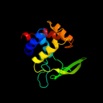
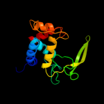
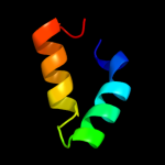
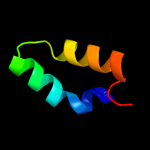
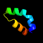
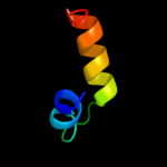
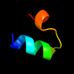
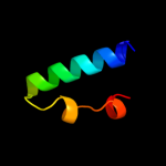
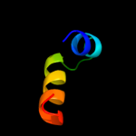
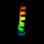
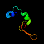
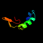
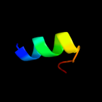
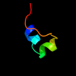
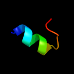
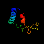
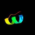
1 c2zycA_
100.0
45
PDB header: hydrolaseChain: A: PDB Molecule: peptidoglycan hydrolase flgj;PDBTitle: crystal structure of peptidoglycan hydrolase from2 sphingomonas sp. a1
2 c3fi7A_
100.0
32
PDB header: hydrolaseChain: A: PDB Molecule: lmo1076 protein;PDBTitle: crystal structure of the autolysin auto (lmo1076) from listeria2 monocytogenes, catalytic domain
3 d1qusa_
96.9
28
Fold: Lysozyme-likeSuperfamily: Lysozyme-likeFamily: Bacterial muramidase, catalytic domain4 c3mgwA_
95.2
18
PDB header: hydrolaseChain: A: PDB Molecule: lysozyme g;PDBTitle: thermodynamics and structure of a salmon cold-active goose-type2 lysozyme
5 c3gxkB_
95.1
18
PDB header: hydrolaseChain: B: PDB Molecule: goose-type lysozyme 1;PDBTitle: the crystal structure of g-type lysozyme from atlantic cod2 (gadus morhua l.) in complex with nag oligomers sheds new3 light on substrate binding and the catalytic mechanism.4 native structure to 1.9
6 d1gbsa_
95.1
14
Fold: Lysozyme-likeSuperfamily: Lysozyme-likeFamily: G-type lysozyme7 c2y8pA_
95.0
33
PDB header: lyaseChain: A: PDB Molecule: endo-type membrane-bound lytic murein transglycosylase a;PDBTitle: crystal structure of an outer membrane-anchored endolytic2 peptidoglycan lytic transglycosylase (mlte) from3 escherichia coli
8 d1qsaa2
94.8
21
Fold: Lysozyme-likeSuperfamily: Lysozyme-likeFamily: Bacterial muramidase, catalytic domain9 c3bkhA_
89.8
20
PDB header: hydrolaseChain: A: PDB Molecule: lytic transglycosylase;PDBTitle: crystal structure of the bacteriophage phikz lytic2 transglycosylase, gp144
10 d1nvma1
60.7
21
Fold: RuvA C-terminal domain-likeSuperfamily: post-HMGL domain-likeFamily: DmpG/LeuA communication domain-like11 d1yt3a2
36.7
6
Fold: SAM domain-likeSuperfamily: HRDC-likeFamily: RNase D C-terminal domains12 c1slyA_
27.3
21
PDB header: glycosyltransferaseChain: A: PDB Molecule: 70-kda soluble lytic transglycosylase;PDBTitle: complex of the 70-kda soluble lytic transglycosylase with2 bulgecin a
13 c2k0nA_
27.1
31
PDB header: transcriptionChain: A: PDB Molecule: mediator of rna polymerase ii transcriptionPDBTitle: solution structure of yeast gal11p kix domain
14 c2fbdB_
25.9
25
PDB header: hydrolaseChain: B: PDB Molecule: lysozyme 1;PDBTitle: the crystallographic structure of the digestive lysozyme 1 from musca2 domestica at 1.90 ang.
15 d1yroa1
20.5
18
Fold: Lysozyme-likeSuperfamily: Lysozyme-likeFamily: C-type lysozyme16 c2eq7C_
18.9
27
PDB header: oxidoreductaseChain: C: PDB Molecule: 2-oxoglutarate dehydrogenase e2 component;PDBTitle: crystal structure of lipoamide dehydrogenase from thermus thermophilus2 hb8 with psbdo
17 c3l87A_
18.8
29
PDB header: hydrolaseChain: A: PDB Molecule: peptide deformylase;PDBTitle: the crystal structure of smu.143c from streptococcus mutans ua159
18 c2eq9C_
18.3
20
PDB header: oxidoreductaseChain: C: PDB Molecule: pyruvate dehydrogenase complex, dihydrolipoamidePDBTitle: crystal structure of lipoamide dehydrogenase from thermus thermophilus2 hb8 with psbdb
19 d1t3ca_
14.6
13
Fold: Zincin-likeSuperfamily: Metalloproteases ("zincins"), catalytic domainFamily: Clostridium neurotoxins, catalytic domain20 c2eq8C_
14.4
13
PDB header: oxidoreductaseChain: C: PDB Molecule: pyruvate dehydrogenase complex, dihydrolipoamidePDBTitle: crystal structure of lipoamide dehydrogenase from thermus thermophilus2 hb8 with psbdp
21 d1bw6a_
not modelled
13.7
8
Fold: DNA/RNA-binding 3-helical bundleSuperfamily: Homeodomain-likeFamily: Centromere-binding22 d1h3ob_
not modelled
13.2
17
Fold: Histone-foldSuperfamily: Histone-foldFamily: TBP-associated factors, TAFs23 c2cooA_
not modelled
12.4
0
PDB header: transferaseChain: A: PDB Molecule: lipoamide acyltransferase component of branched-PDBTitle: solution structure of the e3_binding domain of2 dihydrolipoamide branched chaintransacylase
24 c3dv0I_
not modelled
12.4
13
PDB header: oxidoreductase/transferaseChain: I: PDB Molecule: dihydrolipoyllysine-residue acetyltransferasePDBTitle: snapshots of catalysis in the e1 subunit of the pyruvate2 dehydrogenase multi-enzyme complex
25 d1w85i_
not modelled
12.4
13
Fold: Peripheral subunit-binding domain of 2-oxo acid dehydrogenase complexSuperfamily: Peripheral subunit-binding domain of 2-oxo acid dehydrogenase complexFamily: Peripheral subunit-binding domain of 2-oxo acid dehydrogenase complex26 d1w4ha1
not modelled
12.2
0
Fold: Peripheral subunit-binding domain of 2-oxo acid dehydrogenase complexSuperfamily: Peripheral subunit-binding domain of 2-oxo acid dehydrogenase complexFamily: Peripheral subunit-binding domain of 2-oxo acid dehydrogenase complex27 d1gd6a_
not modelled
12.1
20
Fold: Lysozyme-likeSuperfamily: Lysozyme-likeFamily: C-type lysozyme28 d1mp1a_
not modelled
12.0
20
Fold: PWI domainSuperfamily: PWI domainFamily: PWI domain29 d2p02a2
not modelled
11.6
38
Fold: S-adenosylmethionine synthetaseSuperfamily: S-adenosylmethionine synthetaseFamily: S-adenosylmethionine synthetase30 c3sn6R_
not modelled
11.3
11
PDB header: signaling protein/hydrolaseChain: R: PDB Molecule: lysozyme, beta-2 adrenergic receptor;PDBTitle: crystal structure of the beta2 adrenergic receptor-gs protein complex
31 d2cyua1
not modelled
11.2
0
Fold: Peripheral subunit-binding domain of 2-oxo acid dehydrogenase complexSuperfamily: Peripheral subunit-binding domain of 2-oxo acid dehydrogenase complexFamily: Peripheral subunit-binding domain of 2-oxo acid dehydrogenase complex32 c1ewrA_
not modelled
10.7
21
PDB header: hydrolaseChain: A: PDB Molecule: dna mismatch repair protein muts;PDBTitle: crystal structure of taq muts
33 d1p35a_
not modelled
10.7
67
Fold: Baculovirus p35 proteinSuperfamily: Baculovirus p35 proteinFamily: Baculovirus p35 protein34 d1qm4a2
not modelled
9.7
40
Fold: S-adenosylmethionine synthetaseSuperfamily: S-adenosylmethionine synthetaseFamily: S-adenosylmethionine synthetase35 c1w3dA_
not modelled
9.6
13
PDB header: transferaseChain: A: PDB Molecule: dihydrolipoyllysine-residue acetyltransferasePDBTitle: nmr structure of the peripheral-subunit binding domain of2 bacillus stearothermophilus e2p
36 d1alca_
not modelled
9.5
21
Fold: Lysozyme-likeSuperfamily: Lysozyme-likeFamily: C-type lysozyme37 d1iiza_
not modelled
9.0
23
Fold: Lysozyme-likeSuperfamily: Lysozyme-likeFamily: C-type lysozyme38 d1lm4a_
not modelled
8.9
29
Fold: Peptide deformylaseSuperfamily: Peptide deformylaseFamily: Peptide deformylase39 d1lm6a_
not modelled
8.9
29
Fold: Peptide deformylaseSuperfamily: Peptide deformylaseFamily: Peptide deformylase40 d2bgxa2
not modelled
8.7
12
Fold: N-acetylmuramoyl-L-alanine amidase-likeSuperfamily: N-acetylmuramoyl-L-alanine amidase-likeFamily: N-acetylmuramoyl-L-alanine amidase-like41 d1wuda1
not modelled
8.7
22
Fold: SAM domain-likeSuperfamily: HRDC-likeFamily: HRDC domain from helicases42 c2lmdA_
not modelled
8.6
14
PDB header: transcriptionChain: A: PDB Molecule: prospero homeobox protein 1;PDBTitle: minimal constraints solution nmr structure of prospero homeobox2 protein 1 from homo sapiens, northeast structural genomics consortium3 target hr4660b
43 d1yzma1
not modelled
8.3
16
Fold: Long alpha-hairpinSuperfamily: Rabenosyn-5 Rab-binding domain-likeFamily: Rabenosyn-5 Rab-binding domain-like44 d1f6sa_
not modelled
8.3
24
Fold: Lysozyme-likeSuperfamily: Lysozyme-likeFamily: C-type lysozyme45 c1w4kA_
not modelled
8.3
13
PDB header: transferaseChain: A: PDB Molecule: pyruvate dehydrogenase e2;PDBTitle: peripheral-subunit binding domains from mesophilic,2 thermophilic, and hyperthermophilic bacteria fold by3 ultrafast, apparently two-state transitions
46 d1w7pd2
not modelled
8.2
13
Fold: DNA/RNA-binding 3-helical bundleSuperfamily: "Winged helix" DNA-binding domainFamily: Vacuolar sorting protein domain47 d1mxaa2
not modelled
8.1
38
Fold: S-adenosylmethionine synthetaseSuperfamily: S-adenosylmethionine synthetaseFamily: S-adenosylmethionine synthetase48 d1u5tb1
not modelled
8.1
13
Fold: DNA/RNA-binding 3-helical bundleSuperfamily: "Winged helix" DNA-binding domainFamily: Vacuolar sorting protein domain49 c1zwvA_
not modelled
7.9
0
PDB header: transferaseChain: A: PDB Molecule: lipoamide acyltransferase component of branched-PDBTitle: solution structure of the subunit binding domain (hbsbd) of2 the human mitochondrial branched-chain alpha-ketoacid3 dehydrogenase
50 d1b9oa_
not modelled
7.7
19
Fold: Lysozyme-likeSuperfamily: Lysozyme-likeFamily: C-type lysozyme51 c1u1iC_
not modelled
7.2
33
PDB header: isomeraseChain: C: PDB Molecule: myo-inositol-1-phosphate synthase;PDBTitle: myo-inositol phosphate synthase mips from a. fulgidus
52 c2bh7A_
not modelled
7.2
15
PDB header: hydrolaseChain: A: PDB Molecule: n-acetylmuramoyl-l-alanine amidase;PDBTitle: crystal structure of a semet derivative of amid at 2.22 angstroms
53 d1z0kb1
not modelled
7.0
18
Fold: Long alpha-hairpinSuperfamily: Rabenosyn-5 Rab-binding domain-likeFamily: Rabenosyn-5 Rab-binding domain-like54 d1fkqa_
not modelled
6.8
28
Fold: Lysozyme-likeSuperfamily: Lysozyme-likeFamily: C-type lysozyme55 c1wx4B_
not modelled
6.1
18
PDB header: oxidoreductase/metal transportChain: B: PDB Molecule: melc;PDBTitle: crystal structure of the oxy-form of the copper-bound streptomyces2 castaneoglobisporus tyrosinase complexed with a caddie protein3 prepared by the addition of dithiothreitol
56 d1pjqa3
not modelled
6.1
14
Fold: Siroheme synthase middle domains-likeSuperfamily: Siroheme synthase middle domains-likeFamily: Siroheme synthase middle domains-like57 d2csba2
not modelled
5.7
17
Fold: SAM domain-likeSuperfamily: RuvA domain 2-likeFamily: Topoisomerase V repeat domain58 c2l69A_
not modelled
5.7
20
PDB header: de novo proteinChain: A: PDB Molecule: rossmann 2x3 fold protein;PDBTitle: solution nmr structure of de novo designed protein, p-loop ntpase2 fold, northeast structural genomics consortium target or28
59 d2coba1
not modelled
5.6
29
Fold: DNA/RNA-binding 3-helical bundleSuperfamily: Homeodomain-likeFamily: Psq domain60 d1bala_
not modelled
5.4
0
Fold: Peripheral subunit-binding domain of 2-oxo acid dehydrogenase complexSuperfamily: Peripheral subunit-binding domain of 2-oxo acid dehydrogenase complexFamily: Peripheral subunit-binding domain of 2-oxo acid dehydrogenase complex61 d1tuka1
not modelled
5.3
42
Fold: Bifunctional inhibitor/lipid-transfer protein/seed storage 2S albuminSuperfamily: Bifunctional inhibitor/lipid-transfer protein/seed storage 2S albuminFamily: Plant lipid-transfer and hydrophobic proteins62 d2jn6a1
not modelled
5.3
13
Fold: DNA/RNA-binding 3-helical bundleSuperfamily: Homeodomain-likeFamily: Cgl2762-like63 c3cymA_
not modelled
5.1
9
PDB header: structural genomics, unknown functionChain: A: PDB Molecule: uncharacterized protein bad_0989;PDBTitle: crystal structure of protein bad_0989 from bifidobacterium2 adolescentis


