| 1 |
|
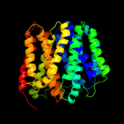
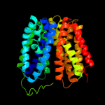
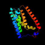
PDB 2gfp chain A
Region: 9 - 383
Aligned: 375
Modelled: 375
Confidence: 100.0%
Identity: 100%
PDB header:membrane protein
Chain: A: PDB Molecule:multidrug resistance protein d;
PDBTitle: structure of the multidrug transporter emrd from2 escherichia coli
Phyre2
| 2 |
|
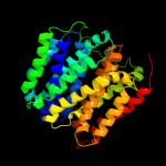
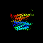
PDB 1pw4 chain A
Region: 3 - 392
Aligned: 389
Modelled: 390
Confidence: 100.0%
Identity: 15%
Fold: MFS general substrate transporter
Superfamily: MFS general substrate transporter
Family: Glycerol-3-phosphate transporter
Phyre2
| 3 |
|
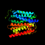
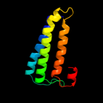
PDB 3o7p chain A
Region: 5 - 387
Aligned: 375
Modelled: 383
Confidence: 100.0%
Identity: 11%
PDB header:transport protein
Chain: A: PDB Molecule:l-fucose-proton symporter;
PDBTitle: crystal structure of the e.coli fucose:proton symporter, fucp (n162a)
Phyre2
| 4 |
|
PDB 1pv7 chain A
Region: 1 - 390
Aligned: 386
Modelled: 390
Confidence: 100.0%
Identity: 11%
Fold: MFS general substrate transporter
Superfamily: MFS general substrate transporter
Family: LacY-like proton/sugar symporter
Phyre2
| 5 |
|
PDB 2xut chain C
Region: 10 - 382
Aligned: 373
Modelled: 373
Confidence: 100.0%
Identity: 11%
PDB header:transport protein
Chain: C: PDB Molecule:proton/peptide symporter family protein;
PDBTitle: crystal structure of a proton dependent oligopeptide (pot)2 family transporter.
Phyre2
| 6 |
|
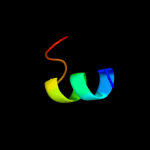
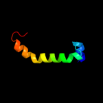
PDB 2g9p chain A
Region: 62 - 75
Aligned: 14
Modelled: 14
Confidence: 21.9%
Identity: 21%
PDB header:antimicrobial protein
Chain: A: PDB Molecule:antimicrobial peptide latarcin 2a;
PDBTitle: nmr structure of a novel antimicrobial peptide, latarcin 2a,2 from spider (lachesana tarabaevi) venom
Phyre2
| 7 |
|
PDB 3rko chain N
Region: 2 - 102
Aligned: 101
Modelled: 101
Confidence: 11.0%
Identity: 14%
PDB header:oxidoreductase
Chain: N: PDB Molecule:nadh-quinone oxidoreductase subunit n;
PDBTitle: crystal structure of the membrane domain of respiratory complex i from2 e. coli at 3.0 angstrom resolution
Phyre2
| 8 |
|
PDB 3pro chain C domain 1
Region: 30 - 71
Aligned: 42
Modelled: 42
Confidence: 8.7%
Identity: 12%
Fold: Alpha-lytic protease prodomain-like
Superfamily: Alpha-lytic protease prodomain
Family: Alpha-lytic protease prodomain
Phyre2
| 9 |
|
PDB 3b9y chain A
Region: 194 - 392
Aligned: 191
Modelled: 199
Confidence: 6.0%
Identity: 9%
PDB header:transport protein
Chain: A: PDB Molecule:ammonium transporter family rh-like protein;
PDBTitle: crystal structure of the nitrosomonas europaea rh protein
Phyre2