1 c3nctC_
100.0
96
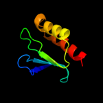
PDB header: dna binding protein, chaperoneChain: C: PDB Molecule: protein psib;PDBTitle: x-ray crystal structure of the bacterial conjugation factor psib, a2 negative regulator of reca
2 c3d30A_
84.1
14
PDB header: peptidoglycan-binding proteinChain: A: PDB Molecule: expansin like protein;PDBTitle: structure of an expansin like protein from bacillus subtilis at 1.9a2 resolution
3 c2w56B_
56.9
24
PDB header: unknown functionChain: B: PDB Molecule: vc0508;PDBTitle: structure of the hypothetical protein vc0508 from vibrio cholerae2 vsp-ii pathogenicity island
4 c2v1lA_
48.6
27
PDB header: unknown functionChain: A: PDB Molecule: hypothetical protein;PDBTitle: structure of the conserved hypothetical protein vc1805 from2 pathogenicity island vpi-2 of vibrio cholerae o1 biovar3 eltor str. n16961 shares structural homology with the4 human p32 protein
5 c2noxP_
33.7
42
PDB header: oxidoreductaseChain: P: PDB Molecule: tryptophan 2,3-dioxygenase;PDBTitle: crystal structure of tryptophan 2,3-dioxygenase from ralstonia2 metallidurans
6 d2nw8a1
19.7
32
Fold: Indolic compounds 2,3-dioxygenase-likeSuperfamily: Indolic compounds 2,3-dioxygenase-likeFamily: Bacterial tryptophan 2,3-dioxygenase7 c2nw7C_
17.8
32
PDB header: oxidoreductaseChain: C: PDB Molecule: tryptophan 2,3-dioxygenase;PDBTitle: crystal structure of tryptophan 2,3-dioxygenase (tdo) from2 xanthomonas campestris in complex with ferric heme.3 northeast structural genomics target xcr13
8 c2hczX_
16.4
14
PDB header: allergenChain: X: PDB Molecule: beta-expansin 1a;PDBTitle: crystal structure of expb1 (zea m 1), a beta-expansin and group-12 pollen allergen from maize
9 d1dx5i3
13.5
27
Fold: Knottins (small inhibitors, toxins, lectins)Superfamily: EGF/LamininFamily: EGF-type module10 d2i0ka1
13.3
21
Fold: Ferredoxin-likeSuperfamily: FAD-linked oxidases, C-terminal domainFamily: Cholesterol oxidase11 d2ftxa1
13.2
18
Fold: Kinetochore globular domain-likeSuperfamily: Kinetochore globular domainFamily: Spc25-like12 d1c3aa_
13.0
21
Fold: C-type lectin-likeSuperfamily: C-type lectin-likeFamily: C-type lectin domain13 c3le4A_
12.5
50
PDB header: nuclear proteinChain: A: PDB Molecule: microprocessor complex subunit dgcr8;PDBTitle: crystal structure of the dgcr8 dimerization domain
14 c3oibB_
12.4
13
PDB header: oxidoreductaseChain: B: PDB Molecule: acyl-coa dehydrogenase;PDBTitle: crystal structure of a putative acyl-coa dehydrogenase from2 mycobacterium smegmatis, iodide soak
15 c1lshB_
11.8
23
PDB header: lipid binding proteinChain: B: PDB Molecule: lipovitellin (lv-2);PDBTitle: lipid-protein interactions in lipovitellin
16 d1lshb_
11.8
23
Fold: Lipovitellin-phosvitin complex; beta-sheet shell regionsSuperfamily: Lipovitellin-phosvitin complex; beta-sheet shell regionsFamily: Lipovitellin-phosvitin complex; beta-sheet shell regions17 c3kbhE_
11.6
31
PDB header: hydrolaseChain: E: PDB Molecule: spike glycoprotein;PDBTitle: crystal structure of nl63 respiratory coronavirus receptor-binding2 domain complexed with its human receptor
18 d1d5va_
11.2
15
Fold: DNA/RNA-binding 3-helical bundleSuperfamily: "Winged helix" DNA-binding domainFamily: Forkhead DNA-binding domain19 d1x6va1
10.6
9
Fold: PUA domain-likeSuperfamily: PUA domain-likeFamily: ATP sulfurylase N-terminal domain20 d1whoa_
10.6
27
Fold: C2 domain-likeSuperfamily: PHL pollen allergenFamily: PHL pollen allergen21 d1v7wa2
not modelled
10.6
19
Fold: SupersandwichSuperfamily: Galactose mutarotase-likeFamily: Glycosyltransferase family 36 N-terminal domain22 c2w0tA_
not modelled
10.4
25
PDB header: transcriptionChain: A: PDB Molecule: lethal(3)malignant brain tumor-like 2 protein;PDBTitle: solution structure of the fcs zinc finger domain of human2 lmbl2
23 d1qo3c_
not modelled
10.3
24
Fold: C-type lectin-likeSuperfamily: C-type lectin-likeFamily: C-type lectin domain24 c2y7eA_
not modelled
10.0
17
PDB header: lyaseChain: A: PDB Molecule: 3-keto-5-aminohexanoate cleavage enzyme;PDBTitle: crystal structure of the 3-keto-5-aminohexanoate cleavage enzyme2 (kce) from candidatus cloacamonas acidaminovorans (tetragonal form)
25 c2c5sA_
not modelled
9.9
23
PDB header: rna-binding proteinChain: A: PDB Molecule: probable thiamine biosynthesis protein thii;PDBTitle: crystal structure of bacillus anthracis thii, a trna-2 modifying enzyme containing the predicted rna-binding3 thump domain
26 c1j1eC_
not modelled
9.8
27
PDB header: contractile proteinChain: C: PDB Molecule: troponin i;PDBTitle: crystal structure of the 52kda domain of human cardiac2 troponin in the ca2+ saturated form
27 c2qjfB_
not modelled
9.6
8
PDB header: transferaseChain: B: PDB Molecule: bifunctional 3'-phosphoadenosine 5'-PDBTitle: crystal structure of atp-sulfurylase domain of human paps2 synthetase 1
28 c2wmhA_
not modelled
9.3
15
PDB header: hydrolaseChain: A: PDB Molecule: fucolectin-related protein;PDBTitle: crystal structure of the catalytic module of a family 982 glycoside hydrolase from streptococcus pneumoniae tigr4 in3 complex with the h-disaccharide blood group antigen.
29 c2j83B_
not modelled
9.1
10
PDB header: hydrolaseChain: B: PDB Molecule: ulilysin;PDBTitle: ulilysin metalloprotease in complex with batimastat.
30 d1j6ra_
not modelled
8.7
14
Fold: Methionine synthase activation domain-likeSuperfamily: Methionine synthase activation domain-likeFamily: Hypothetical protein TM026931 d1s48a_
not modelled
8.5
20
Fold: DNA/RNA polymerasesSuperfamily: DNA/RNA polymerasesFamily: RNA-dependent RNA-polymerase32 c1v47B_
not modelled
8.3
20
PDB header: transferaseChain: B: PDB Molecule: atp sulfurylase;PDBTitle: crystal structure of atp sulfurylase from thermus2 thermophillus hb8 in complex with aps
33 d2a07f1
not modelled
8.1
18
Fold: DNA/RNA-binding 3-helical bundleSuperfamily: "Winged helix" DNA-binding domainFamily: Forkhead DNA-binding domain34 c2zibA_
not modelled
7.8
36
PDB header: antifreeze proteinChain: A: PDB Molecule: type ii antifreeze protein;PDBTitle: crystal structure analysis of calcium-independent type ii2 antifreeze protein
35 c3e4wB_
not modelled
7.7
36
PDB header: oxidoreductaseChain: B: PDB Molecule: putative uncharacterized protein;PDBTitle: crystal structure of a 33kda catalase-related protein from2 mycobacterium avium subsp. paratuberculosis. p2(1)2(1)2(1) crystal3 form.
36 c1vq0A_
not modelled
7.7
30
PDB header: chaperoneChain: A: PDB Molecule: 33 kda chaperonin;PDBTitle: crystal structure of 33 kda chaperonin (heat shock protein 33 homolog)2 (hsp33) (tm1394) from thermotoga maritima at 2.20 a resolution
37 c2p04B_
not modelled
7.6
50
PDB header: transferaseChain: B: PDB Molecule: signal transduction histidine kinase;PDBTitle: 2.1 ang structure of the dimerized pas domain of signal transduction2 histidine kinase from nostoc punctiforme pcc 73102 with homology to3 the h-noxa/h-noba domain of the soluble guanylyl cyclase
38 c1xwjB_
not modelled
7.5
78
PDB header: cell adhesion/protein bindingChain: B: PDB Molecule: talin;PDBTitle: vinculin head (1-258) in complex with the talin vinculin2 binding site 3 (1945-1969)
39 d3bdwa1
not modelled
7.5
9
Fold: C-type lectin-likeSuperfamily: C-type lectin-likeFamily: C-type lectin domain40 c2dlwA_
not modelled
6.9
37
PDB header: signaling proteinChain: A: PDB Molecule: docking protein 2, isoform a;PDBTitle: solution structure of the irs domain of human docking2 protein 2, isoform a
41 c2r5kE_
not modelled
6.6
31
PDB header: viral proteinChain: E: PDB Molecule: major capsid protein l1;PDBTitle: pentamer structure of major capsid protein l1 of human2 papilloma virus type 11
42 d1deca_
not modelled
6.6
33
Fold: Knottins (small inhibitors, toxins, lectins)Superfamily: Leech antihemostatic proteinsFamily: Hirudin-like43 c2yruA_
not modelled
6.6
17
PDB header: apoptosisChain: A: PDB Molecule: steroid receptor rna activator 1;PDBTitle: solution structure of mouse steroid receptor rna activator2 1 (sra1) protein
44 c1rkcB_
not modelled
6.4
78
PDB header: cell adhesion, structural proteinChain: B: PDB Molecule: talin;PDBTitle: human vinculin head (1-258) in complex with talin's2 vinculin binding site 3 (residues 1944-1969)
45 d1atxa_
not modelled
6.4
25
Fold: Defensin-likeSuperfamily: Defensin-likeFamily: Defensin46 c2lc0A_
not modelled
6.3
15
PDB header: protein bindingChain: A: PDB Molecule: putative uncharacterized protein tb39.8;PDBTitle: rv0020c_nter structure
47 d1gyxa_
not modelled
6.3
15
Fold: Tautomerase/MIFSuperfamily: Tautomerase/MIFFamily: 4-oxalocrotonate tautomerase-like48 c3tixB_
not modelled
6.3
21
PDB header: gene regulation/protein bindingChain: B: PDB Molecule: chromo domain-containing protein 1;PDBTitle: crystal structure of the chp1-tas3 complex core
49 c1fm5A_
not modelled
6.3
8
PDB header: immune systemChain: A: PDB Molecule: early activation antigen cd69;PDBTitle: crystal structure of human cd69
50 c2ox8A_
not modelled
6.2
17
PDB header: sugar binding proteinChain: A: PDB Molecule: scavenger receptor with c-type lectin type i;PDBTitle: human scavenger receptor c-type lectin carbohydrate-2 recognition domain.
51 c3m9zA_
not modelled
6.2
23
PDB header: signaling proteinChain: A: PDB Molecule: killer cell lectin-like receptor subfamily b member 1a;PDBTitle: crystal structure of extracellular domain of mouse nkr-p1a
52 d1t3ta1
not modelled
6.0
10
Fold: RuvA C-terminal domain-likeSuperfamily: FGAM synthase PurL, linker domainFamily: FGAM synthase PurL, linker domain53 d1skza1
not modelled
5.9
67
Fold: Knottins (small inhibitors, toxins, lectins)Superfamily: Leech antihemostatic proteinsFamily: Huristasin-like54 c3hj7A_
not modelled
5.8
27
PDB header: ligaseChain: A: PDB Molecule: trna(ile)-lysidine synthase;PDBTitle: crystal structure of tils c-terminal domain
55 d1i8gb_
not modelled
5.7
22
Fold: WW domain-likeSuperfamily: WW domainFamily: WW domain56 d1ib8a2
not modelled
5.6
38
Fold: Alpha-lytic protease prodomain-likeSuperfamily: YhbC-like, N-terminal domainFamily: YhbC-like, N-terminal domain57 d1umra_
not modelled
5.6
38
Fold: C-type lectin-likeSuperfamily: C-type lectin-likeFamily: C-type lectin domain58 d1gpja3
not modelled
5.6
29
Fold: Ferredoxin-likeSuperfamily: Glutamyl tRNA-reductase catalytic, N-terminal domainFamily: Glutamyl tRNA-reductase catalytic, N-terminal domain59 c1n10A_
not modelled
5.5
13
PDB header: allergenChain: A: PDB Molecule: pollen allergen phl p 1;PDBTitle: crystal structure of phl p 1, a major timothy grass pollen allergen
60 c3gprC_
not modelled
5.5
13
PDB header: cell adhesionChain: C: PDB Molecule: rhodocetin subunit gamma;PDBTitle: crystal structure of rhodocetin
61 d1psea_
not modelled
5.3
71
Fold: SH3-like barrelSuperfamily: Electron transport accessory proteinsFamily: Photosystem I accessory protein E (PsaE)62 d1jb0e_
not modelled
5.3
71
Fold: SH3-like barrelSuperfamily: Electron transport accessory proteinsFamily: Photosystem I accessory protein E (PsaE)63 d1dxga_
not modelled
5.2
33
Fold: Rubredoxin-likeSuperfamily: Rubredoxin-likeFamily: Desulforedoxin64 d1qp3a_
not modelled
5.0
71
Fold: SH3-like barrelSuperfamily: Electron transport accessory proteinsFamily: Photosystem I accessory protein E (PsaE)