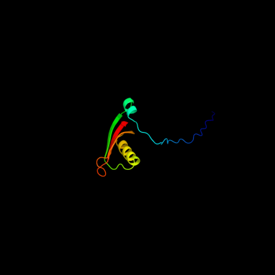
1 d2jnaa1
100.0
32
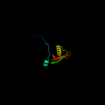
Fold: Dodecin subunit-likeSuperfamily: YdgH-likeFamily: YdgH-like2 d2noca1
99.9
22
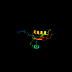
Fold: Dodecin subunit-likeSuperfamily: YdgH-likeFamily: YdgH-like3 c3b9nB_
32.1
8
PDB header: oxidoreductaseChain: B: PDB Molecule: alkane monoxygenase;PDBTitle: crystal structure of long-chain alkane monooxygenase (lada)
4 c2y1bA_
22.5
13
PDB header: membrane proteinChain: A: PDB Molecule: putative outer membrane protein, signal;PDBTitle: crystal structure of the e. coli outer membrane lipoprotein2 rcsf
5 c1hl8B_
20.7
21
PDB header: hydrolaseChain: B: PDB Molecule: putative alpha-l-fucosidase;PDBTitle: crystal structure of thermotoga maritima alpha-fucosidase
6 d1luca_
20.3
18
Fold: TIM beta/alpha-barrelSuperfamily: Bacterial luciferase-likeFamily: Bacterial luciferase (alkanal monooxygenase)7 c3sdoB_
19.7
12
PDB header: oxidoreductaseChain: B: PDB Molecule: nitrilotriacetate monooxygenase;PDBTitle: structure of a nitrilotriacetate monooxygenase from burkholderia2 pseudomallei
8 c1oy8A_
18.7
10
PDB header: membrane proteinChain: A: PDB Molecule: acriflavine resistance protein b;PDBTitle: structural basis of multiple drug binding capacity of the acrb2 multidrug efflux pump
9 c3qkbB_
16.0
15
PDB header: structural genomics, unknown functionChain: B: PDB Molecule: uncharacterized protein;PDBTitle: crystal structure of a protein with unknown function which belongs to2 pfam duf74 family (pepe_0654) from pediococcus pentosaceus atcc 257453 at 2.73 a resolution
10 c3eypB_
15.9
13
PDB header: structural genomics, unknown functionChain: B: PDB Molecule: putative alpha-l-fucosidase;PDBTitle: crystal structure of putative alpha-l-fucosidase from bacteroides2 thetaiotaomicron
11 c3mo4B_
15.3
13
PDB header: hydrolaseChain: B: PDB Molecule: alpha-1,3/4-fucosidase;PDBTitle: the crystal structure of an alpha-(1-3,4)-fucosidase from2 bifidobacterium longum subsp. infantis atcc 15697
12 d2fiqa1
13.0
16
Fold: TIM beta/alpha-barrelSuperfamily: AldolaseFamily: GatZ-like13 d1lucb_
11.1
25
Fold: TIM beta/alpha-barrelSuperfamily: Bacterial luciferase-likeFamily: Bacterial luciferase (alkanal monooxygenase)14 d1hl9a2
10.6
21
Fold: TIM beta/alpha-barrelSuperfamily: (Trans)glycosidasesFamily: Putative alpha-L-fucosidase, catalytic domain15 c1kqfB_
9.8
22
PDB header: oxidoreductaseChain: B: PDB Molecule: formate dehydrogenase, nitrate-inducible, iron-sulfurPDBTitle: formate dehydrogenase n from e. coli
16 c2wvsD_
9.6
13
PDB header: hydrolaseChain: D: PDB Molecule: alpha-l-fucosidase;PDBTitle: crystal structure of an alpha-l-fucosidase gh29 trapped2 covalent intermediate from bacteroides thetaiotaomicron in3 complex with 2-fluoro-fucosyl fluoride using an e288q4 mutant
17 d1xn9a_
9.3
14
Fold: Ribosomal proteins S24e, L23 and L15eSuperfamily: Ribosomal proteins S24e, L23 and L15eFamily: Ribosomal protein S24e18 c1z69D_
9.2
13
PDB header: oxidoreductaseChain: D: PDB Molecule: coenzyme f420-dependent n(5),n(10)-PDBTitle: crystal structure of methylenetetrahydromethanopterin2 reductase (mer) in complex with coenzyme f420
19 d3etja1
8.9
12
Fold: Barrel-sandwich hybridSuperfamily: Rudiment single hybrid motifFamily: BC C-terminal domain-like20 c3k07A_
8.1
9
PDB header: transport proteinChain: A: PDB Molecule: cation efflux system protein cusa;PDBTitle: crystal structure of cusa
21 c1tvlA_
not modelled
8.1
15
PDB header: oxidoreductaseChain: A: PDB Molecule: protein ytnj;PDBTitle: structure of ytnj from bacillus subtilis
22 d1tvla_
not modelled
8.1
15
Fold: TIM beta/alpha-barrelSuperfamily: Bacterial luciferase-likeFamily: Ssud-like monoxygenases23 c3ir9A_
not modelled
7.7
19
PDB header: structural genomics, unknown functionChain: A: PDB Molecule: peptide chain release factor subunit 1;PDBTitle: c-terminal domain of peptide chain release factor from2 methanosarcina mazei.
24 c2k38A_
not modelled
7.0
25
PDB header: antimicrobial proteinChain: A: PDB Molecule: cupiennin-1a;PDBTitle: cupiennin 1a, nmr, minimized average structure
25 c2jz7A_
not modelled
6.4
17
PDB header: selenium-binding proteinChain: A: PDB Molecule: selenium binding protein;PDBTitle: solution nmr structure of selenium-binding protein from2 methanococcus vannielii
26 c1f02T_
not modelled
6.3
7
PDB header: cell adhesionChain: T: PDB Molecule: translocated intimin receptor;PDBTitle: crystal structure of c-terminal 282-residue fragment of2 intimin in complex with translocated intimin receptor3 (tir) intimin-binding domain
27 d1f07a_
not modelled
6.3
20
Fold: TIM beta/alpha-barrelSuperfamily: Bacterial luciferase-likeFamily: F420 dependent oxidoreductases28 c3gzaB_
not modelled
6.3
17
PDB header: hydrolaseChain: B: PDB Molecule: putative alpha-l-fucosidase;PDBTitle: crystal structure of putative alpha-l-fucosidase (np_812709.1) from2 bacteroides thetaiotaomicron vpi-5482 at 1.60 a resolution
29 d1wu7a1
not modelled
6.2
22
Fold: Anticodon-binding domain-likeSuperfamily: Class II aaRS ABD-relatedFamily: Anticodon-binding domain of Class II aaRS30 c2i7gA_
not modelled
6.2
0
PDB header: oxidoreductaseChain: A: PDB Molecule: monooxygenase;PDBTitle: crystal structure of monooxygenase from agrobacterium tumefaciens
31 c2k4qA_
not modelled
6.2
19
PDB header: viral proteinChain: A: PDB Molecule: major tail protein v;PDBTitle: the solution structure of gpv, the major tail protein from2 bacteriophage lambda
32 c3dfeA_
not modelled
5.8
23
PDB header: signaling proteinChain: A: PDB Molecule: putative pii-like signaling protein;PDBTitle: crystal structure of a putative pii-like signaling protein2 (yp_323533.1) from anabaena variabilis atcc 29413 at 2.35 a3 resolution
33 d1ywxa1
not modelled
5.8
18
Fold: Ribosomal proteins S24e, L23 and L15eSuperfamily: Ribosomal proteins S24e, L23 and L15eFamily: Ribosomal protein S24e34 d1kjqa1
not modelled
5.4
17
Fold: Barrel-sandwich hybridSuperfamily: Rudiment single hybrid motifFamily: BC C-terminal domain-like35 c2wgkA_
not modelled
5.3
9
PDB header: oxidoreductaseChain: A: PDB Molecule: 3,6-diketocamphane 1,6 monooxygenase;PDBTitle: type ii baeyer-villiger monooxygenase oxygenating2 constituent of 3,6-diketocamphane 1,6 monooxygenase from3 pseudomonas putida
36 d1nqka_
not modelled
5.2
13
Fold: TIM beta/alpha-barrelSuperfamily: Bacterial luciferase-likeFamily: Ssud-like monoxygenases