Phyre Home Retrieve Phyre Job Id
Emaill.a.kelley@imperial.ac.uk
DescriptionP28915
DateThu Jan 5 11:45:25 GMT 2012
Unique Job ID9be07ed3549734fc

Summary 

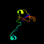
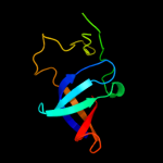
Top model
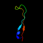
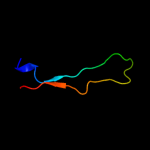
Image coloured by rainbow N → C terminus
Model (left) based on template d1gzsb_
Top template information
Fold:SopE-like GEF domain
Superfamily:SopE-like GEF domain
Family:SopE-like GEF domain
Confidence and coverage
Confidence: 50.3% Coverage: 7%
13 residues ( 7% of your sequence) have been modelled with 50.3% confidence by the single highest scoring template.
You may wish to submit your sequence to Phyrealarm. This will automatically scan your sequence every week for new potential templates as they appear in the Phyre2 library.
Please note: You must be registered and logged in to use Phyrealarm.
3D viewing
Interactive 3D view in Jmol

Sequence analysis 

Secondary structure and disorder prediction 

   1........10.........20.........30.........40.........50.........60
Sequence  MKRVLFFLLMIFVSFGVIADCEIQAKDHDCFTIFAKGTIFSAFPVLNNKAMWRWYQNEDI
Secondary structure 


SS confidence 


Disorder  ??


Disorder confidence 


   .........70.........80.........90.........100.........110.........120
Sequence  GEYYWQTELGTCKNNKFTPSGARLLIRVGSLRLNENHAIKGTLQELINTAEKTAFLGDRF
Secondary structure 


SS confidence 


Disorder 


??


???
?


??


Disorder confidence 


   .........130.........140.........150.........160.........170.........180
Sequence  RSYIRAGIYQKKSSDPVQLLAVLDNSIMVKYFKDEKPTYARMTAHLPNKNESYECLIKIQ
Secondary structure 


SS confidence 


Disorder 


?


?


Disorder confidence 


   .........
Sequence  HELIRSEEK
Secondary structure 


SS confidence 


Disorder 


??????
Disorder confidence 


Confidence Key
High(9)                    Low (0)
?Disordered
Alpha helix
Beta strand

Domain analysis 

Hover over an aligned region to see model and summary info

Please note, only up to the top 20 hits are modelled to reduce computer load

RankAligned region

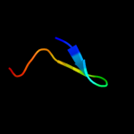
PDB 1gzs chain B

3D model

Region: 70 - 82
Aligned: 13
Modelled: 13
Confidence: 50.3%
Identity: 77%
Fold: SopE-like GEF domain
Superfamily: SopE-like GEF domain
Family: SopE-like GEF domain

Phyre2

PDB 2jok chain A domain 1

3D model

Region: 70 - 82
Aligned: 13
Modelled: 13
Confidence: 48.8%
Identity: 62%
Fold: SopE-like GEF domain
Superfamily: SopE-like GEF domain
Family: SopE-like GEF domain

Phyre2

PDB 1r6e chain A

3D model

Region: 70 - 82
Aligned: 13
Modelled: 13
Confidence: 46.9%
Identity: 62%
Fold: SopE-like GEF domain
Superfamily: SopE-like GEF domain
Family: SopE-like GEF domain

Phyre2

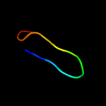
PDB 1jqs chain B

3D model

Region: 141 - 158
Aligned: 18
Modelled: 18
Confidence: 33.4%
Identity: 50%
PDB header:ribosome
Chain: B: PDB Molecule:elongation factor g;
PDBTitle: fitting of l11 protein and elongation factor g (domain g'2 and v) in the cryo-em map of e. coli 70s ribosome bound3 with ef-g and gmppcp, a nonhydrolysable gtp analog

Phyre2

PDB 1bec chain A domain 1

3D model

Region: 56 - 87
Aligned: 32
Modelled: 32
Confidence: 27.9%
Identity: 16%
Fold: Immunoglobulin-like beta-sandwich
Superfamily: Immunoglobulin
Family: V set domains (antibody variable domain-like)

Phyre2

PDB 2nts chain P domain 1

3D model

Region: 56 - 87
Aligned: 32
Modelled: 32
Confidence: 25.2%
Identity: 22%
Fold: Immunoglobulin-like beta-sandwich
Superfamily: Immunoglobulin
Family: V set domains (antibody variable domain-like)

Phyre2

PDB 2cde chain B domain 1

3D model

Region: 56 - 87
Aligned: 32
Modelled: 32
Confidence: 24.2%
Identity: 13%
Fold: Immunoglobulin-like beta-sandwich
Superfamily: Immunoglobulin
Family: V set domains (antibody variable domain-like)

Phyre2

PDB 2oez chain A domain 1

3D model

Region: 125 - 142
Aligned: 17
Modelled: 18
Confidence: 18.4%
Identity: 35%
Fold: YacF-like
Superfamily: YacF-like
Family: YacF-like

Phyre2

PDB 3i5d chain C

3D model

Region: 31 - 91
Aligned: 51
Modelled: 61
Confidence: 17.7%
Identity: 24%
PDB header:transport protein
Chain: C: PDB Molecule:p2x purinoceptor;
PDBTitle: crystal structure of the atp-gated p2x4 ion channel in the closed, apo2 state at 3.5 angstroms (r3)

Phyre2

PDB 1gso chain A domain 1

3D model

Region: 70 - 89
Aligned: 19
Modelled: 20
Confidence: 15.8%
Identity: 32%
Fold: Barrel-sandwich hybrid
Superfamily: Rudiment single hybrid motif
Family: BC C-terminal domain-like

Phyre2

PDB 2rhe chain A

3D model

Region: 56 - 86
Aligned: 30
Modelled: 31
Confidence: 15.1%
Identity: 23%
Fold: Immunoglobulin-like beta-sandwich
Superfamily: Immunoglobulin
Family: V set domains (antibody variable domain-like)

Phyre2

PDB 1j8h chain D domain 1

3D model

Region: 56 - 87
Aligned: 32
Modelled: 32
Confidence: 14.9%
Identity: 31%
Fold: Immunoglobulin-like beta-sandwich
Superfamily: Immunoglobulin
Family: V set domains (antibody variable domain-like)

Phyre2

PDB 2nxy chain C domain 1

3D model

Region: 56 - 87
Aligned: 31
Modelled: 32
Confidence: 13.9%
Identity: 35%
Fold: Immunoglobulin-like beta-sandwich
Superfamily: Immunoglobulin
Family: V set domains (antibody variable domain-like)

Phyre2

PDB 2axh chain A domain 1

3D model

Region: 56 - 87
Aligned: 31
Modelled: 32
Confidence: 13.1%
Identity: 26%
Fold: Immunoglobulin-like beta-sandwich
Superfamily: Immunoglobulin
Family: V set domains (antibody variable domain-like)

Phyre2

PDB 2rcj chain A domain 1

3D model

Region: 56 - 84
Aligned: 28
Modelled: 29
Confidence: 12.7%
Identity: 21%
Fold: Immunoglobulin-like beta-sandwich
Superfamily: Immunoglobulin
Family: V set domains (antibody variable domain-like)

Phyre2

PDB 1nl0 chain L domain 1

3D model

Region: 56 - 87
Aligned: 31
Modelled: 32
Confidence: 12.4%
Identity: 23%
Fold: Immunoglobulin-like beta-sandwich
Superfamily: Immunoglobulin
Family: V set domains (antibody variable domain-like)

Phyre2

PDB 2rf4 chain A

3D model

Region: 16 - 103
Aligned: 81
Modelled: 87
Confidence: 11.2%
Identity: 16%
PDB header:transferase
Chain: A: PDB Molecule:dna-directed rna polymerase i subunit rpa4;
PDBTitle: crystal structure of the rna polymerase i subcomplex a14/43

Phyre2

PDB 1hng chain A domain 1

3D model

Region: 56 - 95
Aligned: 40
Modelled: 40
Confidence: 11.1%
Identity: 28%
Fold: Immunoglobulin-like beta-sandwich
Superfamily: Immunoglobulin
Family: V set domains (antibody variable domain-like)

Phyre2

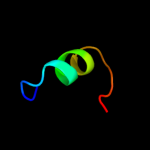
PDB 2rnn chain A

3D model

Region: 107 - 142
Aligned: 35
Modelled: 36
Confidence: 10.2%
Identity: 26%
PDB header:ligase
Chain: A: PDB Molecule:e3 sumo-protein ligase siz1;
PDBTitle: solution structure of the n-terminal sap domain of sumo e3 ligases2 from saccharomyces cerevisiae

Phyre2

PDB 1kgc chain E domain 1

3D model

Region: 56 - 87
Aligned: 31
Modelled: 32
Confidence: 9.4%
Identity: 35%
Fold: Immunoglobulin-like beta-sandwich
Superfamily: Immunoglobulin
Family: V set domains (antibody variable domain-like)

Phyre2
1

d1gzsb_
2

d2joka1
3

d1r6ea_
4

c1jqsB_
5

d1beca1
6

d2ntsp1
7

d2cdeb1
8

d2oeza1
9

c3i5dC_
10

d1gsoa1
11

d2rhea_
12

d1j8hd1
13

d2nxyc1
14

d2axha1
15

d2rcja1
16

d1nl0l1
17

c2rf4A_
18

d1hnga1
19

c2rnnA_
20

d1kgce1
21


22


23


24


25


26


27


28


29


30


31


32


33


34


35


36


37


38


39


40


41


42


43


44


45


46


47


Detailed template information 

#
Template Alignment Coverage3D Model Confidence
% i.d. Template Information
1d1gzsb_


50.3 77 Fold:SopE-like GEF domain
Superfamily:SopE-like GEF domain
Family:SopE-like GEF domain
2d2joka1


48.8 62 Fold:SopE-like GEF domain
Superfamily:SopE-like GEF domain
Family:SopE-like GEF domain
3d1r6ea_


46.9 62 Fold:SopE-like GEF domain
Superfamily:SopE-like GEF domain
Family:SopE-like GEF domain
4c1jqsB_


33.4 50 PDB header:ribosome
Chain: B: PDB Molecule:elongation factor g;
PDBTitle: fitting of l11 protein and elongation factor g (domain g'2 and v) in the cryo-em map of e. coli 70s ribosome bound3 with ef-g and gmppcp, a nonhydrolysable gtp analog
5d1beca1


27.9 16 Fold:Immunoglobulin-like beta-sandwich
Superfamily:Immunoglobulin
Family:V set domains (antibody variable domain-like)
6d2ntsp1


25.2 22 Fold:Immunoglobulin-like beta-sandwich
Superfamily:Immunoglobulin
Family:V set domains (antibody variable domain-like)
7d2cdeb1


24.2 13 Fold:Immunoglobulin-like beta-sandwich
Superfamily:Immunoglobulin
Family:V set domains (antibody variable domain-like)
8d2oeza1


18.4 35 Fold:YacF-like
Superfamily:YacF-like
Family:YacF-like
9c3i5dC_


17.7 24 PDB header:transport protein
Chain: C: PDB Molecule:p2x purinoceptor;
PDBTitle: crystal structure of the atp-gated p2x4 ion channel in the closed, apo2 state at 3.5 angstroms (r3)
10d1gsoa1


15.8 32 Fold:Barrel-sandwich hybrid
Superfamily:Rudiment single hybrid motif
Family:BC C-terminal domain-like
11d2rhea_


15.1 23 Fold:Immunoglobulin-like beta-sandwich
Superfamily:Immunoglobulin
Family:V set domains (antibody variable domain-like)
12d1j8hd1


14.9 31 Fold:Immunoglobulin-like beta-sandwich
Superfamily:Immunoglobulin
Family:V set domains (antibody variable domain-like)
13d2nxyc1


13.9 35 Fold:Immunoglobulin-like beta-sandwich
Superfamily:Immunoglobulin
Family:V set domains (antibody variable domain-like)
14d2axha1


13.1 26 Fold:Immunoglobulin-like beta-sandwich
Superfamily:Immunoglobulin
Family:V set domains (antibody variable domain-like)
15d2rcja1


12.7 21 Fold:Immunoglobulin-like beta-sandwich
Superfamily:Immunoglobulin
Family:V set domains (antibody variable domain-like)
16d1nl0l1


12.4 23 Fold:Immunoglobulin-like beta-sandwich
Superfamily:Immunoglobulin
Family:V set domains (antibody variable domain-like)
17c2rf4A_


11.2 16 PDB header:transferase
Chain: A: PDB Molecule:dna-directed rna polymerase i subunit rpa4;
PDBTitle: crystal structure of the rna polymerase i subcomplex a14/43
18d1hnga1


11.1 28 Fold:Immunoglobulin-like beta-sandwich
Superfamily:Immunoglobulin
Family:V set domains (antibody variable domain-like)
19c2rnnA_


10.2 26 PDB header:ligase
Chain: A: PDB Molecule:e3 sumo-protein ligase siz1;
PDBTitle: solution structure of the n-terminal sap domain of sumo e3 ligases2 from saccharomyces cerevisiae
20d1kgce1


9.4 35 Fold:Immunoglobulin-like beta-sandwich
Superfamily:Immunoglobulin
Family:V set domains (antibody variable domain-like)
21d1aqkl1


not modelled 8.8 29 Fold:Immunoglobulin-like beta-sandwich
Superfamily:Immunoglobulin
Family:V set domains (antibody variable domain-like)
22d1nfdb1


not modelled 8.6 26 Fold:Immunoglobulin-like beta-sandwich
Superfamily:Immunoglobulin
Family:V set domains (antibody variable domain-like)
23d1u3hb1


not modelled 8.5 13 Fold:Immunoglobulin-like beta-sandwich
Superfamily:Immunoglobulin
Family:V set domains (antibody variable domain-like)
24c2ckzB_


not modelled 8.2 15 PDB header:transferase
Chain: B: PDB Molecule:dna-directed rna polymerase iii 25 kd
PDBTitle: x-ray structure of rna polymerase iii subcomplex c17-c25.
25c3l15A_


not modelled 7.6 30 PDB header:transcription
Chain: A: PDB Molecule:transcriptional enhancer factor tef-4;
PDBTitle: human tead2 transcriptional factor
26c2kuyA_


not modelled 7.5 43 PDB header:antimicrobial protein
Chain: A: PDB Molecule:prebacteriocin glycocin f;
PDBTitle: structure of glycocin f
27d1ktke1


not modelled 7.2 22 Fold:Immunoglobulin-like beta-sandwich
Superfamily:Immunoglobulin
Family:V set domains (antibody variable domain-like)
28d1q1jl1


not modelled 7.2 19 Fold:Immunoglobulin-like beta-sandwich
Superfamily:Immunoglobulin
Family:V set domains (antibody variable domain-like)
29d2aw2a1


not modelled 6.9 52 Fold:Immunoglobulin-like beta-sandwich
Superfamily:Immunoglobulin
Family:V set domains (antibody variable domain-like)
30c3fd9C_


not modelled 6.9 25 PDB header:unknown function
Chain: C: PDB Molecule:uncharacterized protein;
PDBTitle: crystal structure of the transcriptional anti-activator exsd2 from pseudomonas aeruginosa
31d1lc0a2


not modelled 6.9 18 Fold:FwdE/GAPDH domain-like
Superfamily:Glyceraldehyde-3-phosphate dehydrogenase-like, C-terminal domain
Family:Biliverdin reductase
32d1nfda1


not modelled 6.8 28 Fold:Immunoglobulin-like beta-sandwich
Superfamily:Immunoglobulin
Family:V set domains (antibody variable domain-like)
33c1ye9E_


not modelled 6.7 35 PDB header:oxidoreductase
Chain: E: PDB Molecule:catalase hpii;
PDBTitle: crystal structure of proteolytically truncated catalase2 hpii from e. coli
34d1xaua_


not modelled 6.6 29 Fold:Immunoglobulin-like beta-sandwich
Superfamily:Immunoglobulin
Family:I set domains
35d1nzpa_


not modelled 6.5 29 Fold:SAM domain-like
Superfamily:DNA polymerase beta, N-terminal domain-like
Family:DNA polymerase beta, N-terminal domain-like
36c2hpvA_


not modelled 6.4 16 PDB header:oxidoreductase
Chain: A: PDB Molecule:fmn-dependent nadh-azoreductase;
PDBTitle: crystal structure of fmn-dependent azoreductase from enterococcus2 faecalis
37d1vmaa1


not modelled 6.2 17 Fold:Four-helical up-and-down bundle
Superfamily:Domain of the SRP/SRP receptor G-proteins
Family:Domain of the SRP/SRP receptor G-proteins
38c3ayhB_


not modelled 6.2 8 PDB header:transcription
Chain: B: PDB Molecule:dna-directed rna polymerase iii subunit rpc8;
PDBTitle: crystal structure of the c17/25 subcomplex from s. pombe rna2 polymerase iii
39d2pxyd2


not modelled 6.1 57 Fold:MHC antigen-recognition domain
Superfamily:MHC antigen-recognition domain
Family:MHC antigen-recognition domain
40d2bnub1


not modelled 5.9 16 Fold:Immunoglobulin-like beta-sandwich
Superfamily:Immunoglobulin
Family:V set domains (antibody variable domain-like)
41c3fe7A_


not modelled 5.8 15 PDB header:cell cycle
Chain: A: PDB Molecule:mdm4 protein;
PDBTitle: crystal structure of hdmx bound to the p53-peptidomimetic2 ac-phe-met-aib-pmp-trp-glu-ac3c-leu-nh2 at 1.35a
42d2ak4d1


not modelled 5.6 19 Fold:Immunoglobulin-like beta-sandwich
Superfamily:Immunoglobulin
Family:V set domains (antibody variable domain-like)
43d2ho2a1


not modelled 5.5 55 Fold:WW domain-like
Superfamily:WW domain
Family:WW domain
44d2j6el1


not modelled 5.4 26 Fold:Immunoglobulin-like beta-sandwich
Superfamily:Immunoglobulin
Family:V set domains (antibody variable domain-like)
45d1w72l1


not modelled 5.2 22 Fold:Immunoglobulin-like beta-sandwich
Superfamily:Immunoglobulin
Family:V set domains (antibody variable domain-like)
46c2l0eA_


not modelled 5.2 67 PDB header:membrane protein
Chain: A: PDB Molecule:sodium/hydrogen exchanger 1;
PDBTitle: structural and functional analysis of tm vi of the nhe1 isoform of the2 na+/h+ exchanger
47d2axia1


not modelled 5.1 15 Fold:SWIB/MDM2 domain
Superfamily:SWIB/MDM2 domain
Family:SWIB/MDM2 domain

Binding site prediction 

Due to computational demand, binding site predictions are not run for batch jobs

If you want to predict binding sites, please manually submit your model of choice to 3DLigandSite


Phyre is for academic use only

Please cite: Protein structure prediction on the web: a case study using the Phyre server
Kelley LA and Sternberg MJE. Nature Protocols 4, 363 - 371 (2009) [pdf] [Import into BibTeX]
 
If you use the binding site predictions from 3DLigandSite, please also cite:
3DLigandSite: predicting ligand-binding sites using similar structures.
Wass MN, Kelley LA and Sternberg MJ Nucleic Acids Research 38, W469-73 (2010) [PubMed]
 
© Structural Bioinformatics Group
Imperial College London
Lawrence Kelley, Benjamin Jefferys
Disclaimer
Terms and Conditions
Component software
Template detection: HHpred 1.51
Secondary structure prediction: Psi-pred 2.5
Disorder prediction: Disopred 2.4
Transmembrane prediction: Memsat_SVM
Multi-template modelling and ab initio: Poing 1.0