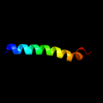
| 1 |
|
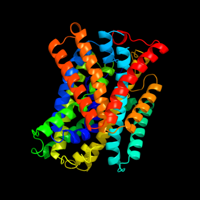
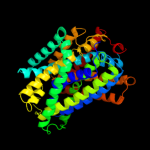
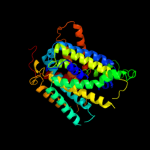
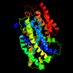
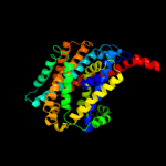
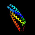
PDB 3gia chain A
Region: 8 - 457
Aligned: 428
Modelled: 428
Confidence: 100.0%
Identity: 15%
PDB header:transport protein
Chain: A: PDB Molecule:uncharacterized protein mj0609;
PDBTitle: crystal structure of apct transporter
Phyre2
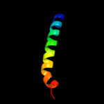
| 2 |
|
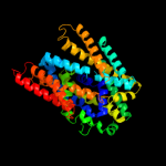
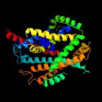
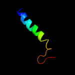
PDB 3lrc chain C
Region: 8 - 454
Aligned: 404
Modelled: 404
Confidence: 100.0%
Identity: 18%
PDB header:transport protein
Chain: C: PDB Molecule:arginine/agmatine antiporter;
PDBTitle: structure of e. coli adic (p1)
Phyre2
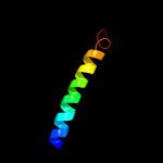
| 3 |
|
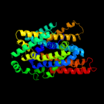
PDB 2jln chain A
Region: 1 - 456
Aligned: 441
Modelled: 441
Confidence: 100.0%
Identity: 9%
PDB header:membrane protein
Chain: A: PDB Molecule:mhp1;
PDBTitle: structure of mhp1, a nucleobase-cation-symport-1 family2 transporter
Phyre2
| 4 |
|
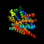
PDB 2xq2 chain A
Region: 9 - 455
Aligned: 430
Modelled: 430
Confidence: 99.7%
Identity: 11%
PDB header:transport protein
Chain: A: PDB Molecule:sodium/glucose cotransporter;
PDBTitle: structure of the k294a mutant of vsglt
Phyre2
| 5 |
|
PDB 3dh4 chain A
Region: 9 - 457
Aligned: 433
Modelled: 433
Confidence: 99.6%
Identity: 8%
PDB header:transport protein
Chain: A: PDB Molecule:sodium/glucose cotransporter;
PDBTitle: crystal structure of sodium/sugar symporter with bound galactose from2 vibrio parahaemolyticus
Phyre2
| 6 |
|
PDB 2a65 chain A domain 1
Region: 12 - 455
Aligned: 436
Modelled: 436
Confidence: 98.5%
Identity: 14%
Fold: SNF-like
Superfamily: SNF-like
Family: SNF-like
Phyre2
| 7 |
|
PDB 2w8a chain C
Region: 4 - 397
Aligned: 385
Modelled: 379
Confidence: 97.7%
Identity: 12%
PDB header:membrane protein
Chain: C: PDB Molecule:glycine betaine transporter betp;
PDBTitle: crystal structure of the sodium-coupled glycine betaine2 symporter betp from corynebacterium glutamicum with bound3 substrate
Phyre2
| 8 |
|
PDB 3hfx chain A
Region: 2 - 389
Aligned: 378
Modelled: 378
Confidence: 94.1%
Identity: 11%
PDB header:transport protein
Chain: A: PDB Molecule:l-carnitine/gamma-butyrobetaine antiporter;
PDBTitle: crystal structure of carnitine transporter
Phyre2
| 9 |
|
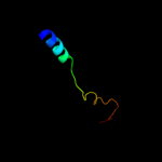
PDB 2knc chain A
Region: 423 - 457
Aligned: 35
Modelled: 35
Confidence: 75.2%
Identity: 17%
PDB header:cell adhesion
Chain: A: PDB Molecule:integrin alpha-iib;
PDBTitle: platelet integrin alfaiib-beta3 transmembrane-cytoplasmic2 heterocomplex
Phyre2
| 10 |
|
PDB 2rdd chain B
Region: 431 - 457
Aligned: 27
Modelled: 27
Confidence: 28.1%
Identity: 15%
PDB header:membrane protein/transport protein
Chain: B: PDB Molecule:upf0092 membrane protein yajc;
PDBTitle: x-ray crystal structure of acrb in complex with a novel2 transmembrane helix.
Phyre2
| 11 |
|
PDB 3m7b chain A
Region: 331 - 457
Aligned: 127
Modelled: 127
Confidence: 26.4%
Identity: 10%
PDB header:structural genomics, unknown function
Chain: A: PDB Molecule:tellurite resistance protein teha homolog;
PDBTitle: crystal structure of plant slac1 homolog teha
Phyre2
| 12 |
|
PDB 2klu chain A
Region: 427 - 456
Aligned: 30
Modelled: 30
Confidence: 15.1%
Identity: 27%
PDB header:immune system, membrane protein
Chain: A: PDB Molecule:t-cell surface glycoprotein cd4;
PDBTitle: nmr structure of the transmembrane and cytoplasmic domains2 of human cd4
Phyre2
| 13 |
|
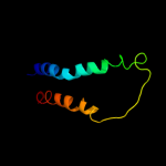
PDB 1fft chain B domain 2
Region: 354 - 421
Aligned: 68
Modelled: 68
Confidence: 9.5%
Identity: 9%
Fold: Transmembrane helix hairpin
Superfamily: Cytochrome c oxidase subunit II-like, transmembrane region
Family: Cytochrome c oxidase subunit II-like, transmembrane region
Phyre2
| 14 |
|
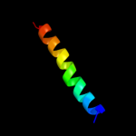
PDB 2l1n chain A
Region: 2 - 13
Aligned: 12
Modelled: 12
Confidence: 6.0%
Identity: 33%
PDB header:structural genomics, unknown function
Chain: A: PDB Molecule:uncharacterized protein;
PDBTitle: solution nmr structure of the protein yp_399305.1
Phyre2
| 15 |
|
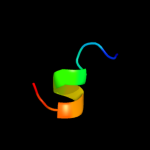
PDB 3cwb chain Q
Region: 427 - 457
Aligned: 31
Modelled: 31
Confidence: 5.9%
Identity: 13%
PDB header:oxidoreductase
Chain: Q: PDB Molecule:mitochondrial cytochrome c1, heme protein;
PDBTitle: chicken cytochrome bc1 complex inhibited by an iodinated analogue of2 the polyketide crocacin-d
Phyre2
| 16 |
|
PDB 1p84 chain D
Region: 427 - 457
Aligned: 31
Modelled: 31
Confidence: 5.4%
Identity: 10%
PDB header:oxidoreductase
Chain: D: PDB Molecule:cytochrome c1, heme protein;
PDBTitle: hdbt inhibited yeast cytochrome bc1 complex
Phyre2
| 17 |
|
PDB 1zrt chain D
Region: 427 - 457
Aligned: 31
Modelled: 31
Confidence: 5.4%
Identity: 6%
PDB header:oxidoreductase/metal transport
Chain: D: PDB Molecule:cytochrome c1;
PDBTitle: rhodobacter capsulatus cytochrome bc1 complex with2 stigmatellin bound
Phyre2