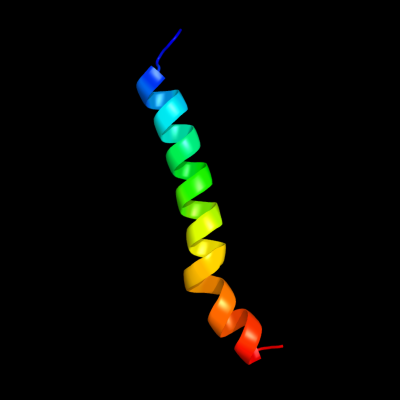
Phyre Home Retrieve Phyre Job Id
Emaill.a.kelley@imperial.ac.uk
DescriptionP0ABA0
DateThu Jan 5 11:15:01 GMT 2012
Unique Job ID844336a0421fe333

Summary 

Top model
Image coloured by rainbow N → C terminus
Model (left) based on template c1b9uA_
Top template information
PDB header:hydrolase
Chain: A: PDB Molecule:protein (atp synthase);
PDBTitle: membrane domain of the subunit b of the e.coli atp synthase
Confidence and coverage
Confidence: 98.3% Coverage: 21%
33 residues ( 21% of your sequence) have been modelled with 98.3% confidence by the single highest scoring template.
Additional confident templates have been detected (see Domain analysis) which cover other regions of your sequence.
154 residues ( 99%) could be modelled at >90% confidence using multiple-templates.
You may wish to try resubmitting your sequence in "intensive" mode to model more of your sequence.
3D viewing
Interactive 3D view in Jmol

Sequence analysis 

Secondary structure and disorder prediction 

   1........10.........20.........30.........40.........50.........60
Sequence  MNLNATILGQAIAFVLFVLFCMKYVWPPLMAAIEKRQKEIADGLASAERAHKDLDLAKAS
Secondary structure 


SS confidence 


Disorder  ????


Disorder confidence 


   .........70.........80.........90.........100.........110.........120
Sequence  ATDQLKKAKAEAQVIIEQANKRRSQILDEAKAEAEQERTKIVAQAQAEIEAERKRAREEL
Secondary structure 
SS confidence 


Disorder 


Disorder confidence 


   .........130.........140.........150......
Sequence  RKQVAILAVAGAEKIIERSVDEAANSDIVDKLVAEL
Secondary structure 


SS confidence 


Disorder 


????


???
Disorder confidence 


Confidence Key
High(9)                    Low (0)
?Disordered
Alpha helix
Beta strand

Domain analysis 

Hover over an aligned region to see model and summary info

Please note, only up to the top 20 hits are modelled to reduce computer load

RankAligned region

PDB 1b9u chain A

3D model

Region: 1 - 33
Aligned: 33
Modelled: 33
Confidence: 98.3%
Identity: 100%
PDB header:hydrolase
Chain: A: PDB Molecule:protein (atp synthase);
PDBTitle: membrane domain of the subunit b of the e.coli atp synthase

Phyre2

PDB 1l2p chain A

3D model

Region: 63 - 121
Aligned: 59
Modelled: 59
Confidence: 97.6%
Identity: 100%
Fold: Single transmembrane helix
Superfamily: F1F0 ATP synthase subunit B, membrane domain
Family: F1F0 ATP synthase subunit B, membrane domain

Phyre2

PDB 3k5b chain E

3D model

Region: 55 - 155
Aligned: 101
Modelled: 101
Confidence: 96.3%
Identity: 19%
PDB header:hydrolase
Chain: E: PDB Molecule:v-type atp synthase subunit e;
PDBTitle: crystal structure of the peripheral stalk of thermus thermophilus h+-2 atpase/synthase

Phyre2

PDB 2oar chain A domain 1

3D model

Region: 3 - 35
Aligned: 33
Modelled: 33
Confidence: 90.9%
Identity: 24%
Fold: Gated mechanosensitive channel
Superfamily: Gated mechanosensitive channel
Family: Gated mechanosensitive channel

Phyre2

PDB 2oar chain A

3D model

Region: 2 - 60
Aligned: 57
Modelled: 59
Confidence: 90.1%
Identity: 16%
PDB header:membrane protein
Chain: A: PDB Molecule:large-conductance mechanosensitive channel;
PDBTitle: mechanosensitive channel of large conductance (mscl)

Phyre2

PDB 2khk chain A

3D model

Region: 33 - 79
Aligned: 47
Modelled: 47
Confidence: 87.9%
Identity: 100%
PDB header:transport protein
Chain: A: PDB Molecule:atp synthase subunit b;
PDBTitle: nmr solution structure of the b30-82 domain of subunit b of2 escherichia coli f1fo atp synthase

Phyre2

PDB 3hzq chain A

3D model

Region: 2 - 31
Aligned: 29
Modelled: 30
Confidence: 81.0%
Identity: 7%
PDB header:membrane protein
Chain: A: PDB Molecule:large-conductance mechanosensitive channel;
PDBTitle: structure of a tetrameric mscl in an expanded intermediate2 state

Phyre2

PDB 2rdd chain B

3D model

Region: 8 - 41
Aligned: 34
Modelled: 34
Confidence: 77.0%
Identity: 15%
PDB header:membrane protein/transport protein
Chain: B: PDB Molecule:upf0092 membrane protein yajc;
PDBTitle: x-ray crystal structure of acrb in complex with a novel2 transmembrane helix.

Phyre2

PDB 3k5b chain B

3D model

Region: 40 - 136
Aligned: 97
Modelled: 97
Confidence: 76.3%
Identity: 21%
PDB header:hydrolase
Chain: B: PDB Molecule:v-type atp synthase, subunit (vapc-therm);
PDBTitle: crystal structure of the peripheral stalk of thermus thermophilus h+-2 atpase/synthase

Phyre2

PDB 1y4c chain A

3D model

Region: 40 - 156
Aligned: 115
Modelled: 117
Confidence: 70.0%
Identity: 18%
PDB header:de novo protein
Chain: A: PDB Molecule:maltose binding protein fused with designed
PDBTitle: designed helical protein fusion mbp

Phyre2

PDB 1qcr chain D

3D model

Region: 7 - 37
Aligned: 31
Modelled: 31
Confidence: 43.6%
Identity: 6%
PDB header:
PDB COMPND:

Phyre2

PDB 3rfr chain I

3D model

Region: 1 - 30
Aligned: 30
Modelled: 30
Confidence: 37.3%
Identity: 3%
PDB header:oxidoreductase
Chain: I: PDB Molecule:pmob;
PDBTitle: crystal structure of particulate methane monooxygenase (pmmo) from2 methylocystis sp. strain m

Phyre2

PDB 2k88 chain A

3D model

Region: 65 - 108
Aligned: 44
Modelled: 44
Confidence: 33.0%
Identity: 34%
PDB header:hydrolase
Chain: A: PDB Molecule:vacuolar proton pump subunit g;
PDBTitle: association of subunit d (vma6p) and e (vma4p) with g2 (vma10p) and the nmr solution structure of subunit g (g1-3 59) of the saccharomyces cerevisiae v1vo atpase

Phyre2

PDB 3dtu chain B domain 2

3D model

Region: 3 - 27
Aligned: 25
Modelled: 25
Confidence: 24.9%
Identity: 16%
Fold: Transmembrane helix hairpin
Superfamily: Cytochrome c oxidase subunit II-like, transmembrane region
Family: Cytochrome c oxidase subunit II-like, transmembrane region

Phyre2

PDB 1jdm chain A

3D model

Region: 1 - 25
Aligned: 25
Modelled: 25
Confidence: 20.4%
Identity: 16%
PDB header:membrane protein
Chain: A: PDB Molecule:sarcolipin;
PDBTitle: nmr structure of sarcolipin

Phyre2

PDB 1ar1 chain B

3D model

Region: 3 - 27
Aligned: 25
Modelled: 25
Confidence: 17.0%
Identity: 4%
PDB header:complex (oxidoreductase/antibody)
Chain: B: PDB Molecule:cytochrome c oxidase;
PDBTitle: structure at 2.7 angstrom resolution of the paracoccus2 denitrificans two-subunit cytochrome c oxidase complexed3 with an antibody fv fragment

Phyre2

PDB 1qle chain B

3D model

Region: 3 - 27
Aligned: 25
Modelled: 25
Confidence: 17.0%
Identity: 4%
PDB header:oxidoreductase/immune system
Chain: B: PDB Molecule:cytochrome c oxidase polypeptide ii;
PDBTitle: cryo-structure of the paracoccus denitrificans four-subunit2 cytochrome c oxidase in the completely oxidized state3 complexed with an antibody fv fragment

Phyre2

PDB 2kk7 chain A

3D model

Region: 62 - 96
Aligned: 35
Modelled: 35
Confidence: 16.5%
Identity: 46%
PDB header:hydrolase
Chain: A: PDB Molecule:v-type atp synthase subunit e;
PDBTitle: nmr solution structure of the n terminal domain of subunit e2 (e1-52) of a1ao atp synthase from methanocaldococcus3 jannaschii

Phyre2

PDB 3ehb chain B domain 2

3D model

Region: 3 - 27
Aligned: 25
Modelled: 25
Confidence: 14.6%
Identity: 4%
Fold: Transmembrane helix hairpin
Superfamily: Cytochrome c oxidase subunit II-like, transmembrane region
Family: Cytochrome c oxidase subunit II-like, transmembrane region

Phyre2

PDB 1fft chain B domain 2

3D model

Region: 3 - 27
Aligned: 25
Modelled: 25
Confidence: 12.7%
Identity: 12%
Fold: Transmembrane helix hairpin
Superfamily: Cytochrome c oxidase subunit II-like, transmembrane region
Family: Cytochrome c oxidase subunit II-like, transmembrane region

Phyre2
1

c1b9uA_
2

d1l2pa_
3

c3k5bE_
4

d2oara1
5

c2oarA_
6

c2khkA_
7

c3hzqA_
8

c2rddB_
9

c3k5bB_
10

c1y4cA_
11

c1qcrD_
12

c3rfrI_
13

c2k88A_
14

d3dtub2
15

c1jdmA_
16

c1ar1B_
17

c1qleB_
18

c2kk7A_
19

d3ehbb2
20

d1fftb2
21


22


23


24


25


26


27


28


29


30


31


32


33


34


35


36


37


38


39


40


41


42


43


44


45


Detailed template information 

#
Template Alignment Coverage3D Model Confidence
% i.d. Template Information
1c1b9uA_


98.3 100 PDB header:hydrolase
Chain: A: PDB Molecule:protein (atp synthase);
PDBTitle: membrane domain of the subunit b of the e.coli atp synthase
2d1l2pa_


97.6 100 Fold:Single transmembrane helix
Superfamily:F1F0 ATP synthase subunit B, membrane domain
Family:F1F0 ATP synthase subunit B, membrane domain
3c3k5bE_


96.3 19 PDB header:hydrolase
Chain: E: PDB Molecule:v-type atp synthase subunit e;
PDBTitle: crystal structure of the peripheral stalk of thermus thermophilus h+-2 atpase/synthase
4d2oara1


90.9 24 Fold:Gated mechanosensitive channel
Superfamily:Gated mechanosensitive channel
Family:Gated mechanosensitive channel
5c2oarA_


90.1 16 PDB header:membrane protein
Chain: A: PDB Molecule:large-conductance mechanosensitive channel;
PDBTitle: mechanosensitive channel of large conductance (mscl)
6c2khkA_


87.9 100 PDB header:transport protein
Chain: A: PDB Molecule:atp synthase subunit b;
PDBTitle: nmr solution structure of the b30-82 domain of subunit b of2 escherichia coli f1fo atp synthase
7c3hzqA_


81.0 7 PDB header:membrane protein
Chain: A: PDB Molecule:large-conductance mechanosensitive channel;
PDBTitle: structure of a tetrameric mscl in an expanded intermediate2 state
8c2rddB_


77.0 15 PDB header:membrane protein/transport protein
Chain: B: PDB Molecule:upf0092 membrane protein yajc;
PDBTitle: x-ray crystal structure of acrb in complex with a novel2 transmembrane helix.
9c3k5bB_


76.3 21 PDB header:hydrolase
Chain: B: PDB Molecule:v-type atp synthase, subunit (vapc-therm);
PDBTitle: crystal structure of the peripheral stalk of thermus thermophilus h+-2 atpase/synthase
10c1y4cA_


70.0 18 PDB header:de novo protein
Chain: A: PDB Molecule:maltose binding protein fused with designed
PDBTitle: designed helical protein fusion mbp
11c1qcrD_


43.6 6 PDB header:
PDB COMPND:
12c3rfrI_


37.3 3 PDB header:oxidoreductase
Chain: I: PDB Molecule:pmob;
PDBTitle: crystal structure of particulate methane monooxygenase (pmmo) from2 methylocystis sp. strain m
13c2k88A_


33.0 34 PDB header:hydrolase
Chain: A: PDB Molecule:vacuolar proton pump subunit g;
PDBTitle: association of subunit d (vma6p) and e (vma4p) with g2 (vma10p) and the nmr solution structure of subunit g (g1-3 59) of the saccharomyces cerevisiae v1vo atpase
14d3dtub2


24.9 16 Fold:Transmembrane helix hairpin
Superfamily:Cytochrome c oxidase subunit II-like, transmembrane region
Family:Cytochrome c oxidase subunit II-like, transmembrane region
15c1jdmA_


20.4 16 PDB header:membrane protein
Chain: A: PDB Molecule:sarcolipin;
PDBTitle: nmr structure of sarcolipin
16c1ar1B_


17.0 4 PDB header:complex (oxidoreductase/antibody)
Chain: B: PDB Molecule:cytochrome c oxidase;
PDBTitle: structure at 2.7 angstrom resolution of the paracoccus2 denitrificans two-subunit cytochrome c oxidase complexed3 with an antibody fv fragment
17c1qleB_


17.0 4 PDB header:oxidoreductase/immune system
Chain: B: PDB Molecule:cytochrome c oxidase polypeptide ii;
PDBTitle: cryo-structure of the paracoccus denitrificans four-subunit2 cytochrome c oxidase in the completely oxidized state3 complexed with an antibody fv fragment
18c2kk7A_


16.5 46 PDB header:hydrolase
Chain: A: PDB Molecule:v-type atp synthase subunit e;
PDBTitle: nmr solution structure of the n terminal domain of subunit e2 (e1-52) of a1ao atp synthase from methanocaldococcus3 jannaschii
19d3ehbb2


14.6 4 Fold:Transmembrane helix hairpin
Superfamily:Cytochrome c oxidase subunit II-like, transmembrane region
Family:Cytochrome c oxidase subunit II-like, transmembrane region
20d1fftb2


12.7 12 Fold:Transmembrane helix hairpin
Superfamily:Cytochrome c oxidase subunit II-like, transmembrane region
Family:Cytochrome c oxidase subunit II-like, transmembrane region
21c1bctA_


not modelled 11.5 15 PDB header:photoreceptor
Chain: A: PDB Molecule:bacteriorhodopsin;
PDBTitle: three-dimensional structure of proteolytic fragment 163-2312 of bacterioopsin determined from nuclear magnetic3 resonance data in solution
22c3mk7F_


not modelled 10.2 8 PDB header:oxidoreductase
Chain: F: PDB Molecule:cytochrome c oxidase, cbb3-type, subunit p;
PDBTitle: the structure of cbb3 cytochrome oxidase
23d1v54d_


not modelled 9.5 17 Fold:Single transmembrane helix
Superfamily:Mitochondrial cytochrome c oxidase subunit IV
Family:Mitochondrial cytochrome c oxidase subunit IV
24c2y69Q_


not modelled 9.3 17 PDB header:electron transport
Chain: Q: PDB Molecule:cytochrome c oxidase subunit 4 isoform 1;
PDBTitle: bovine heart cytochrome c oxidase re-refined with molecular2 oxygen
25c1ei3E_


not modelled 8.9 9 PDB header:
PDB COMPND:
26c3ghgK_


not modelled 8.9 15 PDB header:blood clotting
Chain: K: PDB Molecule:fibrinogen beta chain;
PDBTitle: crystal structure of human fibrinogen
27c1dxzA_


not modelled 8.5 28 PDB header:transmembrane protein
Chain: A: PDB Molecule:acetylcholine receptor protein, alpha chain;
PDBTitle: m2 transmembrane segment of alpha-subunit of nicotinic2 acetylcholine receptor from torpedo californica, nmr, 203 structures
28c2k59B_


not modelled 8.3 30 PDB header:transport protein
Chain: B: PDB Molecule:neuronal acetylcholine receptor subunit beta-2;
PDBTitle: nmr structures of the second transmembrane domain of the2 neuronal acetylcholine receptor beta 2 subunit
29c1m57H_


not modelled 7.9 16 PDB header:oxidoreductase
Chain: H: PDB Molecule:cytochrome c oxidase;
PDBTitle: structure of cytochrome c oxidase from rhodobacter2 sphaeroides (eq(i-286) mutant))
30c1fftG_


not modelled 7.5 12 PDB header:oxidoreductase
Chain: G: PDB Molecule:ubiquinol oxidase;
PDBTitle: the structure of ubiquinol oxidase from escherichia coli
31c2ww9B_


not modelled 7.4 16 PDB header:ribosome
Chain: B: PDB Molecule:protein transport protein sss1;
PDBTitle: cryo-em structure of the active yeast ssh1 complex bound to the2 yeast 80s ribosome
32c1kilE_


not modelled 6.2 28 PDB header:membrane protein
Chain: E: PDB Molecule:complexin i snare-complex binding region;
PDBTitle: three-dimensional structure of the complexin/snare complex
33c3bz1T_


not modelled 5.4 17 PDB header:electron transport
Chain: T: PDB Molecule:photosystem ii reaction center protein t;
PDBTitle: crystal structure of cyanobacterial photosystem ii (part 12 of 2). this file contains first monomer of psii dimer
34c3prrT_


not modelled 5.4 17 PDB header:photosynthesis
Chain: T: PDB Molecule:photosystem ii reaction center protein t;
PDBTitle: crystal structure of cyanobacterial photosystem ii in complex with2 terbutryn (part 2 of 2). this file contains second monomer of psii3 dimer
35c3prqT_


not modelled 5.4 17 PDB header:photosynthesis
Chain: T: PDB Molecule:photosystem ii reaction center protein t;
PDBTitle: crystal structure of cyanobacterial photosystem ii in complex with2 terbutryn (part 1 of 2). this file contains first monomer of psii3 dimer
36c3bz2T_


not modelled 5.4 17 PDB header:electron transport
Chain: T: PDB Molecule:photosystem ii reaction center protein t;
PDBTitle: crystal structure of cyanobacterial photosystem ii (part 22 of 2). this file contains second monomer of psii dimer
37c1s5lT_


not modelled 5.2 16 PDB header:photosynthesis
Chain: T: PDB Molecule:photosystem ii psbt protein;
PDBTitle: architecture of the photosynthetic oxygen evolving center
38c1s5lt_


not modelled 5.2 16 PDB header:photosynthesis
Chain: T: PDB Molecule:photosystem ii psbt protein;
PDBTitle: architecture of the photosynthetic oxygen evolving center
39c2axtt_


not modelled 5.2 17 PDB header:electron transport
Chain: T: PDB Molecule:photosystem ii reaction center t protein;
PDBTitle: crystal structure of photosystem ii from thermosynechococcus elongatus
40c2axtT_


not modelled 5.2 17 PDB header:electron transport
Chain: T: PDB Molecule:photosystem ii reaction center t protein;
PDBTitle: crystal structure of photosystem ii from thermosynechococcus elongatus
41c3a0ht_


not modelled 5.2 17 PDB header:electron transport
Chain: T: PDB Molecule:photosystem ii reaction center protein t;
PDBTitle: crystal structure of i-substituted photosystem ii complex
42c3kziT_


not modelled 5.2 17 PDB header:electron transport
Chain: T: PDB Molecule:photosystem ii reaction center protein t;
PDBTitle: crystal structure of monomeric form of cyanobacterial photosystem ii
43d2axtt1


not modelled 5.2 17 Fold:Single transmembrane helix
Superfamily:Photosystem II reaction center protein T, PsbT
Family:PsbT-like
44c3a0hT_


not modelled 5.2 17 PDB header:electron transport
Chain: T: PDB Molecule:photosystem ii reaction center protein t;
PDBTitle: crystal structure of i-substituted photosystem ii complex
45c3arcT_


not modelled 5.2 17 PDB header:electron transport, photosynthesis
Chain: T: PDB Molecule:photosystem ii reaction center protein t;
PDBTitle: crystal structure of oxygen-evolving photosystem ii at 1.9 angstrom2 resolution

Binding site prediction 

Due to computational demand, binding site predictions are not run for batch jobs

If you want to predict binding sites, please manually submit your model of choice to 3DLigandSite


Phyre is for academic use only

Please cite: Protein structure prediction on the web: a case study using the Phyre server
Kelley LA and Sternberg MJE. Nature Protocols 4, 363 - 371 (2009) [pdf] [Import into BibTeX]
 
If you use the binding site predictions from 3DLigandSite, please also cite:
3DLigandSite: predicting ligand-binding sites using similar structures.
Wass MN, Kelley LA and Sternberg MJ Nucleic Acids Research 38, W469-73 (2010) [PubMed]
 
© Structural Bioinformatics Group
Imperial College London
Lawrence Kelley, Benjamin Jefferys
Disclaimer
Terms and Conditions
Component software
Template detection: HHpred 1.51
Secondary structure prediction: Psi-pred 2.5
Disorder prediction: Disopred 2.4
Transmembrane prediction: Memsat_SVM
Multi-template modelling and ab initio: Poing 1.0