Phyre Home Retrieve Phyre Job Id
Emaill.a.kelley@imperial.ac.uk
DescriptionP27126
DateThu Jan 5 11:43:13 GMT 2012
Unique Job ID825a3895eb5ede76

Summary 

Top model
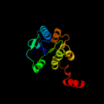
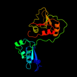
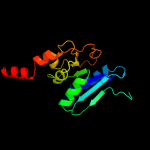
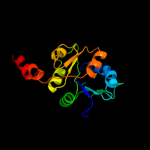
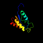
Image coloured by rainbow N → C terminus
Model (left) based on template c3oy2A_
Top template information
PDB header:viral protein,transferase
Chain: A: PDB Molecule:glycosyltransferase b736l;
PDBTitle: crystal structure of a putative glycosyltransferase from paramecium2 bursaria chlorella virus ny2a
Confidence and coverage
Confidence: 93.9% Coverage: 51%
160 residues ( 51% of your sequence) have been modelled with 93.9% confidence by the single highest scoring template.
You may wish to submit your sequence to Phyrealarm. This will automatically scan your sequence every week for new potential templates as they appear in the Phyre2 library.
3D viewing
Interactive 3D view in Jmol

Sequence analysis 

Secondary structure and disorder prediction 

   1........10.........20.........30.........40.........50.........60
Sequence  MTIYFINWVADYELKMIQYLKKKYKIKNITTPKKYNWINKKISKIGMDNAWLGRLFIKHY
Secondary structure 


SS confidence 


Disorder  ?


?


?

??


Disorder confidence 


   .........70.........80.........90.........100.........110.........120
Sequence  LNDIKKDDIIIINDSVVNKGINKQILKNINCHKVLLLRNTVGEDFILDNANYFDIIYDFE
Secondary structure 


SS confidence 


Disorder 


Disorder confidence 


   .........130.........140.........150.........160.........170.........180
Sequence  HRFIGNEKIKAIEQFFPIGMDEIRNYSLSDKNNSQPICFFLGRDKGRLQIINELAERLTT
Secondary structure 


SS confidence 


Disorder 


??
?


?????


Disorder confidence 


   .........190.........200.........210.........220.........230.........240
Sequence  LGCKLDFNVVKDKTSSTTSKYLIEKQISYEENIRRTLNANIIVDITKENQSGWTLRILEA
Secondary structure 


SS confidence 


Disorder 


?????


Disorder confidence 


   .........250.........260.........270.........280.........290.........300
Sequence  LFFNKKLITNNINVFGSEIYSESRFFIIGHDDWDKLEYFINSSVKPMDYDSLYKFSPDKM
Secondary structure 


SS confidence 


Disorder 


Disorder confidence 


   .........310.
Sequence  MSTIVSDFIDK
Secondary structure 

SS confidence 


Disorder 


????
Disorder confidence 


Confidence Key
High(9)                    Low (0)
?Disordered
Alpha helix
Beta strand

Domain analysis 

Hover over an aligned region to see model and summary info

Please note, only up to the top 20 hits are modelled to reduce computer load

RankAligned region

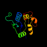
PDB 3oy2 chain A

3D model

Region: 147 - 308
Aligned: 160
Modelled: 162
Confidence: 93.9%
Identity: 19%
PDB header:viral protein,transferase
Chain: A: PDB Molecule:glycosyltransferase b736l;
PDBTitle: crystal structure of a putative glycosyltransferase from paramecium2 bursaria chlorella virus ny2a

Phyre2

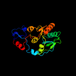
PDB 2gej chain A

3D model

Region: 128 - 306
Aligned: 176
Modelled: 179
Confidence: 91.2%
Identity: 10%
PDB header:transferase
Chain: A: PDB Molecule:phosphatidylinositol mannosyltransferase (pima);
PDBTitle: crystal structure of phosphatidylinositol mannosyltransferase (pima)2 from mycobacterium smegmatis in complex with gdp-man

Phyre2

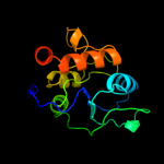
PDB 3c4v chain B

3D model

Region: 148 - 306
Aligned: 157
Modelled: 159
Confidence: 89.4%
Identity: 10%
PDB header:transferase
Chain: B: PDB Molecule:predicted glycosyltransferases;
PDBTitle: structure of the retaining glycosyltransferase msha:the2 first step in mycothiol biosynthesis. organism:3 corynebacterium glutamicum : complex with udp and 1l-ins-1-4 p.

Phyre2

PDB 2x0d chain A

3D model

Region: 144 - 282
Aligned: 136
Modelled: 139
Confidence: 86.2%
Identity: 11%
PDB header:transferase
Chain: A: PDB Molecule:wsaf;
PDBTitle: apo structure of wsaf

Phyre2

PDB 3rhz chain B

3D model

Region: 50 - 251
Aligned: 192
Modelled: 200
Confidence: 85.1%
Identity: 7%
PDB header:transferase
Chain: B: PDB Molecule:nucleotide sugar synthetase-like protein;
PDBTitle: structure and functional analysis of a new subfamily of2 glycosyltransferases required for glycosylation of serine-rich3 streptococcal adhesions

Phyre2

PDB 2xmp chain B

3D model

Region: 151 - 306
Aligned: 154
Modelled: 156
Confidence: 85.1%
Identity: 14%
PDB header:sugar binding protein
Chain: B: PDB Molecule:trehalose-synthase tret;
PDBTitle: crystal structure of trehalose synthase tret mutant e326a2 from p.horishiki in complex with udp

Phyre2

PDB 2r60 chain A

3D model

Region: 169 - 306
Aligned: 122
Modelled: 124
Confidence: 69.6%
Identity: 7%
PDB header:transferase
Chain: A: PDB Molecule:glycosyl transferase, group 1;
PDBTitle: structure of apo sucrose phosphate synthase (sps) of2 halothermothrix orenii

Phyre2

PDB 2bis chain A domain 1

3D model

Region: 152 - 306
Aligned: 152
Modelled: 155
Confidence: 64.4%
Identity: 14%
Fold: UDP-Glycosyltransferase/glycogen phosphorylase
Superfamily: UDP-Glycosyltransferase/glycogen phosphorylase
Family: Glycosyl transferases group 1

Phyre2

PDB 1uqu chain B

3D model

Region: 151 - 307
Aligned: 152
Modelled: 157
Confidence: 63.1%
Identity: 16%
PDB header:synthase
Chain: B: PDB Molecule:alpha, alpha-trehalose-phosphate synthase;
PDBTitle: trehalose-6-phosphate from e. coli bound with udp-glucose.

Phyre2

PDB 2x6r chain A

3D model

Region: 151 - 306
Aligned: 154
Modelled: 155
Confidence: 59.5%
Identity: 13%
PDB header:isomerase
Chain: A: PDB Molecule:trehalose-synthase tret;
PDBTitle: crystal structure of trehalose synthase tret from p.2 horikoshi produced by soaking in trehalose

Phyre2

PDB 2jjm chain H

3D model

Region: 220 - 306
Aligned: 85
Modelled: 87
Confidence: 55.0%
Identity: 14%
PDB header:transferase
Chain: H: PDB Molecule:glycosyl transferase, group 1 family protein;
PDBTitle: crystal structure of a family gt4 glycosyltransferase from2 bacillus anthracis orf ba1558.

Phyre2

PDB 1uqt chain A

3D model

Region: 151 - 307
Aligned: 151
Modelled: 157
Confidence: 47.9%
Identity: 14%
Fold: UDP-Glycosyltransferase/glycogen phosphorylase
Superfamily: UDP-Glycosyltransferase/glycogen phosphorylase
Family: Trehalose-6-phosphate synthase, OtsA

Phyre2

PDB 3oka chain A

3D model

Region: 151 - 306
Aligned: 155
Modelled: 156
Confidence: 34.3%
Identity: 12%
PDB header:transferase
Chain: A: PDB Molecule:gdp-mannose-dependent alpha-(1-6)-phosphatidylinositol
PDBTitle: crystal structure of corynebacterium glutamicum pimb' in complex with2 gdp-man (triclinic crystal form)

Phyre2

PDB 1rzu chain A

3D model

Region: 206 - 306
Aligned: 99
Modelled: 101
Confidence: 30.7%
Identity: 16%
Fold: UDP-Glycosyltransferase/glycogen phosphorylase
Superfamily: UDP-Glycosyltransferase/glycogen phosphorylase
Family: Glycosyl transferases group 1

Phyre2

PDB 3pe3 chain D

3D model

Region: 151 - 250
Aligned: 97
Modelled: 100
Confidence: 29.8%
Identity: 15%
PDB header:transferase
Chain: D: PDB Molecule:udp-n-acetylglucosamine--peptide n-
PDBTitle: structure of human o-glcnac transferase and its complex with a peptide2 substrate

Phyre2

PDB 2xcu chain C

3D model

Region: 109 - 250
Aligned: 140
Modelled: 142
Confidence: 27.0%
Identity: 14%
PDB header:transferase
Chain: C: PDB Molecule:3-deoxy-d-manno-2-octulosonic acid transferase;
PDBTitle: membrane-embedded monofunctional glycosyltransferase waaa of aquifex2 aeolicus, comlex with cmp

Phyre2

PDB 2khz chain B

3D model

Region: 148 - 248
Aligned: 101
Modelled: 101
Confidence: 26.8%
Identity: 17%
PDB header:nuclear protein
Chain: B: PDB Molecule:c-myc-responsive protein rcl;
PDBTitle: solution structure of rcl

Phyre2

PDB 2f9f chain A domain 1

3D model

Region: 152 - 278
Aligned: 124
Modelled: 127
Confidence: 26.6%
Identity: 15%
Fold: UDP-Glycosyltransferase/glycogen phosphorylase
Superfamily: UDP-Glycosyltransferase/glycogen phosphorylase
Family: Glycosyl transferases group 1

Phyre2

PDB 2q6v chain A

3D model

Region: 109 - 307
Aligned: 191
Modelled: 198
Confidence: 26.0%
Identity: 10%
PDB header:transferase
Chain: A: PDB Molecule:glucuronosyltransferase gumk;
PDBTitle: crystal structure of gumk in complex with udp

Phyre2

PDB 1jix chain A

3D model

Region: 145 - 294
Aligned: 149
Modelled: 150
Confidence: 23.4%
Identity: 19%
Fold: UDP-Glycosyltransferase/glycogen phosphorylase
Superfamily: UDP-Glycosyltransferase/glycogen phosphorylase
Family: beta-Glucosyltransferase (DNA-modifying)

Phyre2
1

c3oy2A_
2

c2gejA_
3

c3c4vB_
4

c2x0dA_
5

c3rhzB_
6

c2xmpB_
7

c2r60A_
8

d2bisa1
9

c1uquB_
10

c2x6rA_
11

c2jjmH_
12

d1uqta_
13

c3okaA_
14

d1rzua_
15

c3pe3D_
16

c2xcuC_
17

c2khzB_
18

d2f9fa1
19

c2q6vA_
20

d1jixa_
21


22


23


24


25


26


27


28


29


30


31


32


33


34


Detailed template information 

#
Template Alignment Coverage3D Model Confidence
% i.d. Template Information
1c3oy2A_


93.9 19 PDB header:viral protein,transferase
Chain: A: PDB Molecule:glycosyltransferase b736l;
PDBTitle: crystal structure of a putative glycosyltransferase from paramecium2 bursaria chlorella virus ny2a
2c2gejA_


91.2 10 PDB header:transferase
Chain: A: PDB Molecule:phosphatidylinositol mannosyltransferase (pima);
PDBTitle: crystal structure of phosphatidylinositol mannosyltransferase (pima)2 from mycobacterium smegmatis in complex with gdp-man
3c3c4vB_


89.4 10 PDB header:transferase
Chain: B: PDB Molecule:predicted glycosyltransferases;
PDBTitle: structure of the retaining glycosyltransferase msha:the2 first step in mycothiol biosynthesis. organism:3 corynebacterium glutamicum : complex with udp and 1l-ins-1-4 p.
4c2x0dA_


86.2 11 PDB header:transferase
Chain: A: PDB Molecule:wsaf;
PDBTitle: apo structure of wsaf
5c3rhzB_


85.1 7 PDB header:transferase
Chain: B: PDB Molecule:nucleotide sugar synthetase-like protein;
PDBTitle: structure and functional analysis of a new subfamily of2 glycosyltransferases required for glycosylation of serine-rich3 streptococcal adhesions
6c2xmpB_


85.1 14 PDB header:sugar binding protein
Chain: B: PDB Molecule:trehalose-synthase tret;
PDBTitle: crystal structure of trehalose synthase tret mutant e326a2 from p.horishiki in complex with udp
7c2r60A_


69.6 7 PDB header:transferase
Chain: A: PDB Molecule:glycosyl transferase, group 1;
PDBTitle: structure of apo sucrose phosphate synthase (sps) of2 halothermothrix orenii
8d2bisa1


64.4 14 Fold:UDP-Glycosyltransferase/glycogen phosphorylase
Superfamily:UDP-Glycosyltransferase/glycogen phosphorylase
Family:Glycosyl transferases group 1
9c1uquB_


63.1 16 PDB header:synthase
Chain: B: PDB Molecule:alpha, alpha-trehalose-phosphate synthase;
PDBTitle: trehalose-6-phosphate from e. coli bound with udp-glucose.
10c2x6rA_


59.5 13 PDB header:isomerase
Chain: A: PDB Molecule:trehalose-synthase tret;
PDBTitle: crystal structure of trehalose synthase tret from p.2 horikoshi produced by soaking in trehalose
11c2jjmH_


55.0 14 PDB header:transferase
Chain: H: PDB Molecule:glycosyl transferase, group 1 family protein;
PDBTitle: crystal structure of a family gt4 glycosyltransferase from2 bacillus anthracis orf ba1558.
12d1uqta_


47.9 14 Fold:UDP-Glycosyltransferase/glycogen phosphorylase
Superfamily:UDP-Glycosyltransferase/glycogen phosphorylase
Family:Trehalose-6-phosphate synthase, OtsA
13c3okaA_


34.3 12 PDB header:transferase
Chain: A: PDB Molecule:gdp-mannose-dependent alpha-(1-6)-phosphatidylinositol
PDBTitle: crystal structure of corynebacterium glutamicum pimb' in complex with2 gdp-man (triclinic crystal form)
14d1rzua_


30.7 16 Fold:UDP-Glycosyltransferase/glycogen phosphorylase
Superfamily:UDP-Glycosyltransferase/glycogen phosphorylase
Family:Glycosyl transferases group 1
15c3pe3D_


29.8 15 PDB header:transferase
Chain: D: PDB Molecule:udp-n-acetylglucosamine--peptide n-
PDBTitle: structure of human o-glcnac transferase and its complex with a peptide2 substrate
16c2xcuC_


27.0 14 PDB header:transferase
Chain: C: PDB Molecule:3-deoxy-d-manno-2-octulosonic acid transferase;
PDBTitle: membrane-embedded monofunctional glycosyltransferase waaa of aquifex2 aeolicus, comlex with cmp
17c2khzB_


26.8 17 PDB header:nuclear protein
Chain: B: PDB Molecule:c-myc-responsive protein rcl;
PDBTitle: solution structure of rcl
18d2f9fa1


26.6 15 Fold:UDP-Glycosyltransferase/glycogen phosphorylase
Superfamily:UDP-Glycosyltransferase/glycogen phosphorylase
Family:Glycosyl transferases group 1
19c2q6vA_


26.0 10 PDB header:transferase
Chain: A: PDB Molecule:glucuronosyltransferase gumk;
PDBTitle: crystal structure of gumk in complex with udp
20d1jixa_


23.4 19 Fold:UDP-Glycosyltransferase/glycogen phosphorylase
Superfamily:UDP-Glycosyltransferase/glycogen phosphorylase
Family:beta-Glucosyltransferase (DNA-modifying)
21c2vsnB_


not modelled 21.5 11 PDB header:transferase
Chain: B: PDB Molecule:xcogt;
PDBTitle: structure and topological arrangement of an o-glcnac2 transferase homolog: insight into molecular control of3 intracellular glycosylation
22c3h16A_


not modelled 16.2 14 PDB header:signaling protein
Chain: A: PDB Molecule:tir protein;
PDBTitle: crystal structure of a bacteria tir domain, pdtir from2 paracoccus denitrificans
23d1r0ka2


not modelled 12.9 12 Fold:NAD(P)-binding Rossmann-fold domains
Superfamily:NAD(P)-binding Rossmann-fold domains
Family:Glyceraldehyde-3-phosphate dehydrogenase-like, N-terminal domain
24d1q0qa2


not modelled 12.3 21 Fold:NAD(P)-binding Rossmann-fold domains
Superfamily:NAD(P)-binding Rossmann-fold domains
Family:Glyceraldehyde-3-phosphate dehydrogenase-like, N-terminal domain
25d2iw1a1


not modelled 11.0 14 Fold:UDP-Glycosyltransferase/glycogen phosphorylase
Superfamily:UDP-Glycosyltransferase/glycogen phosphorylase
Family:Glycosyl transferases group 1
26c2eghA_


not modelled 10.1 21 PDB header:oxidoreductase
Chain: A: PDB Molecule:1-deoxy-d-xylulose 5-phosphate reductoisomerase;
PDBTitle: crystal structure of 1-deoxy-d-xylulose 5-phosphate reductoisomerase2 complexed with a magnesium ion, nadph and fosmidomycin
27c3a14B_


not modelled 9.6 14 PDB header:oxidoreductase
Chain: B: PDB Molecule:1-deoxy-d-xylulose 5-phosphate reductoisomerase;
PDBTitle: crystal structure of dxr from thermotoga maritima, in complex with2 nadph
28d1o6ca_


not modelled 7.9 11 Fold:UDP-Glycosyltransferase/glycogen phosphorylase
Superfamily:UDP-Glycosyltransferase/glycogen phosphorylase
Family:UDP-N-acetylglucosamine 2-epimerase
29d1ipaa1


not modelled 7.2 19 Fold:alpha/beta knot
Superfamily:alpha/beta knot
Family:SpoU-like RNA 2'-O ribose methyltransferase
30c1q2lA_


not modelled 6.9 13 PDB header:hydrolase
Chain: A: PDB Molecule:protease iii;
PDBTitle: crystal structure of pitrilysin
31d1m53a1


not modelled 6.2 12 Fold:Glycosyl hydrolase domain
Superfamily:Glycosyl hydrolase domain
Family:alpha-Amylases, C-terminal beta-sheet domain
32c2i6dA_


not modelled 6.1 11 PDB header:transferase
Chain: A: PDB Molecule:rna methyltransferase, trmh family;
PDBTitle: the structure of a putative rna methyltransferase of the trmh family2 from porphyromonas gingivalis.
33c2f06B_


not modelled 5.9 14 PDB header:structural genomics, unknown function
Chain: B: PDB Molecule:conserved hypothetical protein;
PDBTitle: crystal structure of protein bt0572 from bacteroides thetaiotaomicron
34c2hlgA_


not modelled 5.4 44 PDB header:plant protein
Chain: A: PDB Molecule:fruit-specific protein;
PDBTitle: nmr solution structure of a new tomato peptide

Binding site prediction 

Due to computational demand, binding site predictions are not run for batch jobs

If you want to predict binding sites, please manually submit your model of choice to 3DLigandSite


Phyre is for academic use only

Please cite: Protein structure prediction on the web: a case study using the Phyre server
Kelley LA and Sternberg MJE. Nature Protocols 4, 363 - 371 (2009) [pdf] [Import into BibTeX]
 
If you use the binding site predictions from 3DLigandSite, please also cite:
3DLigandSite: predicting ligand-binding sites using similar structures.
Wass MN, Kelley LA and Sternberg MJ Nucleic Acids Research 38, W469-73 (2010) [PubMed]
 
© Structural Bioinformatics Group
Imperial College London
Lawrence Kelley, Benjamin Jefferys
Disclaimer
Terms and Conditions
Component software
Template detection: HHpred 1.51
Secondary structure prediction: Psi-pred 2.5
Disorder prediction: Disopred 2.4
Transmembrane prediction: Memsat_SVM
Multi-template modelling and ab initio: Poing 1.0