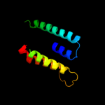
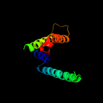
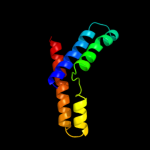
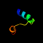
| 1 |
|
PDB 3lrc chain C
Region: 54 - 134
Aligned: 80
Modelled: 81
Confidence: 14.9%
Identity: 10%
PDB header:transport protein
Chain: C: PDB Molecule:arginine/agmatine antiporter;
PDBTitle: structure of e. coli adic (p1)
Phyre2
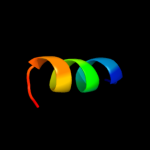
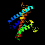
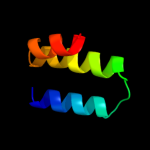
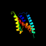
| 2 |
|
PDB 3gia chain A
Region: 36 - 140
Aligned: 105
Modelled: 105
Confidence: 13.6%
Identity: 10%
PDB header:transport protein
Chain: A: PDB Molecule:uncharacterized protein mj0609;
PDBTitle: crystal structure of apct transporter
Phyre2
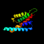
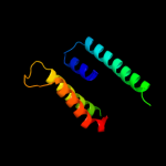
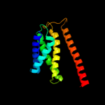
| 3 |
|
PDB 1j4n chain A
Region: 65 - 157
Aligned: 93
Modelled: 93
Confidence: 11.7%
Identity: 11%
Fold: Aquaporin-like
Superfamily: Aquaporin-like
Family: Aquaporin-like
Phyre2
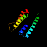
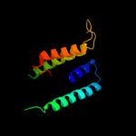
| 4 |
|
PDB 2j83 chain B
Region: 256 - 268
Aligned: 13
Modelled: 13
Confidence: 9.1%
Identity: 38%
PDB header:hydrolase
Chain: B: PDB Molecule:ulilysin;
PDBTitle: ulilysin metalloprotease in complex with batimastat.
Phyre2
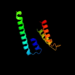
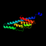
| 5 |
|
PDB 3llq chain B
Region: 65 - 236
Aligned: 138
Modelled: 143
Confidence: 8.9%
Identity: 13%
PDB header:membrane protein
Chain: B: PDB Molecule:aquaporin z 2;
PDBTitle: aquaporin structure from plant pathogen agrobacterium tumerfaciens
Phyre2
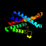
| 6 |
|
PDB 1fx8 chain A
Region: 65 - 154
Aligned: 90
Modelled: 90
Confidence: 8.7%
Identity: 12%
Fold: Aquaporin-like
Superfamily: Aquaporin-like
Family: Aquaporin-like
Phyre2
| 7 |
|
PDB 2d57 chain A
Region: 65 - 163
Aligned: 99
Modelled: 99
Confidence: 7.0%
Identity: 17%
PDB header:transport protein
Chain: A: PDB Molecule:aquaporin-4;
PDBTitle: double layered 2d crystal structure of aquaporin-4 (aqp4m23) at 3.2 a2 resolution by electron crystallography
Phyre2
| 8 |
|
PDB 1lda chain A
Region: 65 - 238
Aligned: 154
Modelled: 168
Confidence: 6.5%
Identity: 18%
PDB header:transport protein
Chain: A: PDB Molecule:glycerol uptake facilitator protein;
PDBTitle: crystal structure of the e. coli glycerol facilitator (glpf) without2 substrate glycerol
Phyre2
| 9 |
|
PDB 1ymg chain A domain 1
Region: 65 - 157
Aligned: 93
Modelled: 93
Confidence: 6.5%
Identity: 15%
Fold: Aquaporin-like
Superfamily: Aquaporin-like
Family: Aquaporin-like
Phyre2
| 10 |
|
PDB 1ymg chain A
Region: 65 - 157
Aligned: 93
Modelled: 93
Confidence: 6.5%
Identity: 15%
PDB header:membrane protein
Chain: A: PDB Molecule:lens fiber major intrinsic protein;
PDBTitle: the channel architecture of aquaporin o at 2.2 angstrom resolution
Phyre2
| 11 |
|
PDB 3klz chain E
Region: 18 - 104
Aligned: 87
Modelled: 87
Confidence: 6.3%
Identity: 17%
PDB header:membrane protein
Chain: E: PDB Molecule:putative formate transporter 1;
PDBTitle: pentameric formate channel with formate bound
Phyre2
| 12 |
|
PDB 1cii chain A
Region: 193 - 235
Aligned: 43
Modelled: 43
Confidence: 6.2%
Identity: 16%
PDB header:transmembrane protein
Chain: A: PDB Molecule:colicin ia;
PDBTitle: colicin ia
Phyre2
| 13 |
|
PDB 1u7g chain A
Region: 50 - 205
Aligned: 155
Modelled: 156
Confidence: 5.9%
Identity: 12%
Fold: Ammonium transporter
Superfamily: Ammonium transporter
Family: Ammonium transporter
Phyre2
| 14 |
|
PDB 3c02 chain A
Region: 65 - 233
Aligned: 152
Modelled: 169
Confidence: 5.8%
Identity: 16%
PDB header:membrane protein
Chain: A: PDB Molecule:aquaglyceroporin;
PDBTitle: x-ray structure of the aquaglyceroporin from plasmodium falciparum
Phyre2
| 15 |
|
PDB 3hfw chain A
Region: 216 - 241
Aligned: 26
Modelled: 26
Confidence: 5.7%
Identity: 8%
PDB header:hydrolase
Chain: A: PDB Molecule:protein adp-ribosylarginine hydrolase;
PDBTitle: crystal structure of human adp-ribosylhydrolase 1 (harh1)
Phyre2
| 16 |
|
PDB 1rc2 chain A
Region: 65 - 201
Aligned: 137
Modelled: 137
Confidence: 5.6%
Identity: 10%
Fold: Aquaporin-like
Superfamily: Aquaporin-like
Family: Aquaporin-like
Phyre2
| 17 |
|
PDB 1h6i chain A
Region: 65 - 163
Aligned: 99
Modelled: 99
Confidence: 5.5%
Identity: 8%
Fold: Aquaporin-like
Superfamily: Aquaporin-like
Family: Aquaporin-like
Phyre2
| 18 |
|
PDB 2ht2 chain B
Region: 71 - 257
Aligned: 165
Modelled: 174
Confidence: 5.5%
Identity: 11%
PDB header:membrane protein
Chain: B: PDB Molecule:h(+)/cl(-) exchange transporter clca;
PDBTitle: structure of the escherichia coli clc chloride channel2 y445h mutant and fab complex
Phyre2
| 19 |
|
PDB 3g9d chain B
Region: 216 - 241
Aligned: 26
Modelled: 26
Confidence: 5.4%
Identity: 19%
PDB header:hydrolase
Chain: B: PDB Molecule:dinitrogenase reductase activacting
PDBTitle: crystal structure glycohydrolase
Phyre2
| 20 |
|
PDB 2jp3 chain A
Region: 111 - 142
Aligned: 32
Modelled: 32
Confidence: 5.3%
Identity: 16%
PDB header:transcription
Chain: A: PDB Molecule:fxyd domain-containing ion transport regulator 4;
PDBTitle: solution structure of the human fxyd4 (chif) protein in sds2 micelles
Phyre2
| 21 |
|