Phyre Home Retrieve Phyre Job Id
Emaill.a.kelley@imperial.ac.uk
DescriptionP58037
DateWed Jan 25 15:20:58 GMT 2012
Unique Job ID7c5a023f81ca0846

Summary 

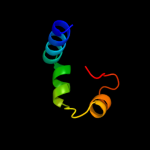
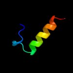
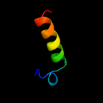
Top model
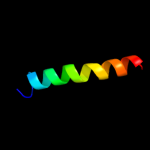
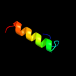
Image coloured by rainbow N → C terminus
Model (left) based on template d1s4ka_
Top template information
Fold:lambda repressor-like DNA-binding domains
Superfamily:lambda repressor-like DNA-binding domains
Family:YdiL-like
Confidence and coverage
Confidence: 21.4% Coverage: 84%
43 residues ( 84% of your sequence) have been modelled with 21.4% confidence by the single highest scoring template.
You may wish to submit your sequence to Phyrealarm. This will automatically scan your sequence every week for new potential templates as they appear in the Phyre2 library.
Please note: You must be registered and logged in to use Phyrealarm.
3D viewing
Interactive 3D view in Jmol

Sequence analysis 

Secondary structure and disorder prediction 

   1........10.........20.........30.........40.........50.
Sequence  MEKLTTELHSLSEMDRRHVVSILAEIANGYDDFNDMLIYLEFYPNHKILIS
Secondary structure 


SS confidence 


Disorder  ???


????
Disorder confidence 


Confidence Key
High(9)                    Low (0)
?Disordered
Alpha helix
Beta strand

Domain analysis 

Hover over an aligned region to see model and summary info

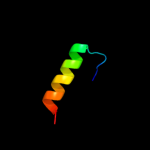
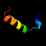
Please note, only up to the top 20 hits are modelled to reduce computer load

RankAligned region

PDB 1s4k chain A

3D model

Region: 7 - 49
Aligned: 43
Modelled: 43
Confidence: 21.4%
Identity: 21%
Fold: lambda repressor-like DNA-binding domains
Superfamily: lambda repressor-like DNA-binding domains
Family: YdiL-like

Phyre2

PDB 3pue chain A

3D model

Region: 5 - 29
Aligned: 25
Modelled: 25
Confidence: 11.7%
Identity: 28%
PDB header:lyase
Chain: A: PDB Molecule:dihydrodipicolinate synthase;
PDBTitle: crystal structure of the complex of dhydrodipicolinate synthase from2 acinetobacter baumannii with lysine at 2.6a resolution

Phyre2

PDB 1plo chain A

3D model

Region: 31 - 49
Aligned: 19
Modelled: 19
Confidence: 10.8%
Identity: 32%
Fold: Snake toxin-like
Superfamily: Snake toxin-like
Family: Extracellular domain of cell surface receptors

Phyre2

PDB 3fkk chain A

3D model

Region: 5 - 29
Aligned: 25
Modelled: 25
Confidence: 10.6%
Identity: 16%
PDB header:lyase
Chain: A: PDB Molecule:l-2-keto-3-deoxyarabonate dehydratase;
PDBTitle: structure of l-2-keto-3-deoxyarabonate dehydratase

Phyre2

PDB 2gsc chain A domain 1

3D model

Region: 7 - 31
Aligned: 25
Modelled: 25
Confidence: 10.3%
Identity: 40%
Fold: Bromodomain-like
Superfamily: IVS-encoded protein-like
Family: IVS-encoded protein-like

Phyre2

PDB 3h5d chain D

3D model

Region: 5 - 29
Aligned: 25
Modelled: 25
Confidence: 9.5%
Identity: 20%
PDB header:lyase
Chain: D: PDB Molecule:dihydrodipicolinate synthase;
PDBTitle: dihydrodipicolinate synthase from drug-resistant streptococcus2 pneumoniae

Phyre2

PDB 3g0s chain A

3D model

Region: 5 - 29
Aligned: 25
Modelled: 25
Confidence: 9.4%
Identity: 36%
PDB header:lyase
Chain: A: PDB Molecule:dihydrodipicolinate synthase;
PDBTitle: dihydrodipicolinate synthase from salmonella typhimurium lt2

Phyre2

PDB 3qfe chain B

3D model

Region: 5 - 29
Aligned: 25
Modelled: 25
Confidence: 9.1%
Identity: 12%
PDB header:lyase
Chain: B: PDB Molecule:putative dihydrodipicolinate synthase family protein;
PDBTitle: crystal structures of a putative dihydrodipicolinate synthase family2 protein from coccidioides immitis

Phyre2

PDB 2a6n chain A domain 1

3D model

Region: 5 - 29
Aligned: 25
Modelled: 25
Confidence: 9.0%
Identity: 28%
Fold: TIM beta/alpha-barrel
Superfamily: Aldolase
Family: Class I aldolase

Phyre2

PDB 1f74 chain A

3D model

Region: 5 - 29
Aligned: 25
Modelled: 25
Confidence: 8.8%
Identity: 24%
Fold: TIM beta/alpha-barrel
Superfamily: Aldolase
Family: Class I aldolase

Phyre2

PDB 3d0c chain B

3D model

Region: 5 - 29
Aligned: 25
Modelled: 25
Confidence: 8.7%
Identity: 28%
PDB header:lyase
Chain: B: PDB Molecule:dihydrodipicolinate synthase;
PDBTitle: crystal structure of dihydrodipicolinate synthase from2 oceanobacillus iheyensis at 1.9 a resolution

Phyre2

PDB 3bi8 chain A

3D model

Region: 5 - 29
Aligned: 25
Modelled: 25
Confidence: 8.5%
Identity: 20%
PDB header:lyase
Chain: A: PDB Molecule:dihydrodipicolinate synthase;
PDBTitle: structure of dihydrodipicolinate synthase from clostridium2 botulinum

Phyre2

PDB 3na8 chain A

3D model

Region: 5 - 29
Aligned: 25
Modelled: 25
Confidence: 8.3%
Identity: 24%
PDB header:lyase
Chain: A: PDB Molecule:putative dihydrodipicolinate synthetase;
PDBTitle: crystal structure of a putative dihydrodipicolinate synthetase from2 pseudomonas aeruginosa

Phyre2

PDB 1hl2 chain A

3D model

Region: 5 - 29
Aligned: 25
Modelled: 25
Confidence: 8.2%
Identity: 40%
Fold: TIM beta/alpha-barrel
Superfamily: Aldolase
Family: Class I aldolase

Phyre2

PDB 2v9d chain B

3D model

Region: 5 - 29
Aligned: 25
Modelled: 25
Confidence: 7.9%
Identity: 12%
PDB header:lyase
Chain: B: PDB Molecule:yage;
PDBTitle: crystal structure of yage, a prophage protein belonging to2 the dihydrodipicolinic acid synthase family from e. coli3 k12

Phyre2

PDB 3n2x chain B

3D model

Region: 5 - 29
Aligned: 25
Modelled: 25
Confidence: 7.9%
Identity: 12%
PDB header:lyase
Chain: B: PDB Molecule:uncharacterized protein yage;
PDBTitle: crystal structure of yage, a prophage protein belonging to the2 dihydrodipicolinic acid synthase family from e. coli k12 in complex3 with pyruvate

Phyre2

PDB 3b4u chain B

3D model

Region: 5 - 29
Aligned: 25
Modelled: 25
Confidence: 7.7%
Identity: 20%
PDB header:lyase
Chain: B: PDB Molecule:dihydrodipicolinate synthase;
PDBTitle: crystal structure of dihydrodipicolinate synthase from agrobacterium2 tumefaciens str. c58

Phyre2

PDB 1z3x chain A domain 1

3D model

Region: 1 - 29
Aligned: 29
Modelled: 29
Confidence: 7.5%
Identity: 17%
Fold: alpha-alpha superhelix
Superfamily: ARM repeat
Family: GUN4-associated domain

Phyre2

PDB 1ki1 chain B domain 2

3D model

Region: 33 - 39
Aligned: 7
Modelled: 7
Confidence: 7.2%
Identity: 57%
Fold: PH domain-like barrel
Superfamily: PH domain-like
Family: Pleckstrin-homology domain (PH domain)

Phyre2

PDB 3si9 chain B

3D model

Region: 5 - 29
Aligned: 25
Modelled: 25
Confidence: 7.1%
Identity: 16%
PDB header:lyase
Chain: B: PDB Molecule:dihydrodipicolinate synthase;
PDBTitle: crystal structure of dihydrodipicolinate synthase from bartonella2 henselae

Phyre2
1

d1s4ka_
2

c3pueA_
3

d1ploa_
4

c3fkkA_
5

d2gsca1
6

c3h5dD_
7

c3g0sA_
8

c3qfeB_
9

d2a6na1
10

d1f74a_
11

c3d0cB_
12

c3bi8A_
13

c3na8A_
14

d1hl2a_
15

c2v9dB_
16

c3n2xB_
17

c3b4uB_
18

d1z3xa1
19

d1ki1b2
20

c3si9B_
21


22


23


24


25


26


27


28


29


30


31


Detailed template information 

#
Template Alignment Coverage3D Model Confidence
% i.d. Template Information
1d1s4ka_


21.4 21 Fold:lambda repressor-like DNA-binding domains
Superfamily:lambda repressor-like DNA-binding domains
Family:YdiL-like
2c3pueA_


11.7 28 PDB header:lyase
Chain: A: PDB Molecule:dihydrodipicolinate synthase;
PDBTitle: crystal structure of the complex of dhydrodipicolinate synthase from2 acinetobacter baumannii with lysine at 2.6a resolution
3d1ploa_


10.8 32 Fold:Snake toxin-like
Superfamily:Snake toxin-like
Family:Extracellular domain of cell surface receptors
4c3fkkA_


10.6 16 PDB header:lyase
Chain: A: PDB Molecule:l-2-keto-3-deoxyarabonate dehydratase;
PDBTitle: structure of l-2-keto-3-deoxyarabonate dehydratase
5d2gsca1


10.3 40 Fold:Bromodomain-like
Superfamily:IVS-encoded protein-like
Family:IVS-encoded protein-like
6c3h5dD_


9.5 20 PDB header:lyase
Chain: D: PDB Molecule:dihydrodipicolinate synthase;
PDBTitle: dihydrodipicolinate synthase from drug-resistant streptococcus2 pneumoniae
7c3g0sA_


9.4 36 PDB header:lyase
Chain: A: PDB Molecule:dihydrodipicolinate synthase;
PDBTitle: dihydrodipicolinate synthase from salmonella typhimurium lt2
8c3qfeB_


9.1 12 PDB header:lyase
Chain: B: PDB Molecule:putative dihydrodipicolinate synthase family protein;
PDBTitle: crystal structures of a putative dihydrodipicolinate synthase family2 protein from coccidioides immitis
9d2a6na1


9.0 28 Fold:TIM beta/alpha-barrel
Superfamily:Aldolase
Family:Class I aldolase
10d1f74a_


8.8 24 Fold:TIM beta/alpha-barrel
Superfamily:Aldolase
Family:Class I aldolase
11c3d0cB_


8.7 28 PDB header:lyase
Chain: B: PDB Molecule:dihydrodipicolinate synthase;
PDBTitle: crystal structure of dihydrodipicolinate synthase from2 oceanobacillus iheyensis at 1.9 a resolution
12c3bi8A_


8.5 20 PDB header:lyase
Chain: A: PDB Molecule:dihydrodipicolinate synthase;
PDBTitle: structure of dihydrodipicolinate synthase from clostridium2 botulinum
13c3na8A_


8.3 24 PDB header:lyase
Chain: A: PDB Molecule:putative dihydrodipicolinate synthetase;
PDBTitle: crystal structure of a putative dihydrodipicolinate synthetase from2 pseudomonas aeruginosa
14d1hl2a_


8.2 40 Fold:TIM beta/alpha-barrel
Superfamily:Aldolase
Family:Class I aldolase
15c2v9dB_


7.9 12 PDB header:lyase
Chain: B: PDB Molecule:yage;
PDBTitle: crystal structure of yage, a prophage protein belonging to2 the dihydrodipicolinic acid synthase family from e. coli3 k12
16c3n2xB_


7.9 12 PDB header:lyase
Chain: B: PDB Molecule:uncharacterized protein yage;
PDBTitle: crystal structure of yage, a prophage protein belonging to the2 dihydrodipicolinic acid synthase family from e. coli k12 in complex3 with pyruvate
17c3b4uB_


7.7 20 PDB header:lyase
Chain: B: PDB Molecule:dihydrodipicolinate synthase;
PDBTitle: crystal structure of dihydrodipicolinate synthase from agrobacterium2 tumefaciens str. c58
18d1z3xa1


7.5 17 Fold:alpha-alpha superhelix
Superfamily:ARM repeat
Family:GUN4-associated domain
19d1ki1b2


7.2 57 Fold:PH domain-like barrel
Superfamily:PH domain-like
Family:Pleckstrin-homology domain (PH domain)
20c3si9B_


7.1 16 PDB header:lyase
Chain: B: PDB Molecule:dihydrodipicolinate synthase;
PDBTitle: crystal structure of dihydrodipicolinate synthase from bartonella2 henselae
21c3qwmA_


not modelled 6.9 57 PDB header:signaling protein
Chain: A: PDB Molecule:iq motif and sec7 domain-containing protein 1;
PDBTitle: crystal structure of gep100, the plextrin homology domain of iq motif2 and sec7 domain-containing protein 1 isoform a
22d1xkya1


not modelled 6.9 12 Fold:TIM beta/alpha-barrel
Superfamily:Aldolase
Family:Class I aldolase
23c3eb2A_


not modelled 6.8 32 PDB header:lyase
Chain: A: PDB Molecule:putative dihydrodipicolinate synthetase;
PDBTitle: crystal structure of dihydrodipicolinate synthase from2 rhodopseudomonas palustris at 2.0a resolution
24c3fluD_


not modelled 6.3 20 PDB header:lyase
Chain: D: PDB Molecule:dihydrodipicolinate synthase;
PDBTitle: crystal structure of dihydrodipicolinate synthase from the pathogen2 neisseria meningitidis
25c3noeA_


not modelled 6.2 24 PDB header:lyase
Chain: A: PDB Molecule:dihydrodipicolinate synthase;
PDBTitle: crystal structure of dihydrodipicolinate synthase from pseudomonas2 aeruginosa
26c2yxgD_


not modelled 5.6 28 PDB header:lyase
Chain: D: PDB Molecule:dihydrodipicolinate synthase;
PDBTitle: crystal structure of dihyrodipicolinate synthase (dapa)
27c2vc6A_


not modelled 5.6 40 PDB header:lyase
Chain: A: PDB Molecule:dihydrodipicolinate synthase;
PDBTitle: structure of mosa from s. meliloti with pyruvate bound
28c2jnsA_


not modelled 5.6 21 PDB header:unknown function
Chain: A: PDB Molecule:bromodomain-containing protein 4;
PDBTitle: solution structure of the bromodomain-containing protein 42 et domain
29d1o5ka_


not modelled 5.6 36 Fold:TIM beta/alpha-barrel
Superfamily:Aldolase
Family:Class I aldolase
30d1txda2


not modelled 5.6 33 Fold:PH domain-like barrel
Superfamily:PH domain-like
Family:Pleckstrin-homology domain (PH domain)
31d1xcga2


not modelled 5.6 33 Fold:PH domain-like barrel
Superfamily:PH domain-like
Family:Pleckstrin-homology domain (PH domain)

Binding site prediction 

Due to computational demand, binding site predictions are not run for batch jobs

If you want to predict binding sites, please manually submit your model of choice to 3DLigandSite


Phyre is for academic use only

Please cite: Protein structure prediction on the web: a case study using the Phyre server
Kelley LA and Sternberg MJE. Nature Protocols 4, 363 - 371 (2009) [pdf] [Import into BibTeX]
 
If you use the binding site predictions from 3DLigandSite, please also cite:
3DLigandSite: predicting ligand-binding sites using similar structures.
Wass MN, Kelley LA and Sternberg MJ Nucleic Acids Research 38, W469-73 (2010) [PubMed]
 
© Structural Bioinformatics Group
Imperial College London
Lawrence Kelley, Benjamin Jefferys
Disclaimer
Terms and Conditions
Component software
Template detection: HHpred 1.51
Secondary structure prediction: Psi-pred 2.5
Disorder prediction: Disopred 2.4
Transmembrane prediction: Memsat_SVM
Multi-template modelling and ab initio: Poing 1.0