1 c2g8yB_
100.0
97
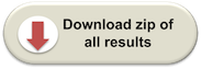
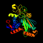
PDB header: oxidoreductaseChain: B: PDB Molecule: malate/l-lactate dehydrogenases;PDBTitle: the structure of a putative malate/lactate dehydrogenase from e. coli.
2 c3i0pA_
100.0
26
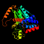
PDB header: oxidoreductaseChain: A: PDB Molecule: malate dehydrogenase;PDBTitle: crystal structure of malate dehydrogenase from entamoeba histolytica
3 c1z2iA_
100.0
28
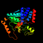
PDB header: oxidoreductaseChain: A: PDB Molecule: malate dehydrogenase;PDBTitle: crystal structure of agrobacterium tumefaciens malate2 dehydrogenase, new york structural genomics consortium
4 c1vbiA_
100.0
31
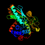
PDB header: oxidoreductaseChain: A: PDB Molecule: type 2 malate/lactate dehydrogenase;PDBTitle: crystal structure of type 2 malate/lactate dehydrogenase from thermus2 thermophilus hb8
5 d1rfma_
100.0
32
Fold: L-sulfolactate dehydrogenase-likeSuperfamily: L-sulfolactate dehydrogenase-likeFamily: L-sulfolactate dehydrogenase-like6 d1xrha_
100.0
28
Fold: L-sulfolactate dehydrogenase-likeSuperfamily: L-sulfolactate dehydrogenase-likeFamily: L-sulfolactate dehydrogenase-like7 c1wtjB_
100.0
28
PDB header: oxidoreductaseChain: B: PDB Molecule: ureidoglycolate dehydrogenase;PDBTitle: crystal structure of delta1-piperideine-2-carboxylate2 reductase from pseudomonas syringae pvar.tomato
8 d1nxua_
100.0
24
Fold: L-sulfolactate dehydrogenase-likeSuperfamily: L-sulfolactate dehydrogenase-likeFamily: L-sulfolactate dehydrogenase-like9 c1v9nA_
100.0
32
PDB header: oxidoreductaseChain: A: PDB Molecule: malate dehydrogenase;PDBTitle: structure of malate dehydrogenase from pyrococcus horikoshii ot3
10 c3uoeB_
100.0
26
PDB header: oxidoreductaseChain: B: PDB Molecule: dehydrogenase;PDBTitle: the crystal structure of dehydrogenase from sinorhizobium meliloti
11 d1jmxa1
40.9
14
Fold: Cytochrome cSuperfamily: Cytochrome cFamily: Quinohemoprotein amine dehydrogenase A chain, domains 1 and 212 c3irvA_
38.1
9
PDB header: hydrolaseChain: A: PDB Molecule: cysteine hydrolase;PDBTitle: crystal structure of cysteine hydrolase pspph_2384 from pseudomonas2 syringae pv. phaseolicola 1448a
13 c3eefA_
34.6
22
PDB header: hydrolaseChain: A: PDB Molecule: n-carbamoylsarcosine amidase related protein;PDBTitle: crystal structure of n-carbamoylsarcosine amidase from thermoplasma2 acidophilum
14 c3kl2K_
34.5
13
PDB header: structural genomics, unknown functionChain: K: PDB Molecule: putative isochorismatase;PDBTitle: crystal structure of a putative isochorismatase from streptomyces2 avermitilis
15 c2qiwA_
34.2
15
PDB header: transferaseChain: A: PDB Molecule: pep phosphonomutase;PDBTitle: crystal structure of a putative phosphoenolpyruvate phosphonomutase2 (ncgl1015, cgl1060) from corynebacterium glutamicum atcc 13032 at3 1.80 a resolution
16 c2kebA_
31.3
19
PDB header: dna binding proteinChain: A: PDB Molecule: dna polymerase subunit alpha b;PDBTitle: nmr solution structure of the n-terminal domain of the dna polymerase2 alpha p68 subunit
17 c2ze3A_
30.4
20
PDB header: isomeraseChain: A: PDB Molecule: dfa0005;PDBTitle: crystal structure of dfa0005 complexed with alpha-ketoglutarate: a2 novel member of the icl/pepm superfamily from alkali-tolerant3 deinococcus ficus
18 d3bzra1
28.4
20
Fold: EscU C-terminal domain-likeSuperfamily: EscU C-terminal domain-likeFamily: EscU C-terminal domain-like19 c3bzrA_
28.4
20
PDB header: membrane protein, protein transportChain: A: PDB Molecule: escu;PDBTitle: crystal structure of escu c-terminal domain with n262d mutation, space2 group p 41 21 2
20 c1e0mA_
28.2
36
PDB header: de novo proteinChain: A: PDB Molecule: wwprototype;PDBTitle: prototype ww domain
21 c2k29A_
not modelled
27.8
12
PDB header: transcriptionChain: A: PDB Molecule: antitoxin relb;PDBTitle: structure of the dbd domain of e. coli antitoxin relb
22 c1yiuA_
not modelled
27.3
50
PDB header: ligaseChain: A: PDB Molecule: itchy e3 ubiquitin protein ligase;PDBTitle: itch e3 ubiquitin ligase ww3 domain
23 c3b8iF_
not modelled
26.9
14
PDB header: lyaseChain: F: PDB Molecule: pa4872 oxaloacetate decarboxylase;PDBTitle: crystal structure of oxaloacetate decarboxylase from pseudomonas2 aeruginosa (pa4872) in complex with oxalate and mg2+.
24 c3ih1A_
not modelled
26.4
21
PDB header: lyaseChain: A: PDB Molecule: methylisocitrate lyase;PDBTitle: crystal structure of carboxyvinyl-carboxyphosphonate phosphorylmutase2 from bacillus anthracis
25 c3ot4F_
not modelled
26.2
20
PDB header: hydrolaseChain: F: PDB Molecule: putative isochorismatase;PDBTitle: structure and catalytic mechanism of bordetella bronchiseptica nicf
26 c2b34C_
not modelled
25.9
13
PDB header: hydrolaseChain: C: PDB Molecule: mar1 ribonuclease;PDBTitle: structure of mar1 ribonuclease from caenorhabditis elegans
27 c2ysbA_
not modelled
25.9
29
PDB header: protein bindingChain: A: PDB Molecule: salvador homolog 1 protein;PDBTitle: solution structure of the first ww domain from the mouse2 salvador homolog 1 protein (sav1)
28 c2jlhA_
not modelled
25.7
23
PDB header: protein transportChain: A: PDB Molecule: yop proteins translocation protein u;PDBTitle: crystal structure of the cytoplasmic domain of yersinia2 pestis yscu n263a mutant
29 c3l4hA_
not modelled
25.5
17
PDB header: protein bindingChain: A: PDB Molecule: e3 ubiquitin-protein ligase hecw1;PDBTitle: helical box domain and second ww domain of the human e3 ubiquitin-2 protein ligase hecw1
30 d1nf9a_
not modelled
25.5
11
Fold: Isochorismatase-like hydrolasesSuperfamily: Isochorismatase-like hydrolasesFamily: Isochorismatase-like hydrolases31 d2ebfx2
not modelled
25.5
16
Fold: EreA/ChaN-likeSuperfamily: EreA/ChaN-likeFamily: PMT domain-like32 c3t7yB_
not modelled
24.9
23
PDB header: protein transportChain: B: PDB Molecule: yop proteins translocation protein u;PDBTitle: structure of an autocleavage-inactive mutant of the cytoplasmic domain2 of ct091, the yscu homologue of chlamydia trachomatis
33 d1nbaa_
not modelled
24.8
20
Fold: Isochorismatase-like hydrolasesSuperfamily: Isochorismatase-like hydrolasesFamily: Isochorismatase-like hydrolases34 d1vp8a_
not modelled
24.3
24
Fold: Pyruvate kinase C-terminal domain-likeSuperfamily: PK C-terminal domain-likeFamily: MTH1675-like35 c1ymzA_
not modelled
24.1
50
PDB header: unknown functionChain: A: PDB Molecule: cc45;PDBTitle: cc45, an artificial ww domain designed using statistical2 coupling analysis
36 d1qusa_
not modelled
23.6
17
Fold: Lysozyme-likeSuperfamily: Lysozyme-likeFamily: Bacterial muramidase, catalytic domain37 d1t57a_
not modelled
23.2
23
Fold: Pyruvate kinase C-terminal domain-likeSuperfamily: PK C-terminal domain-likeFamily: MTH1675-like38 c3mcwA_
not modelled
23.0
12
PDB header: hydrolaseChain: A: PDB Molecule: putative hydrolase;PDBTitle: crystal structure of an a putative hydrolase of the isochorismatase2 family (cv_1320) from chromobacterium violaceum atcc 12472 at 1.06 a3 resolution
39 c2ysdA_
not modelled
22.7
27
PDB header: protein bindingChain: A: PDB Molecule: membrane-associated guanylate kinase, ww and pdzPDBTitle: solution structure of the first ww domain from the human2 membrane-associated guanylate kinase, ww and pdz domain-3 containing protein 1. magi-1
40 c3i44A_
not modelled
22.0
18
PDB header: oxidoreductaseChain: A: PDB Molecule: aldehyde dehydrogenase;PDBTitle: crystal structure of aldehyde dehydrogenase from bartonella2 henselae at 2.0a resolution
41 d1yaca_
not modelled
21.5
8
Fold: Isochorismatase-like hydrolasesSuperfamily: Isochorismatase-like hydrolasesFamily: Isochorismatase-like hydrolases42 d1j2ra_
not modelled
21.4
18
Fold: Isochorismatase-like hydrolasesSuperfamily: Isochorismatase-like hydrolasesFamily: Isochorismatase-like hydrolases43 d1f8ab1
not modelled
21.3
27
Fold: WW domain-likeSuperfamily: WW domainFamily: WW domain44 c2djyA_
not modelled
21.0
36
PDB header: ligase/signaling proteinChain: A: PDB Molecule: smad ubiquitination regulatory factor 2;PDBTitle: solution structure of smurf2 ww3 domain-smad7 py peptide2 complex
45 c2hjpA_
not modelled
20.5
12
PDB header: hydrolaseChain: A: PDB Molecule: phosphonopyruvate hydrolase;PDBTitle: crystal structure of phosphonopyruvate hydrolase complex with2 phosphonopyruvate and mg++
46 d1wh7a_
not modelled
20.4
17
Fold: DNA/RNA-binding 3-helical bundleSuperfamily: Homeodomain-likeFamily: Homeodomain47 d1k9ra_
not modelled
20.0
29
Fold: WW domain-likeSuperfamily: WW domainFamily: WW domain48 c1wr7A_
not modelled
20.0
47
PDB header: ligaseChain: A: PDB Molecule: nedd4-2;PDBTitle: solution structure of the third ww domain of nedd4-2
49 c3hb7G_
not modelled
19.9
13
PDB header: hydrolaseChain: G: PDB Molecule: isochorismatase hydrolase;PDBTitle: the crystal structure of an isochorismatase-like hydrolase from2 alkaliphilus metalliredigens to 2.3a
50 d1pbya1
not modelled
19.8
17
Fold: Cytochrome cSuperfamily: Cytochrome cFamily: Quinohemoprotein amine dehydrogenase A chain, domains 1 and 251 c2lawA_
not modelled
19.5
29
PDB header: signaling protein/transcriptionChain: A: PDB Molecule: yorkie homolog;PDBTitle: structure of the second ww domain from human yap in complex with a2 human smad1 derived peptide
52 d1lqaa_
not modelled
19.4
13
Fold: TIM beta/alpha-barrelSuperfamily: NAD(P)-linked oxidoreductaseFamily: Aldo-keto reductases (NADP)53 c2gbbA_
not modelled
19.2
11
PDB header: isomeraseChain: A: PDB Molecule: putative chorismate mutase;PDBTitle: crystal structure of secreted chorismate mutase from2 yersinia pestis
54 c1wr4A_
not modelled
19.0
29
PDB header: ligaseChain: A: PDB Molecule: ubiquitin-protein ligase nedd4-2;PDBTitle: solution structure of the second ww domain of nedd4-2
55 c2kykA_
not modelled
18.9
43
PDB header: ligaseChain: A: PDB Molecule: e3 ubiquitin-protein ligase itchy homolog;PDBTitle: the sandwich region between two lmp2a py motif regulates the2 interaction between aip4ww2domain and py motif
56 d3ci0k2
not modelled
18.5
19
Fold: SAM domain-likeSuperfamily: GspK insert domain-likeFamily: GspK insert domain-like57 c3iwfA_
not modelled
18.2
9
PDB header: transcription regulatorChain: A: PDB Molecule: transcription regulator rpir family;PDBTitle: the crystal structure of the n-terminal domain of a rpir2 transcriptional regulator from staphylococcus epidermidis to 1.4a
58 c2jmfA_
not modelled
18.2
33
PDB header: ligase/signaling proteinChain: A: PDB Molecule: e3 ubiquitin-protein ligase suppressor of deltex;PDBTitle: solution structure of the su(dx) ww4- notch py peptide2 complex
59 c1wmvA_
not modelled
18.2
28
PDB header: oxidoreductase, apoptosisChain: A: PDB Molecule: ww domain containing oxidoreductase;PDBTitle: solution structure of the second ww domain of wwox
60 c2p7vA_
not modelled
17.9
29
PDB header: transcriptionChain: A: PDB Molecule: regulator of sigma d;PDBTitle: crystal structure of the escherichia coli regulator of sigma 70, rsd,2 in complex with sigma 70 domain 4
61 d1tk7a1
not modelled
17.8
29
Fold: WW domain-likeSuperfamily: WW domainFamily: WW domain62 c2pmzF_
not modelled
17.6
20
PDB header: translation, transferaseChain: F: PDB Molecule: dna-directed rna polymerase subunit f;PDBTitle: archaeal rna polymerase from sulfolobus solfataricus
63 d1i5hw_
not modelled
17.0
36
Fold: WW domain-likeSuperfamily: WW domainFamily: WW domain64 d2jmfa1
not modelled
16.8
36
Fold: WW domain-likeSuperfamily: WW domainFamily: WW domain65 d1bdga1
not modelled
16.6
17
Fold: Ribonuclease H-like motifSuperfamily: Actin-like ATPase domainFamily: Hexokinase66 c2uygF_
not modelled
16.0
24
PDB header: lyaseChain: F: PDB Molecule: 3-dehydroquinate dehydratase;PDBTitle: crystallogaphic structure of the typeii 3-dehydroquinase2 from thermus thermophilus
67 c1zlpA_
not modelled
15.9
22
PDB header: lyaseChain: A: PDB Molecule: petal death protein;PDBTitle: petal death protein psr132 with cysteine-linked glutaraldehyde forming2 a thiohemiacetal adduct
68 c3e0kA_
not modelled
15.9
13
PDB header: transferaseChain: A: PDB Molecule: amino-acid acetyltransferase;PDBTitle: crystal structure of c-termianl domain of n-acetylglutamate synthase2 from vibrio parahaemolyticus
69 d2cfua2
not modelled
15.5
17
Fold: Metallo-hydrolase/oxidoreductaseSuperfamily: Metallo-hydrolase/oxidoreductaseFamily: Alkylsulfatase-like70 c3eooL_
not modelled
15.1
19
PDB header: lyaseChain: L: PDB Molecule: methylisocitrate lyase;PDBTitle: 2.9a crystal structure of methyl-isocitrate lyase from2 burkholderia pseudomallei
71 c2ez5W_
not modelled
14.9
33
PDB header: signalling protein,ligaseChain: W: PDB Molecule: e3 ubiquitin-protein ligase nedd4;PDBTitle: solution structure of the dnedd4 ww3* domain- comm lpsy2 peptide complex
72 c2js5B_
not modelled
14.8
26
PDB header: structural genomics, unknown functionChain: B: PDB Molecule: uncharacterized protein;PDBTitle: nmr structure of protein q60c73_metca. northeast structural2 genomics consortium target mcr1
73 c3hn2A_
not modelled
14.2
11
PDB header: oxidoreductaseChain: A: PDB Molecule: 2-dehydropantoate 2-reductase;PDBTitle: crystal structure of 2-dehydropantoate 2-reductase from geobacter2 metallireducens gs-15
74 c2yshA_
not modelled
14.1
27
PDB header: protein bindingChain: A: PDB Molecule: growth-arrest-specific protein 7;PDBTitle: solution structure of the ww domain from the human growth-2 arrest-specific protein 7, gas-7
75 c2yy9A_
not modelled
14.0
31
PDB header: transcriptionChain: A: PDB Molecule: zinc finger and btb domain-containing protein 48;PDBTitle: crystal structure of btb domain from mouse hkr3
76 d2csua3
not modelled
14.0
17
Fold: Flavodoxin-likeSuperfamily: Succinyl-CoA synthetase domainsFamily: Succinyl-CoA synthetase domains77 c2q81C_
not modelled
13.6
19
PDB header: transcriptionChain: C: PDB Molecule: miz-1 protein;PDBTitle: crystal structure of the miz-1 btb/poz domain
78 c3fa4D_
not modelled
13.6
20
PDB header: lyaseChain: D: PDB Molecule: 2,3-dimethylmalate lyase;PDBTitle: crystal structure of 2,3-dimethylmalate lyase, a pep mutase/isocitrate2 lyase superfamily member, triclinic crystal form
79 d3djba1
not modelled
13.5
17
Fold: HD-domain/PDEase-likeSuperfamily: HD-domain/PDEase-likeFamily: HD domain80 d1xn7a_
not modelled
13.1
24
Fold: DNA/RNA-binding 3-helical bundleSuperfamily: "Winged helix" DNA-binding domainFamily: Hypothetical protein YhgG81 d1dq3a2
not modelled
13.1
32
Fold: dsRBD-likeSuperfamily: YcfA/nrd intein domainFamily: PI-Pfui intein middle domain82 c3le4A_
not modelled
12.9
33
PDB header: nuclear proteinChain: A: PDB Molecule: microprocessor complex subunit dgcr8;PDBTitle: crystal structure of the dgcr8 dimerization domain
83 c1ks9A_
not modelled
12.8
11
PDB header: oxidoreductaseChain: A: PDB Molecule: 2-dehydropantoate 2-reductase;PDBTitle: ketopantoate reductase from escherichia coli
84 d1j9ia_
not modelled
12.8
25
Fold: Putative DNA-binding domainSuperfamily: Putative DNA-binding domainFamily: Terminase gpNU1 subunit domain85 c2r73C_
not modelled
12.7
13
PDB header: transport proteinChain: C: PDB Molecule: trichosurin;PDBTitle: crystal structure of the possum milk whey lipocalin2 trichosurin at ph 8.2
86 c2l4jA_
not modelled
12.5
33
PDB header: transcriptionChain: A: PDB Molecule: yes-associated protein 2 (yap2);PDBTitle: yap ww2
87 d2ieca1
not modelled
12.5
17
Fold: MK0786-likeSuperfamily: MK0786-likeFamily: MK0786-like88 d1dzka_
not modelled
12.3
23
Fold: LipocalinsSuperfamily: LipocalinsFamily: Retinol binding protein-like89 c2jt1A_
not modelled
12.2
21
PDB header: transcriptionChain: A: PDB Molecule: pefi protein;PDBTitle: solution nmr structure of pefi (plasmid-encoded fimbriae regulatory)2 protein from salmonella typhimurium. northeast structural genomics3 target str82
90 c2h0rD_
not modelled
12.2
12
PDB header: hydrolaseChain: D: PDB Molecule: nicotinamidase;PDBTitle: structure of the yeast nicotinamidase pnc1p
91 c3fiqA_
not modelled
12.1
21
PDB header: transport proteinChain: A: PDB Molecule: odorant-binding protein 1f;PDBTitle: odorant binding protein obp1
92 c2px0D_
not modelled
12.1
11
PDB header: biosynthetic proteinChain: D: PDB Molecule: flagellar biosynthesis protein flhf;PDBTitle: crystal structure of flhf complexed with gmppnp/mg(2+)
93 c2lfcA_
not modelled
11.9
29
PDB header: oxidoreductaseChain: A: PDB Molecule: fumarate reductase, flavoprotein subunit;PDBTitle: solution nmr structure of fumarate reductase flavoprotein subunit from2 lactobacillus plantarum, northeast structural genomics consortium3 target lpr145j
94 c3drzE_
not modelled
11.9
13
PDB header: unknown functionChain: E: PDB Molecule: btb/poz domain-containing protein kctd5;PDBTitle: x-ray crystal structure of the n-terminal btb domain of human kctd52 protein
95 c3fpvC_
not modelled
11.9
21
PDB header: heme binding proteinChain: C: PDB Molecule: extracellular haem-binding protein;PDBTitle: crystal structure of hbps
96 d2i52a1
not modelled
11.6
17
Fold: MK0786-likeSuperfamily: MK0786-likeFamily: MK0786-like97 d1bj7a_
not modelled
11.4
13
Fold: LipocalinsSuperfamily: LipocalinsFamily: Retinol binding protein-like98 c2fq1A_
not modelled
11.4
15
PDB header: hydrolaseChain: A: PDB Molecule: isochorismatase;PDBTitle: crystal structure of the two-domain non-ribosomal peptide synthetase2 entb containing isochorismate lyase and aryl-carrier protein domains
99 c2lazA_
not modelled
11.4
29
PDB header: signaling protein/transcriptionChain: A: PDB Molecule: e3 ubiquitin-protein ligase smurf1;PDBTitle: structure of the first ww domain of human smurf1 in complex with a2 mono-phosphorylated human smad1 derived peptide