Phyre Home Retrieve Phyre Job Id
Emaill.a.kelley@imperial.ac.uk
DescriptionP0AEA2
DateWed Jan 25 15:20:30 GMT 2012
Unique Job ID6fe70837e1bd5b72

Summary 

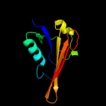
Top model
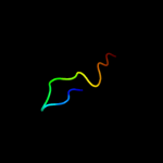
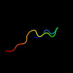
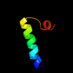
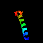
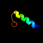
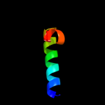
Image coloured by rainbow N → C terminus
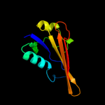
Model (left) based on template d2hqsa2
Top template information
Fold:Anticodon-binding domain-like
Superfamily:TolB, N-terminal domain
Family:TolB, N-terminal domain
Confidence and coverage
Confidence: 98.3% Coverage: 36%
101 residues ( 36% of your sequence) have been modelled with 98.3% confidence by the single highest scoring template.
You may wish to submit your sequence to Phyrealarm. This will automatically scan your sequence every week for new potential templates as they appear in the Phyre2 library.
3D viewing
Interactive 3D view in Jmol

Sequence analysis 

Secondary structure and disorder prediction 

   1........10.........20.........30.........40.........50.........60
Sequence  MQRLFLLVAVMLLSGCLTAPPKEAARPTLMPRAQSYKDLTHLPAPTGKIFVSVYNIQDET
Secondary structure 


SS confidence 


Disorder  ???


??????????????????????


???????


Disorder confidence 


   .........70.........80.........90.........100.........110.........120
Sequence  GQFKPYPASNFSTAVPQSATAMLVTALKDSRWFIPLERQGLQNLLNERKIIRAAQENGTV
Secondary structure 


SS confidence 


Disorder 

??????????????


?????????
Disorder confidence 


   .........130.........140.........150.........160.........170.........180
Sequence  AINNRIPLQSLTAANIMVEGSIIGYESNVKSGGVGARYFGIGADTQYQLDQIAVNLRVVN
Secondary structure 


SS confidence 


Disorder  ????????

?
??


??????????????
????????


Disorder confidence 


   .........190.........200.........210.........220.........230.........240
Sequence  VSTGEILSSVNTSKTILSYEVQAGVFRFIDYQRLLEGEVGYTSNEPVMLCLMSAIETGVI
Secondary structure 


SS confidence 


Disorder 


?


??
???


???????????????


Disorder confidence 


   .........250.........260.........270.......
Sequence  FLINDGIDRGLWDLQNKAERQNDILVKYRHMSVPPES
Secondary structure 


SS confidence 


Disorder 


????


????????
Disorder confidence 


Confidence Key
High(9)                    Low (0)
?Disordered
Alpha helix
Beta strand

Domain analysis 

Hover over an aligned region to see model and summary info

Please note, only up to the top 20 hits are modelled to reduce computer load

RankAligned region

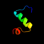
PDB 2hqs chain A domain 2

3D model

Region: 47 - 193
Aligned: 101
Modelled: 101
Confidence: 98.3%
Identity: 21%
Fold: Anticodon-binding domain-like
Superfamily: TolB, N-terminal domain
Family: TolB, N-terminal domain

Phyre2

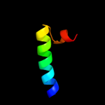
PDB 2iqi chain A domain 1

3D model

Region: 56 - 196
Aligned: 94
Modelled: 94
Confidence: 96.4%
Identity: 15%
Fold: Anticodon-binding domain-like
Superfamily: XCC0632-like
Family: XCC0632-like

Phyre2

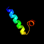
PDB 3ly8 chain A

3D model

Region: 60 - 192
Aligned: 89
Modelled: 93
Confidence: 87.5%
Identity: 13%
PDB header:signaling protein
Chain: A: PDB Molecule:transcriptional activator cadc;
PDBTitle: crystal structure of mutant d471e of the periplasmic domain of cadc

Phyre2

PDB 1wue chain A domain 2

3D model

Region: 230 - 257
Aligned: 28
Modelled: 28
Confidence: 41.0%
Identity: 18%
Fold: Enolase N-terminal domain-like
Superfamily: Enolase N-terminal domain-like
Family: Enolase N-terminal domain-like

Phyre2

PDB 2w8b chain B

3D model

Region: 48 - 193
Aligned: 100
Modelled: 100
Confidence: 32.6%
Identity: 20%
PDB header:protein transport/membrane protein
Chain: B: PDB Molecule:protein tolb;
PDBTitle: crystal structure of processed tolb in complex with pal

Phyre2

PDB 2czr chain A domain 1

3D model

Region: 90 - 138
Aligned: 40
Modelled: 49
Confidence: 21.3%
Identity: 23%
Fold: Restriction endonuclease-like
Superfamily: TBP-interacting protein-like
Family: TBP-interacting protein-like

Phyre2

PDB 2gdq chain A domain 2

3D model

Region: 226 - 256
Aligned: 31
Modelled: 31
Confidence: 20.5%
Identity: 13%
Fold: Enolase N-terminal domain-like
Superfamily: Enolase N-terminal domain-like
Family: Enolase N-terminal domain-like

Phyre2

PDB 2mnr chain A domain 2

3D model

Region: 226 - 256
Aligned: 31
Modelled: 31
Confidence: 20.5%
Identity: 10%
Fold: Enolase N-terminal domain-like
Superfamily: Enolase N-terminal domain-like
Family: Enolase N-terminal domain-like

Phyre2

PDB 1sjd chain A domain 2

3D model

Region: 230 - 256
Aligned: 27
Modelled: 27
Confidence: 19.7%
Identity: 15%
Fold: Enolase N-terminal domain-like
Superfamily: Enolase N-terminal domain-like
Family: Enolase N-terminal domain-like

Phyre2

PDB 3lo7 chain A

3D model

Region: 178 - 192
Aligned: 15
Modelled: 15
Confidence: 18.6%
Identity: 40%
PDB header:transferase
Chain: A: PDB Molecule:penicillin-binding protein a;
PDBTitle: crystal structure of pbpa from mycobacterium tuberculosis

Phyre2

PDB 1wuf chain A domain 2

3D model

Region: 230 - 256
Aligned: 27
Modelled: 27
Confidence: 17.8%
Identity: 19%
Fold: Enolase N-terminal domain-like
Superfamily: Enolase N-terminal domain-like
Family: Enolase N-terminal domain-like

Phyre2

PDB 1jpm chain A domain 2

3D model

Region: 230 - 256
Aligned: 27
Modelled: 27
Confidence: 17.8%
Identity: 11%
Fold: Enolase N-terminal domain-like
Superfamily: Enolase N-terminal domain-like
Family: Enolase N-terminal domain-like

Phyre2

PDB 1jpd chain X domain 2

3D model

Region: 230 - 256
Aligned: 27
Modelled: 27
Confidence: 17.3%
Identity: 19%
Fold: Enolase N-terminal domain-like
Superfamily: Enolase N-terminal domain-like
Family: Enolase N-terminal domain-like

Phyre2

PDB 3ue3 chain A

3D model

Region: 178 - 192
Aligned: 15
Modelled: 15
Confidence: 16.9%
Identity: 47%
PDB header:transferase
Chain: A: PDB Molecule:septum formation, penicillin binding protein 3,
PDBTitle: crystal structure of acinetobacter baumanni pbp3

Phyre2

PDB 1jdf chain A domain 2

3D model

Region: 228 - 256
Aligned: 29
Modelled: 29
Confidence: 16.6%
Identity: 14%
Fold: Enolase N-terminal domain-like
Superfamily: Enolase N-terminal domain-like
Family: Enolase N-terminal domain-like

Phyre2

PDB 1r0m chain A domain 2

3D model

Region: 230 - 256
Aligned: 27
Modelled: 27
Confidence: 16.5%
Identity: 15%
Fold: Enolase N-terminal domain-like
Superfamily: Enolase N-terminal domain-like
Family: Enolase N-terminal domain-like

Phyre2

PDB 2gl5 chain A domain 2

3D model

Region: 226 - 256
Aligned: 31
Modelled: 31
Confidence: 16.3%
Identity: 19%
Fold: Enolase N-terminal domain-like
Superfamily: Enolase N-terminal domain-like
Family: Enolase N-terminal domain-like

Phyre2

PDB 2chr chain A domain 2

3D model

Region: 230 - 256
Aligned: 27
Modelled: 27
Confidence: 16.2%
Identity: 15%
Fold: Enolase N-terminal domain-like
Superfamily: Enolase N-terminal domain-like
Family: Enolase N-terminal domain-like

Phyre2

PDB 1nu5 chain A domain 2

3D model

Region: 230 - 256
Aligned: 27
Modelled: 27
Confidence: 15.0%
Identity: 19%
Fold: Enolase N-terminal domain-like
Superfamily: Enolase N-terminal domain-like
Family: Enolase N-terminal domain-like

Phyre2

PDB 1rp5 chain A domain 4

3D model

Region: 178 - 192
Aligned: 15
Modelled: 15
Confidence: 14.5%
Identity: 40%
Fold: beta-lactamase/transpeptidase-like
Superfamily: beta-lactamase/transpeptidase-like
Family: beta-Lactamase/D-ala carboxypeptidase

Phyre2
1

d2hqsa2
2

d2iqia1
3

c3ly8A_
4

d1wuea2
5

c2w8bB_
6

d2czra1
7

d2gdqa2
8

d2mnra2
9

d1sjda2
10

c3lo7A_
11

d1wufa2
12

d1jpma2
13

d1jpdx2
14

c3ue3A_
15

d1jdfa2
16

d1r0ma2
17

d2gl5a2
18

d2chra2
19

d1nu5a2
20

d1rp5a4
21


22


23


24


25


26


27


28


29


30


31


32


33


34


35


36


37


38


39


40


41


42


43


44


45


46


Detailed template information 

#
Template Alignment Coverage3D Model Confidence
% i.d. Template Information
1d2hqsa2


98.3 21 Fold:Anticodon-binding domain-like
Superfamily:TolB, N-terminal domain
Family:TolB, N-terminal domain
2d2iqia1


96.4 15 Fold:Anticodon-binding domain-like
Superfamily:XCC0632-like
Family:XCC0632-like
3c3ly8A_


87.5 13 PDB header:signaling protein
Chain: A: PDB Molecule:transcriptional activator cadc;
PDBTitle: crystal structure of mutant d471e of the periplasmic domain of cadc
4d1wuea2


41.0 18 Fold:Enolase N-terminal domain-like
Superfamily:Enolase N-terminal domain-like
Family:Enolase N-terminal domain-like
5c2w8bB_


32.6 20 PDB header:protein transport/membrane protein
Chain: B: PDB Molecule:protein tolb;
PDBTitle: crystal structure of processed tolb in complex with pal
6d2czra1


21.3 23 Fold:Restriction endonuclease-like
Superfamily:TBP-interacting protein-like
Family:TBP-interacting protein-like
7d2gdqa2


20.5 13 Fold:Enolase N-terminal domain-like
Superfamily:Enolase N-terminal domain-like
Family:Enolase N-terminal domain-like
8d2mnra2


20.5 10 Fold:Enolase N-terminal domain-like
Superfamily:Enolase N-terminal domain-like
Family:Enolase N-terminal domain-like
9d1sjda2


19.7 15 Fold:Enolase N-terminal domain-like
Superfamily:Enolase N-terminal domain-like
Family:Enolase N-terminal domain-like
10c3lo7A_


18.6 40 PDB header:transferase
Chain: A: PDB Molecule:penicillin-binding protein a;
PDBTitle: crystal structure of pbpa from mycobacterium tuberculosis
11d1wufa2


17.8 19 Fold:Enolase N-terminal domain-like
Superfamily:Enolase N-terminal domain-like
Family:Enolase N-terminal domain-like
12d1jpma2


17.8 11 Fold:Enolase N-terminal domain-like
Superfamily:Enolase N-terminal domain-like
Family:Enolase N-terminal domain-like
13d1jpdx2


17.3 19 Fold:Enolase N-terminal domain-like
Superfamily:Enolase N-terminal domain-like
Family:Enolase N-terminal domain-like
14c3ue3A_


16.9 47 PDB header:transferase
Chain: A: PDB Molecule:septum formation, penicillin binding protein 3,
PDBTitle: crystal structure of acinetobacter baumanni pbp3
15d1jdfa2


16.6 14 Fold:Enolase N-terminal domain-like
Superfamily:Enolase N-terminal domain-like
Family:Enolase N-terminal domain-like
16d1r0ma2


16.5 15 Fold:Enolase N-terminal domain-like
Superfamily:Enolase N-terminal domain-like
Family:Enolase N-terminal domain-like
17d2gl5a2


16.3 19 Fold:Enolase N-terminal domain-like
Superfamily:Enolase N-terminal domain-like
Family:Enolase N-terminal domain-like
18d2chra2


16.2 15 Fold:Enolase N-terminal domain-like
Superfamily:Enolase N-terminal domain-like
Family:Enolase N-terminal domain-like
19d1nu5a2


15.0 19 Fold:Enolase N-terminal domain-like
Superfamily:Enolase N-terminal domain-like
Family:Enolase N-terminal domain-like
20d1rp5a4


14.5 40 Fold:beta-lactamase/transpeptidase-like
Superfamily:beta-lactamase/transpeptidase-like
Family:beta-Lactamase/D-ala carboxypeptidase
21d1vqqa3


not modelled 14.4 40 Fold:beta-lactamase/transpeptidase-like
Superfamily:beta-lactamase/transpeptidase-like
Family:beta-Lactamase/D-ala carboxypeptidase
22d1k25a4


not modelled 14.2 40 Fold:beta-lactamase/transpeptidase-like
Superfamily:beta-lactamase/transpeptidase-like
Family:beta-Lactamase/D-ala carboxypeptidase
23d1muca2


not modelled 12.4 15 Fold:Enolase N-terminal domain-like
Superfamily:Enolase N-terminal domain-like
Family:Enolase N-terminal domain-like
24c3equB_


not modelled 11.0 53 PDB header:biosynthetic protein
Chain: B: PDB Molecule:penicillin-binding protein 2;
PDBTitle: crystal structure of penicillin-binding protein 2 from neisseria2 gonorrhoeae
25d1bqga2


not modelled 10.2 13 Fold:Enolase N-terminal domain-like
Superfamily:Enolase N-terminal domain-like
Family:Enolase N-terminal domain-like
26c3pbqA_


not modelled 9.6 47 PDB header:hydrolase/antibiotic
Chain: A: PDB Molecule:penicillin-binding protein 3;
PDBTitle: crystal structure of pbp3 complexed with imipenem
27c3mbrX_


not modelled 9.3 25 PDB header:transferase
Chain: X: PDB Molecule:glutamine cyclotransferase;
PDBTitle: crystal structure of the glutaminyl cyclase from xanthomonas2 campestris
28d1pyya4


not modelled 9.2 40 Fold:beta-lactamase/transpeptidase-like
Superfamily:beta-lactamase/transpeptidase-like
Family:beta-Lactamase/D-ala carboxypeptidase
29c1mwuA_


not modelled 9.0 40 PDB header:biosynthetic protein
Chain: A: PDB Molecule:penicillin-binding protein 2a;
PDBTitle: structure of methicillin acyl-penicillin binding protein 2a2 from methicillin resistant staphylococcus aureus strain3 27r at 2.60 a resolution.
30d2c5wb1


not modelled 8.7 29 Fold:beta-lactamase/transpeptidase-like
Superfamily:beta-lactamase/transpeptidase-like
Family:beta-Lactamase/D-ala carboxypeptidase
31d1tw3a1


not modelled 8.2 16 Fold:DNA/RNA-binding 3-helical bundle
Superfamily:"Winged helix" DNA-binding domain
Family:Plant O-methyltransferase, N-terminal domain
32c2xzmE_


not modelled 8.1 17 PDB header:ribosome
Chain: E: PDB Molecule:ribosomal protein s5 containing protein;
PDBTitle: crystal structure of the eukaryotic 40s ribosomal2 subunit in complex with initiation factor 1. this file3 contains the 40s subunit and initiation factor for4 molecule 1
33c3nqyA_


not modelled 8.0 42 PDB header:hydrolase
Chain: A: PDB Molecule:secreted metalloprotease mcp02;
PDBTitle: crystal structure of the autoprocessed complex of vibriolysin mcp-022 with a single point mutation e346a
34c3oc2A_


not modelled 7.8 47 PDB header:penicillin-binding protein
Chain: A: PDB Molecule:penicillin-binding protein 3;
PDBTitle: crystal structure of penicillin-binding protein 3 from pseudomonas2 aeruginosa
35c2wadB_


not modelled 6.6 33 PDB header:peptide binding protein
Chain: B: PDB Molecule:penicillin-binding protein 2b;
PDBTitle: penicillin-binding protein 2b (pbp-2b) from streptococcus2 pneumoniae (strain 5204)
36d2bg1a1


not modelled 6.2 36 Fold:beta-lactamase/transpeptidase-like
Superfamily:beta-lactamase/transpeptidase-like
Family:beta-Lactamase/D-ala carboxypeptidase
37d1svda1


not modelled 6.2 20 Fold:TIM beta/alpha-barrel
Superfamily:RuBisCo, C-terminal domain
Family:RuBisCo, large subunit, C-terminal domain
38c1x37A_


not modelled 6.0 21 PDB header:hydrolase
Chain: A: PDB Molecule:atp-dependent protease la 1;
PDBTitle: structure of bacillus subtilis lon protease ssd domain
39c2kbxA_


not modelled 5.8 11 PDB header:cell adhesion
Chain: A: PDB Molecule:integrin-linked protein kinase;
PDBTitle: solution structure of ilk-pinch complex
40c3cb3B_


not modelled 5.7 12 PDB header:isomerase
Chain: B: PDB Molecule:mandelate racemase/muconate lactonizing enzyme;
PDBTitle: crystal structure of l-talarate dehydratase from polaromonas sp. js6662 complexed with mg and l-glucarate
41d1bwva1


not modelled 5.6 17 Fold:TIM beta/alpha-barrel
Superfamily:RuBisCo, C-terminal domain
Family:RuBisCo, large subunit, C-terminal domain
42c1qmfA_


not modelled 5.5 40 PDB header:peptidoglycan synthesis
Chain: A: PDB Molecule:penicillin-binding protein 2x;
PDBTitle: penicillin-binding protein 2x (pbp-2x) acyl-enzyme complex
43d2hc5a1


not modelled 5.5 29 Fold:FlaG-like
Superfamily:FlaG-like
Family:FlaG-like
44d1qzza1


not modelled 5.5 17 Fold:DNA/RNA-binding 3-helical bundle
Superfamily:"Winged helix" DNA-binding domain
Family:Plant O-methyltransferase, N-terminal domain
45d2olua2


not modelled 5.3 14 Fold:beta-lactamase/transpeptidase-like
Superfamily:beta-lactamase/transpeptidase-like
Family:beta-Lactamase/D-ala carboxypeptidase
46c3onrI_


not modelled 5.2 14 PDB header:metal binding protein
Chain: I: PDB Molecule:protein transport protein sece2;
PDBTitle: crystal structure of the calcium chelating immunodominant antigen,2 calcium dodecin (rv0379),from mycobacterium tuberculosis with a novel3 calcium-binding site

Binding site prediction 

Due to computational demand, binding site predictions are not run for batch jobs

If you want to predict binding sites, please manually submit your model of choice to 3DLigandSite


Phyre is for academic use only

Please cite: Protein structure prediction on the web: a case study using the Phyre server
Kelley LA and Sternberg MJE. Nature Protocols 4, 363 - 371 (2009) [pdf] [Import into BibTeX]
 
If you use the binding site predictions from 3DLigandSite, please also cite:
3DLigandSite: predicting ligand-binding sites using similar structures.
Wass MN, Kelley LA and Sternberg MJ Nucleic Acids Research 38, W469-73 (2010) [PubMed]
 
© Structural Bioinformatics Group
Imperial College London
Lawrence Kelley, Benjamin Jefferys
Disclaimer
Terms and Conditions
Component software
Template detection: HHpred 1.51
Secondary structure prediction: Psi-pred 2.5
Disorder prediction: Disopred 2.4
Transmembrane prediction: Memsat_SVM
Multi-template modelling and ab initio: Poing 1.0