Phyre Home Retrieve Phyre Job Id
Emaill.a.kelley@imperial.ac.uk
DescriptionP0A8C4
DateThu Jan 5 11:07:25 GMT 2012
Unique Job ID6f4f66d8076197cd

Summary 

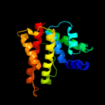
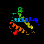
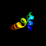
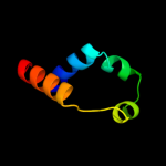
Top model
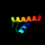
Image coloured by rainbow N → C terminus
Model (left) based on template d1izma_
Top template information
Fold:YgfB-like
Superfamily:YgfB-like
Family:YgfB-like
Confidence and coverage
Confidence:100.0% Coverage: 88%
169 residues ( 88% of your sequence) have been modelled with 100.0% confidence by the single highest scoring template.
3D viewing
Interactive 3D view in Jmol

Sequence analysis 

Secondary structure and disorder prediction 

   1........10.........20.........30.........40.........50.........60
Sequence  MSIQNEMPGYNEMNQYLNQQGTGLTPAEMHGLISGMICGGNDDSSWLPLLHDLTNEGMAF
Secondary structure 


SS confidence 


Disorder  ???????


????


?????
Disorder confidence 


   .........70.........80.........90.........100.........110.........120
Sequence  GHELAQALRKMHSATSDALQDDGFLFQLYLPDGDDVSVFDRADALAGWVNHFLLGLGVTQ
Secondary structure 


SS confidence 


Disorder 


??
?


Disorder confidence 


   .........130.........140.........150.........160.........170.........180
Sequence  PKLDKVTGETGEAIDDLRNIAQLGYDEDEDQEELEMSLEEIIEYVRVAALLCHDTFTHPQ
Secondary structure 


SS confidence 


Disorder  ?
?


??
????????


????
Disorder confidence 


   .........190..
Sequence  PTAPEVQKPTLH
Secondary structure 


SS confidence 


Disorder  ????????????
Disorder confidence 


Confidence Key
High(9)                    Low (0)
?Disordered
Alpha helix
Beta strand

Domain analysis 

Hover over an aligned region to see model and summary info

Please note, only up to the top 20 hits are modelled to reduce computer load

RankAligned region

PDB 1izm chain A

3D model

Region: 7 - 177
Aligned: 169
Modelled: 171
Confidence: 100.0%
Identity: 47%
Fold: YgfB-like
Superfamily: YgfB-like
Family: YgfB-like

Phyre2

PDB 2fbq chain A domain 2

3D model

Region: 59 - 165
Aligned: 83
Modelled: 107
Confidence: 33.0%
Identity: 13%
Fold: Tetracyclin repressor-like, C-terminal domain
Superfamily: Tetracyclin repressor-like, C-terminal domain
Family: Tetracyclin repressor-like, C-terminal domain

Phyre2

PDB 3fqi chain A

3D model

Region: 99 - 116
Aligned: 18
Modelled: 18
Confidence: 27.5%
Identity: 39%
PDB header:protein binding
Chain: A: PDB Molecule:protein dom3z;
PDBTitle: crystal structure of the mouse dom3z

Phyre2

PDB 3fqg chain A

3D model

Region: 101 - 116
Aligned: 16
Modelled: 16
Confidence: 27.1%
Identity: 44%
PDB header:protein binding
Chain: A: PDB Molecule:protein din1;
PDBTitle: crystal structure of the s. pombe rai1

Phyre2

PDB 2au5 chain A domain 1

3D model

Region: 56 - 151
Aligned: 95
Modelled: 96
Confidence: 19.6%
Identity: 12%
Fold: EF2947-like
Superfamily: EF2947-like
Family: EF2947-like

Phyre2

PDB 1zgh chain A domain 1

3D model

Region: 147 - 167
Aligned: 21
Modelled: 20
Confidence: 18.3%
Identity: 24%
Fold: FMT C-terminal domain-like
Superfamily: FMT C-terminal domain-like
Family: Post formyltransferase domain

Phyre2

PDB 2f2a chain C domain 1

3D model

Region: 131 - 166
Aligned: 35
Modelled: 36
Confidence: 15.7%
Identity: 29%
Fold: Non-globular all-alpha subunits of globular proteins
Superfamily: Glu-tRNAGln amidotransferase C subunit
Family: Glu-tRNAGln amidotransferase C subunit

Phyre2

PDB 3h0m chain U

3D model

Region: 131 - 166
Aligned: 35
Modelled: 36
Confidence: 15.2%
Identity: 23%
PDB header:ligase
Chain: U: PDB Molecule:glutamyl-trna(gln) amidotransferase subunit c;
PDBTitle: structure of trna-dependent amidotransferase gatcab from2 aquifex aeolicus

Phyre2

PDB 1ci4 chain A

3D model

Region: 102 - 113
Aligned: 12
Modelled: 12
Confidence: 14.9%
Identity: 33%
Fold: SAM domain-like
Superfamily: Barrier-to-autointegration factor, BAF
Family: Barrier-to-autointegration factor, BAF

Phyre2

PDB 3kfu chain J

3D model

Region: 131 - 165
Aligned: 34
Modelled: 35
Confidence: 14.8%
Identity: 24%
PDB header:ligase/rna
Chain: J: PDB Molecule:glutamyl-trna(gln) amidotransferase subunit c;
PDBTitle: crystal structure of the transamidosome

Phyre2

PDB 2csb chain A domain 1

3D model

Region: 127 - 160
Aligned: 31
Modelled: 33
Confidence: 14.7%
Identity: 32%
Fold: SAM domain-like
Superfamily: RuvA domain 2-like
Family: Topoisomerase V repeat domain

Phyre2

PDB 3a0h chain X

3D model

Region: 29 - 39
Aligned: 11
Modelled: 11
Confidence: 14.1%
Identity: 27%
PDB header:electron transport
Chain: X: PDB Molecule:photosystem ii reaction center protein x;
PDBTitle: crystal structure of i-substituted photosystem ii complex

Phyre2

PDB 3a0b chain X

3D model

Region: 29 - 39
Aligned: 11
Modelled: 11
Confidence: 14.1%
Identity: 27%
PDB header:electron transport
Chain: X: PDB Molecule:photosystem ii reaction center protein x;
PDBTitle: crystal structure of br-substituted photosystem ii complex

Phyre2

PDB 3a0b chain X

3D model

Region: 29 - 39
Aligned: 11
Modelled: 11
Confidence: 14.1%
Identity: 27%
PDB header:electron transport
Chain: X: PDB Molecule:photosystem ii reaction center protein x;
PDBTitle: crystal structure of br-substituted photosystem ii complex

Phyre2

PDB 3a0h chain X

3D model

Region: 29 - 39
Aligned: 11
Modelled: 11
Confidence: 14.1%
Identity: 27%
PDB header:electron transport
Chain: X: PDB Molecule:photosystem ii reaction center protein x;
PDBTitle: crystal structure of i-substituted photosystem ii complex

Phyre2

PDB 1xn8 chain A

3D model

Region: 129 - 177
Aligned: 41
Modelled: 49
Confidence: 12.9%
Identity: 24%
Fold: Hypothetical protein YqbG
Superfamily: Hypothetical protein YqbG
Family: Hypothetical protein YqbG

Phyre2

PDB 1s5l chain X

3D model

Region: 29 - 39
Aligned: 11
Modelled: 11
Confidence: 12.6%
Identity: 27%
PDB header:photosynthesis
Chain: X: PDB Molecule:photosystem ii psbx protein;
PDBTitle: architecture of the photosynthetic oxygen evolving center

Phyre2

PDB 1lgh chain A

3D model

Region: 41 - 51
Aligned: 11
Modelled: 11
Confidence: 12.2%
Identity: 36%
Fold: Light-harvesting complex subunits
Superfamily: Light-harvesting complex subunits
Family: Light-harvesting complex subunits

Phyre2

PDB 1riq chain A domain 1

3D model

Region: 131 - 172
Aligned: 39
Modelled: 42
Confidence: 11.8%
Identity: 15%
Fold: Putative anticodon-binding domain of alanyl-tRNA synthetase (AlaRS)
Superfamily: Putative anticodon-binding domain of alanyl-tRNA synthetase (AlaRS)
Family: Putative anticodon-binding domain of alanyl-tRNA synthetase (AlaRS)

Phyre2

PDB 2ee7 chain A

3D model

Region: 2 - 35
Aligned: 34
Modelled: 34
Confidence: 11.2%
Identity: 12%
PDB header:structural protein
Chain: A: PDB Molecule:sperm flagellar protein 1;
PDBTitle: solution structure of the ch domain from human sperm2 flagellar protein 1

Phyre2
1

d1izma_
2

d2fbqa2
3

c3fqiA_
4

c3fqgA_
5

d2au5a1
6

d1zgha1
7

d2f2ac1
8

c3h0mU_
9

d1ci4a_
10

c3kfuJ_
11

d2csba1
12

c3a0hX_
13

c3a0bx_
14

c3a0bX_
15

c3a0hx_
16

d1xn8a_
17

c1s5lx_
18

d1lgha_
19

d1riqa1
20

c2ee7A_
21


22


23


24


25


26


27


Detailed template information 

#
Template Alignment Coverage3D Model Confidence
% i.d. Template Information
1d1izma_


100.0 47 Fold:YgfB-like
Superfamily:YgfB-like
Family:YgfB-like
2d2fbqa2


33.0 13 Fold:Tetracyclin repressor-like, C-terminal domain
Superfamily:Tetracyclin repressor-like, C-terminal domain
Family:Tetracyclin repressor-like, C-terminal domain
3c3fqiA_


27.5 39 PDB header:protein binding
Chain: A: PDB Molecule:protein dom3z;
PDBTitle: crystal structure of the mouse dom3z
4c3fqgA_


27.1 44 PDB header:protein binding
Chain: A: PDB Molecule:protein din1;
PDBTitle: crystal structure of the s. pombe rai1
5d2au5a1


19.6 12 Fold:EF2947-like
Superfamily:EF2947-like
Family:EF2947-like
6d1zgha1


18.3 24 Fold:FMT C-terminal domain-like
Superfamily:FMT C-terminal domain-like
Family:Post formyltransferase domain
7d2f2ac1


15.7 29 Fold:Non-globular all-alpha subunits of globular proteins
Superfamily:Glu-tRNAGln amidotransferase C subunit
Family:Glu-tRNAGln amidotransferase C subunit
8c3h0mU_


15.2 23 PDB header:ligase
Chain: U: PDB Molecule:glutamyl-trna(gln) amidotransferase subunit c;
PDBTitle: structure of trna-dependent amidotransferase gatcab from2 aquifex aeolicus
9d1ci4a_


14.9 33 Fold:SAM domain-like
Superfamily:Barrier-to-autointegration factor, BAF
Family:Barrier-to-autointegration factor, BAF
10c3kfuJ_


14.8 24 PDB header:ligase/rna
Chain: J: PDB Molecule:glutamyl-trna(gln) amidotransferase subunit c;
PDBTitle: crystal structure of the transamidosome
11d2csba1


14.7 32 Fold:SAM domain-like
Superfamily:RuvA domain 2-like
Family:Topoisomerase V repeat domain
12c3a0hX_


14.1 27 PDB header:electron transport
Chain: X: PDB Molecule:photosystem ii reaction center protein x;
PDBTitle: crystal structure of i-substituted photosystem ii complex
13c3a0bx_


14.1 27 PDB header:electron transport
Chain: X: PDB Molecule:photosystem ii reaction center protein x;
PDBTitle: crystal structure of br-substituted photosystem ii complex
14c3a0bX_


14.1 27 PDB header:electron transport
Chain: X: PDB Molecule:photosystem ii reaction center protein x;
PDBTitle: crystal structure of br-substituted photosystem ii complex
15c3a0hx_


14.1 27 PDB header:electron transport
Chain: X: PDB Molecule:photosystem ii reaction center protein x;
PDBTitle: crystal structure of i-substituted photosystem ii complex
16d1xn8a_


12.9 24 Fold:Hypothetical protein YqbG
Superfamily:Hypothetical protein YqbG
Family:Hypothetical protein YqbG
17c1s5lx_


12.6 27 PDB header:photosynthesis
Chain: X: PDB Molecule:photosystem ii psbx protein;
PDBTitle: architecture of the photosynthetic oxygen evolving center
18d1lgha_


12.2 36 Fold:Light-harvesting complex subunits
Superfamily:Light-harvesting complex subunits
Family:Light-harvesting complex subunits
19d1riqa1


11.8 15 Fold:Putative anticodon-binding domain of alanyl-tRNA synthetase (AlaRS)
Superfamily:Putative anticodon-binding domain of alanyl-tRNA synthetase (AlaRS)
Family:Putative anticodon-binding domain of alanyl-tRNA synthetase (AlaRS)
20c2ee7A_


11.2 12 PDB header:structural protein
Chain: A: PDB Molecule:sperm flagellar protein 1;
PDBTitle: solution structure of the ch domain from human sperm2 flagellar protein 1
21d1dq3a3


not modelled 10.8 36 Fold:Homing endonuclease-like
Superfamily:Homing endonucleases
Family:Intein endonuclease
22c2w84B_


not modelled 10.3 42 PDB header:protein transport
Chain: B: PDB Molecule:peroxisomal targeting signal 1 receptor;
PDBTitle: structure of pex14 in compex with pex5
23c4a4zA_


not modelled 8.6 17 PDB header:hydrolase
Chain: A: PDB Molecule:antiviral helicase ski2;
PDBTitle: crystal structure of the s. cerevisiae dexh helicase ski2 bound to2 amppnp
24c3aa0C_


not modelled 8.4 32 PDB header:protein binding
Chain: C: PDB Molecule:21mer peptide from leucine-rich repeat-containing protein
PDBTitle: crystal structure of actin capping protein in complex with the cp-2 binding motif derived from carmil
25d1zs4a1


not modelled 7.0 8 Fold:lambda repressor-like DNA-binding domains
Superfamily:lambda repressor-like DNA-binding domains
Family:Bacteriophage CII protein
26c2zzrA_


not modelled 5.5 17 PDB header:hydrolase
Chain: A: PDB Molecule:unsaturated glucuronyl hydrolase;
PDBTitle: crystal structure of unsaturated glucuronyl hydrolase from2 streptcoccus agalactiae
27c2l0eA_


not modelled 5.5 25 PDB header:membrane protein
Chain: A: PDB Molecule:sodium/hydrogen exchanger 1;
PDBTitle: structural and functional analysis of tm vi of the nhe1 isoform of the2 na+/h+ exchanger

Binding site prediction 

Due to computational demand, binding site predictions are not run for batch jobs

If you want to predict binding sites, please manually submit your model of choice to 3DLigandSite


Phyre is for academic use only

Please cite: Protein structure prediction on the web: a case study using the Phyre server
Kelley LA and Sternberg MJE. Nature Protocols 4, 363 - 371 (2009) [pdf] [Import into BibTeX]
 
If you use the binding site predictions from 3DLigandSite, please also cite:
3DLigandSite: predicting ligand-binding sites using similar structures.
Wass MN, Kelley LA and Sternberg MJ Nucleic Acids Research 38, W469-73 (2010) [PubMed]
 
© Structural Bioinformatics Group
Imperial College London
Lawrence Kelley, Benjamin Jefferys
Disclaimer
Terms and Conditions
Component software
Template detection: HHpred 1.51
Secondary structure prediction: Psi-pred 2.5
Disorder prediction: Disopred 2.4
Transmembrane prediction: Memsat_SVM
Multi-template modelling and ab initio: Poing 1.0