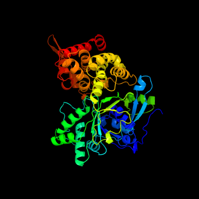
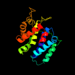
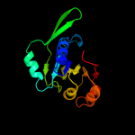
| 1 | c1m2wA_
|
|
 |
100.0 |
23 |
PDB header:oxidoreductase
Chain: A: PDB Molecule:mannitol dehydrogenase;
PDBTitle: pseudomonas fluorescens mannitol 2-dehydrogenase ternary complex with2 nad and d-mannitol
|
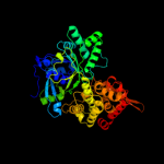
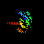
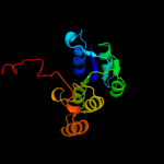
| 2 | c3h2zA_
|
|
 |
100.0 |
22 |
PDB header:oxidoreductase
Chain: A: PDB Molecule:mannitol-1-phosphate 5-dehydrogenase;
PDBTitle: the crystal structure of mannitol-1-phosphate dehydrogenase from2 shigella flexneri
|
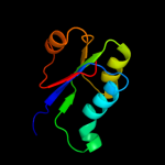
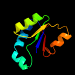
| 3 | d1lj8a4
|
|
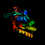 |
100.0 |
21 |
Fold:NAD(P)-binding Rossmann-fold domains
Superfamily:NAD(P)-binding Rossmann-fold domains
Family:6-phosphogluconate dehydrogenase-like, N-terminal domain |
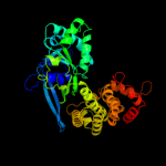
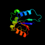
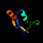
| 4 | d1lj8a3
|
|
 |
100.0 |
25 |
Fold:6-phosphogluconate dehydrogenase C-terminal domain-like
Superfamily:6-phosphogluconate dehydrogenase C-terminal domain-like
Family:Mannitol 2-dehydrogenase |
| 5 | c2ph5A_
|
|
 |
88.1 |
13 |
PDB header:transferase
Chain: A: PDB Molecule:homospermidine synthase;
PDBTitle: crystal structure of the homospermidine synthase hss from legionella2 pneumophila in complex with nad, northeast structural genomics target3 lgr54
|
| 6 | c3ceaA_
|
|
 |
87.8 |
16 |
PDB header:oxidoreductase
Chain: A: PDB Molecule:myo-inositol 2-dehydrogenase;
PDBTitle: crystal structure of myo-inositol 2-dehydrogenase (np_786804.1) from2 lactobacillus plantarum at 2.40 a resolution
|
| 7 | c3mtjA_
|
|
 |
83.1 |
18 |
PDB header:oxidoreductase
Chain: A: PDB Molecule:homoserine dehydrogenase;
PDBTitle: the crystal structure of a homoserine dehydrogenase from thiobacillus2 denitrificans to 2.15a
|
| 8 | c3c7cB_
|
|
 |
78.2 |
12 |
PDB header:oxidoreductase
Chain: B: PDB Molecule:octopine dehydrogenase;
PDBTitle: a structural basis for substrate and stereo selectivity in2 octopine dehydrogenase (odh-nadh-l-arginine)
|
| 9 | c3euwB_
|
|
 |
77.2 |
17 |
PDB header:oxidoreductase
Chain: B: PDB Molecule:myo-inositol dehydrogenase;
PDBTitle: crystal structure of a myo-inositol dehydrogenase from corynebacterium2 glutamicum atcc 13032
|
| 10 | c2axqA_
|
|
 |
66.6 |
21 |
PDB header:oxidoreductase
Chain: A: PDB Molecule:saccharopine dehydrogenase;
PDBTitle: apo histidine-tagged saccharopine dehydrogenase (l-glu2 forming) from saccharomyces cerevisiae
|
| 11 | c3q2kB_
|
|
 |
65.3 |
10 |
PDB header:oxidoreductase
Chain: B: PDB Molecule:oxidoreductase;
PDBTitle: crystal structure of the wlba dehydrogenase from bordetella pertussis2 in complex with nadh and udp-glcnaca
|
| 12 | c3ketA_
|
|
 |
64.7 |
15 |
PDB header:transcription/dna
Chain: A: PDB Molecule:redox-sensing transcriptional repressor rex;
PDBTitle: crystal structure of a rex-family transcriptional regulatory protein2 from streptococcus agalactiae bound to a palindromic operator
|
| 13 | c3e18A_
|
|
 |
60.1 |
23 |
PDB header:oxidoreductase
Chain: A: PDB Molecule:oxidoreductase;
PDBTitle: crystal structure of nad-binding protein from listeria innocua
|
| 14 | c2q4eB_
|
|
 |
60.0 |
17 |
PDB header:oxidoreductase
Chain: B: PDB Molecule:probable oxidoreductase at4g09670;
PDBTitle: ensemble refinement of the protein crystal structure of gene product2 from arabidopsis thaliana at4g09670
|
| 15 | c1e5lA_
|
|
 |
58.1 |
18 |
PDB header:oxidoreductase
Chain: A: PDB Molecule:saccharopine reductase;
PDBTitle: apo saccharopine reductase from magnaporthe grisea
|
| 16 | c3fd8A_
|
|
 |
57.9 |
10 |
PDB header:oxidoreductase
Chain: A: PDB Molecule:oxidoreductase, gfo/idh/moca family;
PDBTitle: crystal structure of an oxidoreductase from enterococcus2 faecalis
|
| 17 | c3moiA_
|
|
 |
55.4 |
17 |
PDB header:oxidoreductase
Chain: A: PDB Molecule:probable dehydrogenase;
PDBTitle: the crystal structure of the putative dehydrogenase from bordetella2 bronchiseptica rb50
|
| 18 | c3c1aB_
|
|
 |
50.5 |
10 |
PDB header:oxidoreductase
Chain: B: PDB Molecule:putative oxidoreductase;
PDBTitle: crystal structure of a putative oxidoreductase (zp_00056571.1) from2 magnetospirillum magnetotacticum ms-1 at 1.85 a resolution
|
| 19 | c3db2C_
|
|
 |
48.6 |
11 |
PDB header:oxidoreductase
Chain: C: PDB Molecule:putative nadph-dependent oxidoreductase;
PDBTitle: crystal structure of a putative nadph-dependent oxidoreductase2 (dhaf_2064) from desulfitobacterium hafniense dcb-2 at 1.70 a3 resolution
|
| 20 | c1ofgF_
|
|
 |
45.8 |
6 |
PDB header:oxidoreductase
Chain: F: PDB Molecule:glucose-fructose oxidoreductase;
PDBTitle: glucose-fructose oxidoreductase
|
| 21 | c1evjC_ |
|
not modelled |
45.4 |
6 |
PDB header:oxidoreductase
Chain: C: PDB Molecule:glucose-fructose oxidoreductase;
PDBTitle: crystal structure of glucose-fructose oxidoreductase (gfor)2 delta1-22 s64d
|
| 22 | c1zh8B_ |
|
not modelled |
41.1 |
15 |
PDB header:oxidoreductase
Chain: B: PDB Molecule:oxidoreductase;
PDBTitle: crystal structure of oxidoreductase (tm0312) from thermotoga maritima2 at 2.50 a resolution
|
| 23 | c1h6dL_ |
|
not modelled |
40.8 |
8 |
PDB header:protein translocation
Chain: L: PDB Molecule:precursor form of glucose-fructose
PDBTitle: oxidized precursor form of glucose-fructose oxidoreductase2 from zymomonas mobilis complexed with glycerol
|
| 24 | c3ezyB_ |
|
not modelled |
39.5 |
14 |
PDB header:structural genomics, unknown function
Chain: B: PDB Molecule:dehydrogenase;
PDBTitle: crystal structure of probable dehydrogenase tm_0414 from2 thermotoga maritima
|
| 25 | c3nt5B_ |
|
not modelled |
38.6 |
16 |
PDB header:oxidoreductase
Chain: B: PDB Molecule:inositol 2-dehydrogenase/d-chiro-inositol 3-dehydrogenase;
PDBTitle: crystal structure of myo-inositol dehydrogenase from bacillus subtilis2 with bound cofactor and product inosose
|
| 26 | d1q0qa2 |
|
not modelled |
38.3 |
13 |
Fold:NAD(P)-binding Rossmann-fold domains
Superfamily:NAD(P)-binding Rossmann-fold domains
Family:Glyceraldehyde-3-phosphate dehydrogenase-like, N-terminal domain |
| 27 | d2nvwa1 |
|
not modelled |
38.2 |
13 |
Fold:NAD(P)-binding Rossmann-fold domains
Superfamily:NAD(P)-binding Rossmann-fold domains
Family:Glyceraldehyde-3-phosphate dehydrogenase-like, N-terminal domain |
| 28 | d1zh8a1 |
|
not modelled |
37.7 |
15 |
Fold:NAD(P)-binding Rossmann-fold domains
Superfamily:NAD(P)-binding Rossmann-fold domains
Family:Glyceraldehyde-3-phosphate dehydrogenase-like, N-terminal domain |
| 29 | d1ydwa1 |
|
not modelled |
36.3 |
20 |
Fold:NAD(P)-binding Rossmann-fold domains
Superfamily:NAD(P)-binding Rossmann-fold domains
Family:Glyceraldehyde-3-phosphate dehydrogenase-like, N-terminal domain |
| 30 | d1k4ia_ |
|
not modelled |
36.3 |
20 |
Fold:YrdC/RibB
Superfamily:YrdC/RibB
Family:3,4-dihydroxy-2-butanone 4-phosphate synthase, DHBP synthase, RibB |
| 31 | d1gzsb_ |
|
not modelled |
34.9 |
18 |
Fold:SopE-like GEF domain
Superfamily:SopE-like GEF domain
Family:SopE-like GEF domain |
| 32 | c2ixaA_ |
|
not modelled |
30.5 |
12 |
PDB header:hydrolase
Chain: A: PDB Molecule:alpha-n-acetylgalactosaminidase;
PDBTitle: a-zyme, n-acetylgalactosaminidase
|
| 33 | c3rbvA_ |
|
not modelled |
29.8 |
13 |
PDB header:sugar binding protein
Chain: A: PDB Molecule:sugar 3-ketoreductase;
PDBTitle: crystal structure of kijd10, a 3-ketoreductase from actinomadura2 kijaniata incomplex with nadp
|
| 34 | d1snna_ |
|
not modelled |
29.2 |
29 |
Fold:YrdC/RibB
Superfamily:YrdC/RibB
Family:3,4-dihydroxy-2-butanone 4-phosphate synthase, DHBP synthase, RibB |
| 35 | d2dt5a2 |
|
not modelled |
27.9 |
10 |
Fold:NAD(P)-binding Rossmann-fold domains
Superfamily:NAD(P)-binding Rossmann-fold domains
Family:Transcriptional repressor Rex, C-terminal domain |
| 36 | c1z9bA_ |
|
not modelled |
27.8 |
11 |
PDB header:translation
Chain: A: PDB Molecule:translation initiation factor if-2;
PDBTitle: solution structure of the c1-subdomain of bacillus2 stearothermophilus translation initiation factor if2
|
| 37 | c3mioA_ |
|
not modelled |
27.4 |
22 |
PDB header:lyase
Chain: A: PDB Molecule:3,4-dihydroxy-2-butanone 4-phosphate synthase;
PDBTitle: crystal structure of 3,4-dihydroxy-2-butanone 4-phosphate synthase2 domain from mycobacterium tuberculosis at ph 6.00
|
| 38 | c3e9mC_ |
|
not modelled |
25.8 |
9 |
PDB header:oxidoreductase
Chain: C: PDB Molecule:oxidoreductase, gfo/idh/moca family;
PDBTitle: crystal structure of an oxidoreductase from enterococcus2 faecalis
|
| 39 | c2dt5A_ |
|
not modelled |
24.6 |
13 |
PDB header:dna binding protein
Chain: A: PDB Molecule:at-rich dna-binding protein;
PDBTitle: crystal structure of ttha1657 (at-rich dna-binding protein) from2 thermus thermophilus hb8
|
| 40 | c1r0lD_ |
|
not modelled |
23.9 |
14 |
PDB header:oxidoreductase
Chain: D: PDB Molecule:1-deoxy-d-xylulose 5-phosphate reductoisomerase;
PDBTitle: 1-deoxy-d-xylulose 5-phosphate reductoisomerase from2 zymomonas mobilis in complex with nadph
|
| 41 | d1f06a1 |
|
not modelled |
23.7 |
19 |
Fold:NAD(P)-binding Rossmann-fold domains
Superfamily:NAD(P)-binding Rossmann-fold domains
Family:Glyceraldehyde-3-phosphate dehydrogenase-like, N-terminal domain |
| 42 | c3dapB_ |
|
not modelled |
23.3 |
19 |
PDB header:oxidoreductase
Chain: B: PDB Molecule:diaminopimelic acid dehydrogenase;
PDBTitle: c. glutamicum dap dehydrogenase in complex with nadp+ and2 the inhibitor 5s-isoxazoline
|
| 43 | c2eghA_ |
|
not modelled |
22.4 |
13 |
PDB header:oxidoreductase
Chain: A: PDB Molecule:1-deoxy-d-xylulose 5-phosphate reductoisomerase;
PDBTitle: crystal structure of 1-deoxy-d-xylulose 5-phosphate reductoisomerase2 complexed with a magnesium ion, nadph and fosmidomycin
|
| 44 | d2o8ra3 |
|
not modelled |
22.3 |
33 |
Fold:Phospholipase D/nuclease
Superfamily:Phospholipase D/nuclease
Family:Polyphosphate kinase C-terminal domain |
| 45 | d1yqga2 |
|
not modelled |
21.1 |
10 |
Fold:NAD(P)-binding Rossmann-fold domains
Superfamily:NAD(P)-binding Rossmann-fold domains
Family:6-phosphogluconate dehydrogenase-like, N-terminal domain |
| 46 | c3kuxA_ |
|
not modelled |
21.1 |
19 |
PDB header:oxidoreductase
Chain: A: PDB Molecule:putative oxidoreductase;
PDBTitle: structure of the ypo2259 putative oxidoreductase from yersinia pestis
|
| 47 | c2glxD_ |
|
not modelled |
20.6 |
20 |
PDB header:oxidoreductase
Chain: D: PDB Molecule:1,5-anhydro-d-fructose reductase;
PDBTitle: crystal structure analysis of bacterial 1,5-af reductase
|
| 48 | c3ff6D_ |
|
not modelled |
20.5 |
16 |
PDB header:ligase
Chain: D: PDB Molecule:acetyl-coa carboxylase 2;
PDBTitle: human acc2 ct domain with cp-640186
|
| 49 | c2ho3D_ |
|
not modelled |
18.8 |
8 |
PDB header:oxidoreductase
Chain: D: PDB Molecule:oxidoreductase, gfo/idh/moca family;
PDBTitle: crystal structure of oxidoreductase, gfo/idh/moca family from2 streptococcus pneumoniae
|
| 50 | c2nvwB_ |
|
not modelled |
18.3 |
13 |
PDB header:transcription
Chain: B: PDB Molecule:galactose/lactose metabolism regulatory protein
PDBTitle: crystal sctucture of transcriptional regulator gal80p from2 kluyveromymes lactis
|
| 51 | c2o48X_ |
|
not modelled |
16.8 |
17 |
PDB header:oxidoreductase
Chain: X: PDB Molecule:dimeric dihydrodiol dehydrogenase;
PDBTitle: crystal structure of mammalian dimeric dihydrodiol dehydrogenase
|
| 52 | c3ec7C_ |
|
not modelled |
15.7 |
15 |
PDB header:oxidoreductase
Chain: C: PDB Molecule:putative dehydrogenase;
PDBTitle: crystal structure of putative dehydrogenase from salmonella2 typhimurium lt2
|
| 53 | c3uuwB_ |
|
not modelled |
15.7 |
11 |
PDB header:oxidoreductase
Chain: B: PDB Molecule:putative oxidoreductase with nad(p)-binding rossmann-fold
PDBTitle: 1.63 angstrom resolution crystal structure of dehydrogenase (mvim)2 from clostridium difficile.
|
| 54 | c3updA_ |
|
not modelled |
15.0 |
12 |
PDB header:transferase
Chain: A: PDB Molecule:ornithine carbamoyltransferase;
PDBTitle: 2.9 angstrom crystal structure of ornithine carbamoyltransferase2 (argf) from vibrio vulnificus
|
| 55 | d1v29a_ |
|
not modelled |
14.4 |
33 |
Fold:Nitrile hydratase alpha chain
Superfamily:Nitrile hydratase alpha chain
Family:Nitrile hydratase alpha chain |
| 56 | c1ceuA_ |
|
not modelled |
14.1 |
11 |
PDB header:viral protein
Chain: A: PDB Molecule:protein (hiv-1 regulatory protein n-terminal
PDBTitle: nmr structure of the (1-51) n-terminal domain of the hiv-12 regulatory protein
|
| 57 | d1im8a_ |
|
not modelled |
13.9 |
11 |
Fold:S-adenosyl-L-methionine-dependent methyltransferases
Superfamily:S-adenosyl-L-methionine-dependent methyltransferases
Family:Hypothetical protein HI0319 (YecO) |
| 58 | d1ebfa1 |
|
not modelled |
13.8 |
24 |
Fold:NAD(P)-binding Rossmann-fold domains
Superfamily:NAD(P)-binding Rossmann-fold domains
Family:Glyceraldehyde-3-phosphate dehydrogenase-like, N-terminal domain |
| 59 | d1tlta1 |
|
not modelled |
13.6 |
12 |
Fold:NAD(P)-binding Rossmann-fold domains
Superfamily:NAD(P)-binding Rossmann-fold domains
Family:Glyceraldehyde-3-phosphate dehydrogenase-like, N-terminal domain |
| 60 | c2ejwB_ |
|
not modelled |
13.6 |
14 |
PDB header:oxidoreductase
Chain: B: PDB Molecule:homoserine dehydrogenase;
PDBTitle: homoserine dehydrogenase from thermus thermophilus hb8
|
| 61 | c3qyhG_ |
|
not modelled |
13.2 |
29 |
PDB header:lyase
Chain: G: PDB Molecule:co-type nitrile hydratase alpha subunit;
PDBTitle: crystal structure of co-type nitrile hydratase beta-h71l from2 pseudomonas putida.
|
| 62 | d2v0fa1 |
|
not modelled |
13.1 |
33 |
Fold:GYF/BRK domain-like
Superfamily:BRK domain-like
Family:BRK domain-like |
| 63 | d1ugpa_ |
|
not modelled |
13.0 |
21 |
Fold:Nitrile hydratase alpha chain
Superfamily:Nitrile hydratase alpha chain
Family:Nitrile hydratase alpha chain |
| 64 | c3v5nA_ |
|
not modelled |
13.0 |
19 |
PDB header:oxidoreductase
Chain: A: PDB Molecule:oxidoreductase;
PDBTitle: the crystal structure of oxidoreductase from sinorhizobium meliloti
|
| 65 | c3eywA_ |
|
not modelled |
12.0 |
18 |
PDB header:transport protein
Chain: A: PDB Molecule:c-terminal domain of glutathione-regulated potassium-efflux
PDBTitle: crystal structure of the c-terminal domain of e. coli kefc in complex2 with keff
|
| 66 | c3btuD_ |
|
not modelled |
11.9 |
10 |
PDB header:transcription
Chain: D: PDB Molecule:galactose/lactose metabolism regulatory protein
PDBTitle: crystal structure of the super-repressor mutant of gal80p2 from saccharomyces cerevisiae; gal80(s2) [e351k]
|
| 67 | c3e82A_ |
|
not modelled |
11.8 |
12 |
PDB header:oxidoreductase
Chain: A: PDB Molecule:putative oxidoreductase;
PDBTitle: crystal structure of a putative oxidoreductase from2 klebsiella pneumoniae
|
| 68 | c3m2tA_ |
|
not modelled |
11.8 |
11 |
PDB header:oxidoreductase
Chain: A: PDB Molecule:probable dehydrogenase;
PDBTitle: the crystal structure of dehydrogenase from chromobacterium2 violaceum
|
| 69 | d2qdya1 |
|
not modelled |
11.6 |
38 |
Fold:Nitrile hydratase alpha chain
Superfamily:Nitrile hydratase alpha chain
Family:Nitrile hydratase alpha chain |
| 70 | d1p32a_ |
|
not modelled |
11.4 |
14 |
Fold:Mitochondrial glycoprotein MAM33-like
Superfamily:Mitochondrial glycoprotein MAM33-like
Family:Mitochondrial glycoprotein MAM33-like |
| 71 | d2py6a1 |
|
not modelled |
11.2 |
17 |
Fold:S-adenosyl-L-methionine-dependent methyltransferases
Superfamily:S-adenosyl-L-methionine-dependent methyltransferases
Family:FkbM-like |
| 72 | c3qv0A_ |
|
not modelled |
11.2 |
15 |
PDB header:protein binding
Chain: A: PDB Molecule:mitochondrial acidic protein mam33;
PDBTitle: crystal structure of saccharomyces cerevisiae mam33
|
| 73 | c3ogiC_ |
|
not modelled |
10.6 |
26 |
PDB header:structural genomics, unknown function
Chain: C: PDB Molecule:putative esat-6-like protein 6;
PDBTitle: crystal structure of the mycobacterium tuberculosis h37rv esxop2 complex (rv2346c-rv2347c)
|
| 74 | c3tsuA_ |
|
not modelled |
10.6 |
5 |
PDB header:transferase
Chain: A: PDB Molecule:transcriptional regulatory protein;
PDBTitle: crystal structure of e. coli hypf with amp-pnp and carbamoyl phosphate
|
| 75 | d2atca2 |
|
not modelled |
10.6 |
26 |
Fold:ATC-like
Superfamily:Aspartate/ornithine carbamoyltransferase
Family:Aspartate/ornithine carbamoyltransferase |
| 76 | c3gcgB_ |
|
not modelled |
10.5 |
21 |
PDB header:signaling protein/transcription
Chain: B: PDB Molecule:l0028 (mitochondria associated protein);
PDBTitle: crystal structure of map and cdc42 complex
|
| 77 | c3ingA_ |
|
not modelled |
10.2 |
26 |
PDB header:oxidoreductase
Chain: A: PDB Molecule:homoserine dehydrogenase;
PDBTitle: crystal structure of homoserine dehydrogenase (np_394635.1) from2 thermoplasma acidophilum at 1.95 a resolution
|
| 78 | c3fggA_ |
|
not modelled |
10.2 |
18 |
PDB header:structural genomics, unknown function
Chain: A: PDB Molecule:uncharacterized protein bce2196;
PDBTitle: crystal structure of putative ecf-type sigma factor negative effector2 from bacillus cereus
|
| 79 | d1ekxa2 |
|
not modelled |
10.0 |
20 |
Fold:ATC-like
Superfamily:Aspartate/ornithine carbamoyltransferase
Family:Aspartate/ornithine carbamoyltransferase |
| 80 | c3guzB_ |
|
not modelled |
9.5 |
18 |
PDB header:ligase
Chain: B: PDB Molecule:pantothenate synthetase;
PDBTitle: structural and substrate-binding studies of pantothenate2 synthenate (ps)provide insights into homotropic inhibition3 by pantoate in ps's
|
| 81 | c1iq5B_ |
|
not modelled |
9.4 |
57 |
PDB header:metal binding protein/protein binding
Chain: B: PDB Molecule:ca2+/calmodulin dependent kinase kinase;
PDBTitle: calmodulin/nematode ca2+/calmodulin dependent kinase kinase2 fragment
|
| 82 | d1hcia1 |
|
not modelled |
9.2 |
19 |
Fold:Spectrin repeat-like
Superfamily:Spectrin repeat
Family:Spectrin repeat |
| 83 | d1yqfa1 |
|
not modelled |
9.0 |
15 |
Fold:Mitochondrial glycoprotein MAM33-like
Superfamily:Mitochondrial glycoprotein MAM33-like
Family:Mitochondrial glycoprotein MAM33-like |
| 84 | c3jv1A_ |
|
not modelled |
8.9 |
19 |
PDB header:hydrolase
Chain: A: PDB Molecule:p22 protein;
PDBTitle: crystal structure of the trypanosoma brucei p22 protein
|
| 85 | c2x24B_ |
|
not modelled |
8.6 |
13 |
PDB header:ligase
Chain: B: PDB Molecule:acetyl-coa carboxylase;
PDBTitle: bovine acc2 ct domain in complex with inhibitor
|
| 86 | c3ip3D_ |
|
not modelled |
8.6 |
14 |
PDB header:oxidoreductase
Chain: D: PDB Molecule:oxidoreductase, putative;
PDBTitle: structure of putative oxidoreductase (tm_0425) from2 thermotoga maritima
|
| 87 | c3b20R_ |
|
not modelled |
8.5 |
17 |
PDB header:oxidoreductase
Chain: R: PDB Molecule:glyceraldehyde 3-phosphate dehydrogenase (nadp+);
PDBTitle: crystal structure analysis of dehydrogenase complexed with nad
|
| 88 | d1mv8a2 |
|
not modelled |
8.3 |
11 |
Fold:NAD(P)-binding Rossmann-fold domains
Superfamily:NAD(P)-binding Rossmann-fold domains
Family:6-phosphogluconate dehydrogenase-like, N-terminal domain |
| 89 | c2dxbR_ |
|
not modelled |
8.1 |
25 |
PDB header:hydrolase
Chain: R: PDB Molecule:thiocyanate hydrolase subunit gamma;
PDBTitle: recombinant thiocyanate hydrolase comprising partially-modified cobalt2 centers
|
| 90 | d1xdpa3 |
|
not modelled |
8.0 |
50 |
Fold:Phospholipase D/nuclease
Superfamily:Phospholipase D/nuclease
Family:Polyphosphate kinase C-terminal domain |
| 91 | c2z8jA_ |
|
not modelled |
7.9 |
26 |
PDB header:transferase
Chain: A: PDB Molecule:gamma-glutamyltranspeptidase;
PDBTitle: crystal structure of escherichia coli gamma-2 glutamyltranspeptidase in complex with azaserine prepared3 in the dark
|
| 92 | c2ew2B_ |
|
not modelled |
7.9 |
12 |
PDB header:oxidoreductase
Chain: B: PDB Molecule:2-dehydropantoate 2-reductase, putative;
PDBTitle: crystal structure of the putative 2-dehydropantoate 2-reductase from2 enterococcus faecalis
|
| 93 | c3d1lB_ |
|
not modelled |
7.8 |
15 |
PDB header:oxidoreductase
Chain: B: PDB Molecule:putative nadp oxidoreductase bf3122;
PDBTitle: crystal structure of putative nadp oxidoreductase bf3122 from2 bacteroides fragilis
|
| 94 | d2ouxa1 |
|
not modelled |
7.8 |
17 |
Fold:alpha-alpha superhelix
Superfamily:MgtE N-terminal domain-like
Family:MgtE N-terminal domain-like |
| 95 | d1ryda1 |
|
not modelled |
7.8 |
6 |
Fold:NAD(P)-binding Rossmann-fold domains
Superfamily:NAD(P)-binding Rossmann-fold domains
Family:Glyceraldehyde-3-phosphate dehydrogenase-like, N-terminal domain |
| 96 | c2jz1A_ |
|
not modelled |
7.8 |
23 |
PDB header:transcription
Chain: A: PDB Molecule:protein doublesex;
PDBTitle: dsx_long
|
| 97 | d1g7sa3 |
|
not modelled |
7.7 |
7 |
Fold:Initiation factor IF2/eIF5b, domain 3
Superfamily:Initiation factor IF2/eIF5b, domain 3
Family:Initiation factor IF2/eIF5b, domain 3 |
| 98 | c3pu6A_ |
|
not modelled |
7.7 |
18 |
PDB header:structural genomics, unknown function
Chain: A: PDB Molecule:uncharacterized protein;
PDBTitle: the crystal structure of an uncharacterized protein from wolinella2 succinogenes
|
| 99 | d1ebda2 |
|
not modelled |
7.7 |
13 |
Fold:FAD/NAD(P)-binding domain
Superfamily:FAD/NAD(P)-binding domain
Family:FAD/NAD-linked reductases, N-terminal and central domains |