| 1 |
|
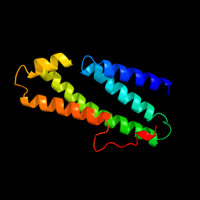
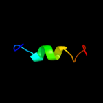
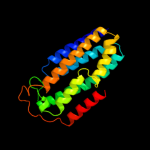
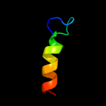
PDB 3rko chain L
Region: 84 - 210
Aligned: 127
Modelled: 127
Confidence: 91.3%
Identity: 13%
PDB header:oxidoreductase
Chain: L: PDB Molecule:nadh-quinone oxidoreductase subunit l;
PDBTitle: crystal structure of the membrane domain of respiratory complex i from2 e. coli at 3.0 angstrom resolution
Phyre2
| 2 |
|
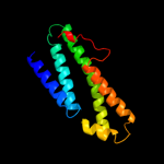
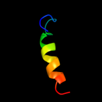
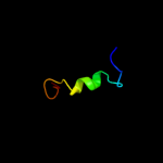
PDB 1q2i chain A
Region: 45 - 60
Aligned: 16
Modelled: 16
Confidence: 39.9%
Identity: 38%
PDB header:antitumor protein
Chain: A: PDB Molecule:pnc27;
PDBTitle: nmr solution structure of a peptide from the mdm-2 binding2 domain of the p53 protein that is selectively cytotoxic to3 cancer cells
Phyre2
| 3 |
|
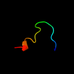
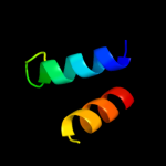
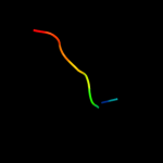
PDB 2jpm chain A
Region: 46 - 63
Aligned: 18
Modelled: 18
Confidence: 22.9%
Identity: 11%
PDB header:antimicrobial protein
Chain: A: PDB Molecule:bacteriocin lactococcin-g subunit beta;
PDBTitle: lactococcin g-b in tfe
Phyre2
| 4 |
|
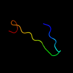
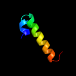
PDB 1lbq chain A
Region: 40 - 61
Aligned: 22
Modelled: 22
Confidence: 17.9%
Identity: 18%
Fold: Chelatase-like
Superfamily: Chelatase
Family: Ferrochelatase
Phyre2
| 5 |
|
PDB 1xn8 chain A
Region: 202 - 232
Aligned: 31
Modelled: 31
Confidence: 14.2%
Identity: 26%
Fold: Hypothetical protein YqbG
Superfamily: Hypothetical protein YqbG
Family: Hypothetical protein YqbG
Phyre2
| 6 |
|
PDB 1ufr chain A
Region: 202 - 221
Aligned: 20
Modelled: 20
Confidence: 9.8%
Identity: 20%
Fold: PRTase-like
Superfamily: PRTase-like
Family: Phosphoribosyltransferases (PRTases)
Phyre2
| 7 |
|
PDB 3rko chain M
Region: 79 - 280
Aligned: 194
Modelled: 195
Confidence: 9.2%
Identity: 16%
PDB header:oxidoreductase
Chain: M: PDB Molecule:nadh-quinone oxidoreductase subunit m;
PDBTitle: crystal structure of the membrane domain of respiratory complex i from2 e. coli at 3.0 angstrom resolution
Phyre2
| 8 |
|
PDB 2k8f chain B
Region: 43 - 65
Aligned: 23
Modelled: 23
Confidence: 6.8%
Identity: 26%
PDB header:transferase/transcription
Chain: B: PDB Molecule:cellular tumor antigen p53;
PDBTitle: structural basis for the regulation of p53 function by p300
Phyre2
| 9 |
|
PDB 2vxs chain B
Region: 198 - 208
Aligned: 11
Modelled: 10
Confidence: 6.7%
Identity: 27%
PDB header:cytokine
Chain: B: PDB Molecule:interleukin-17a;
PDBTitle: structure of il-17a in complex with a potent, fully human2 neutralising antibody
Phyre2
| 10 |
|
PDB 2rmr chain A
Region: 33 - 63
Aligned: 29
Modelled: 31
Confidence: 5.7%
Identity: 34%
PDB header:transcription
Chain: A: PDB Molecule:paired amphipathic helix protein sin3a;
PDBTitle: solution structure of msin3a pah1 domain
Phyre2
| 11 |
|
PDB 2hrc chain A domain 1
Region: 40 - 61
Aligned: 22
Modelled: 22
Confidence: 5.7%
Identity: 18%
Fold: Chelatase-like
Superfamily: Chelatase
Family: Ferrochelatase
Phyre2