Phyre Home Retrieve Phyre Job Id
Emaill.a.kelley@imperial.ac.uk
DescriptionP05050
DateThu Jan 5 10:58:38 GMT 2012
Unique Job ID651b94aedeafc13f

Summary 

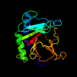
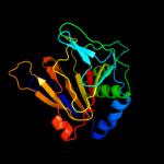
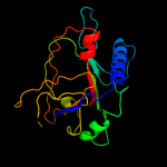
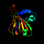
Top model
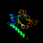
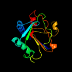
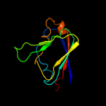
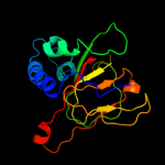
Image coloured by rainbow N → C terminus
Model (left) based on template d2fdia1
Top template information
Fold:Double-stranded beta-helix
Superfamily:Clavaminate synthase-like
Family:AlkB-like
Confidence and coverage
Confidence:100.0% Coverage: 92%
198 residues ( 92% of your sequence) have been modelled with 100.0% confidence by the single highest scoring template.
3D viewing
Interactive 3D view in Jmol

Sequence analysis 

Secondary structure and disorder prediction 

   1........10.........20.........30.........40.........50.........60
Sequence  MLDLFADAEPWQEPLAAGAVILRRFAFNAAEQLIRDINDVASQSPFRQMVTPGGYTMSVA
Secondary structure 


SS confidence 


Disorder  ???????
????


?
??????????


Disorder confidence 


   .........70.........80.........90.........100.........110.........120
Sequence  MTNCGHLGWTTHRQGYLYSPIDPQTNKPWPAMPQSFHNLCQRAATAAGYPDFQPDACLIN
Secondary structure 


SS confidence 


Disorder  ?


????????


Disorder confidence 


   .........130.........140.........150.........160.........170.........180
Sequence  RYAPGAKLSLHQDKDEPDLRAPIVSVSLGLPAIFQFGGLKRNDPLKRLLLEHGDVVVWGG
Secondary structure 


SS confidence 


Disorder 


?


?


Disorder confidence 


   .........190.........200.........210......
Sequence  ESRLFYHGIQPLKAGFHPLTIDCRYNLTFRQAGKKE
Secondary structure 


SS confidence 


Disorder 


?????????


??????
Disorder confidence 


Confidence Key
High(9)                    Low (0)
?Disordered
Alpha helix
Beta strand

Domain analysis 

Hover over an aligned region to see model and summary info

Please note, only up to the top 20 hits are modelled to reduce computer load

RankAligned region

PDB 2fdi chain A domain 1

3D model

Region: 15 - 213
Aligned: 198
Modelled: 199
Confidence: 100.0%
Identity: 100%
Fold: Double-stranded beta-helix
Superfamily: Clavaminate synthase-like
Family: AlkB-like

Phyre2

PDB 3tht chain B

3D model

Region: 6 - 216
Aligned: 177
Modelled: 184
Confidence: 100.0%
Identity: 24%
PDB header:oxidoreductase
Chain: B: PDB Molecule:alkylated dna repair protein alkb homolog 8;
PDBTitle: crystal structure and rna binding properties of the rrm/alkb domains2 in human abh8, an enzyme catalyzing trna hypermodification, northeast3 structural genomics consortium target hr5601b

Phyre2

PDB 2iuw chain A domain 1

3D model

Region: 17 - 212
Aligned: 178
Modelled: 191
Confidence: 100.0%
Identity: 20%
Fold: Double-stranded beta-helix
Superfamily: Clavaminate synthase-like
Family: AlkB-like

Phyre2

PDB 2iuw chain A

3D model

Region: 17 - 212
Aligned: 178
Modelled: 196
Confidence: 100.0%
Identity: 20%
PDB header:oxidoreductase
Chain: A: PDB Molecule:alkylated repair protein alkb homolog 3;
PDBTitle: crystal structure of human abh3 in complex with iron ion2 and 2-oxoglutarate

Phyre2

PDB 3btz chain A

3D model

Region: 11 - 212
Aligned: 188
Modelled: 202
Confidence: 100.0%
Identity: 22%
PDB header:oxidoreductase/dna
Chain: A: PDB Molecule:alpha-ketoglutarate-dependent dioxygenase alkb homolog 2;
PDBTitle: crystal structure of human abh2 cross-linked to dsdna

Phyre2

PDB 3dkq chain B

3D model

Region: 15 - 209
Aligned: 147
Modelled: 164
Confidence: 98.5%
Identity: 19%
PDB header:oxidoreductase
Chain: B: PDB Molecule:pkhd-type hydroxylase sbal_3634;
PDBTitle: crystal structure of putative oxygenase (yp_001051978.1) from2 shewanella baltica os155 at 2.26 a resolution

Phyre2

PDB 2g19 chain A

3D model

Region: 18 - 208
Aligned: 174
Modelled: 191
Confidence: 98.4%
Identity: 14%
PDB header:oxidoreductase
Chain: A: PDB Molecule:egl nine homolog 1;
PDBTitle: cellular oxygen sensing: crystal structure of hypoxia-2 inducible factor prolyl hydroxylase (phd2)

Phyre2

PDB 3itq chain B

3D model

Region: 17 - 215
Aligned: 157
Modelled: 164
Confidence: 97.9%
Identity: 14%
PDB header:oxidoreductase
Chain: B: PDB Molecule:prolyl 4-hydroxylase, alpha subunit domain protein;
PDBTitle: crystal structure of a prolyl 4-hydroxylase from bacillus anthracis

Phyre2

PDB 3oui chain A

3D model

Region: 18 - 208
Aligned: 155
Modelled: 168
Confidence: 97.9%
Identity: 17%
PDB header:oxidoreductase
Chain: A: PDB Molecule:egl nine homolog 1;
PDBTitle: phd2-r717 with 40787422

Phyre2

PDB 2jij chain A

3D model

Region: 15 - 208
Aligned: 151
Modelled: 158
Confidence: 97.9%
Identity: 19%
PDB header:hydrolase
Chain: A: PDB Molecule:prolyl-4 hydroxylase;
PDBTitle: crystal structure of the apo form of chlamydomonas2 reinhardtii prolyl-4 hydroxylase type i

Phyre2

PDB 3lfm chain A

3D model

Region: 113 - 216
Aligned: 95
Modelled: 104
Confidence: 96.7%
Identity: 23%
PDB header:oxidoreductase
Chain: A: PDB Molecule:protein fto;
PDBTitle: crystal structure of the fat mass and obesity associated (fto) protein2 reveals basis for its substrate specificity

Phyre2

PDB 3gjb chain A

3D model

Region: 18 - 210
Aligned: 160
Modelled: 168
Confidence: 95.8%
Identity: 14%
PDB header:biosynthetic protein
Chain: A: PDB Molecule:cytc3;
PDBTitle: cytc3 with fe(ii) and alpha-ketoglutarate

Phyre2

PDB 2a1x chain A domain 1

3D model

Region: 18 - 210
Aligned: 163
Modelled: 174
Confidence: 92.8%
Identity: 18%
Fold: Double-stranded beta-helix
Superfamily: Clavaminate synthase-like
Family: PhyH-like

Phyre2

PDB 2opw chain A

3D model

Region: 18 - 210
Aligned: 184
Modelled: 192
Confidence: 92.2%
Identity: 15%
PDB header:oxidoreductase
Chain: A: PDB Molecule:phyhd1 protein;
PDBTitle: crystal structure of human phytanoyl-coa dioxygenase phyhd1 (apo)

Phyre2

PDB 2rds chain A

3D model

Region: 18 - 216
Aligned: 177
Modelled: 183
Confidence: 92.1%
Identity: 13%
PDB header:oxidoreductase
Chain: A: PDB Molecule:1-deoxypentalenic acid 11-beta hydroxylase; fe(ii)/alpha-
PDBTitle: crystal structure of ptlh with fe/oxalylglycine and ent-1-2 deoxypentalenic acid bound

Phyre2

PDB 3emr chain A

3D model

Region: 18 - 213
Aligned: 166
Modelled: 175
Confidence: 89.6%
Identity: 18%
PDB header:oxidoreductase
Chain: A: PDB Molecule:ectd;
PDBTitle: crystal structure analysis of the ectoine hydroxylase ectd from2 salibacillus salexigens

Phyre2

PDB 2fct chain A domain 1

3D model

Region: 18 - 212
Aligned: 166
Modelled: 173
Confidence: 88.9%
Identity: 16%
Fold: Double-stranded beta-helix
Superfamily: Clavaminate synthase-like
Family: PhyH-like

Phyre2

PDB 3nnl chain B

3D model

Region: 18 - 210
Aligned: 144
Modelled: 151
Confidence: 88.7%
Identity: 15%
PDB header:biosynthetic protein
Chain: B: PDB Molecule:cura;
PDBTitle: halogenase domain from cura module (crystal form iii)

Phyre2

PDB 3kt4 chain A

3D model

Region: 18 - 208
Aligned: 159
Modelled: 168
Confidence: 83.9%
Identity: 16%
PDB header:oxidoreductase
Chain: A: PDB Molecule:pkhd-type hydroxylase tpa1;
PDBTitle: crystal structure of tpa1 from saccharomyces cerevisiae, a2 component of the messenger ribonucleoprotein complex

Phyre2

PDB 3pl0 chain B

3D model

Region: 129 - 213
Aligned: 81
Modelled: 81
Confidence: 79.6%
Identity: 20%
PDB header:biosynthetic protein
Chain: B: PDB Molecule:uncharacterized protein;
PDBTitle: crystal structure of a bsma homolog (mpe_a2762) from methylobium2 petroleophilum pm1 at 1.91 a resolution

Phyre2
1

d2fdia1
2

c3thtB_
3

d2iuwa1
4

c2iuwA_
5

c3btzA_
6

c3dkqB_
7

c2g19A_
8

c3itqB_
9

c3ouiA_
10

c2jijA_
11

c3lfmA_
12

c3gjbA_
13

d2a1xa1
14

c2opwA_
15

c2rdsA_
16

c3emrA_
17

d2fcta1
18

c3nnlB_
19

c3kt4A_
20

c3pl0B_
21


22


23


24


25


26


27


28


29


30


31


32


33


34


35


36


37


38


39


40


41


42


43


44


45


46


47


48


49


50


51


Detailed template information 

#
Template Alignment Coverage3D Model Confidence
% i.d. Template Information
1d2fdia1


100.0 100 Fold:Double-stranded beta-helix
Superfamily:Clavaminate synthase-like
Family:AlkB-like
2c3thtB_


100.0 24 PDB header:oxidoreductase
Chain: B: PDB Molecule:alkylated dna repair protein alkb homolog 8;
PDBTitle: crystal structure and rna binding properties of the rrm/alkb domains2 in human abh8, an enzyme catalyzing trna hypermodification, northeast3 structural genomics consortium target hr5601b
3d2iuwa1


100.0 20 Fold:Double-stranded beta-helix
Superfamily:Clavaminate synthase-like
Family:AlkB-like
4c2iuwA_


100.0 20 PDB header:oxidoreductase
Chain: A: PDB Molecule:alkylated repair protein alkb homolog 3;
PDBTitle: crystal structure of human abh3 in complex with iron ion2 and 2-oxoglutarate
5c3btzA_


100.0 22 PDB header:oxidoreductase/dna
Chain: A: PDB Molecule:alpha-ketoglutarate-dependent dioxygenase alkb homolog 2;
PDBTitle: crystal structure of human abh2 cross-linked to dsdna
6c3dkqB_


98.5 19 PDB header:oxidoreductase
Chain: B: PDB Molecule:pkhd-type hydroxylase sbal_3634;
PDBTitle: crystal structure of putative oxygenase (yp_001051978.1) from2 shewanella baltica os155 at 2.26 a resolution
7c2g19A_


98.4 14 PDB header:oxidoreductase
Chain: A: PDB Molecule:egl nine homolog 1;
PDBTitle: cellular oxygen sensing: crystal structure of hypoxia-2 inducible factor prolyl hydroxylase (phd2)
8c3itqB_


97.9 14 PDB header:oxidoreductase
Chain: B: PDB Molecule:prolyl 4-hydroxylase, alpha subunit domain protein;
PDBTitle: crystal structure of a prolyl 4-hydroxylase from bacillus anthracis
9c3ouiA_


97.9 17 PDB header:oxidoreductase
Chain: A: PDB Molecule:egl nine homolog 1;
PDBTitle: phd2-r717 with 40787422
10c2jijA_


97.9 19 PDB header:hydrolase
Chain: A: PDB Molecule:prolyl-4 hydroxylase;
PDBTitle: crystal structure of the apo form of chlamydomonas2 reinhardtii prolyl-4 hydroxylase type i
11c3lfmA_


96.7 23 PDB header:oxidoreductase
Chain: A: PDB Molecule:protein fto;
PDBTitle: crystal structure of the fat mass and obesity associated (fto) protein2 reveals basis for its substrate specificity
12c3gjbA_


95.8 14 PDB header:biosynthetic protein
Chain: A: PDB Molecule:cytc3;
PDBTitle: cytc3 with fe(ii) and alpha-ketoglutarate
13d2a1xa1


92.8 18 Fold:Double-stranded beta-helix
Superfamily:Clavaminate synthase-like
Family:PhyH-like
14c2opwA_


92.2 15 PDB header:oxidoreductase
Chain: A: PDB Molecule:phyhd1 protein;
PDBTitle: crystal structure of human phytanoyl-coa dioxygenase phyhd1 (apo)
15c2rdsA_


92.1 13 PDB header:oxidoreductase
Chain: A: PDB Molecule:1-deoxypentalenic acid 11-beta hydroxylase; fe(ii)/alpha-
PDBTitle: crystal structure of ptlh with fe/oxalylglycine and ent-1-2 deoxypentalenic acid bound
16c3emrA_


89.6 18 PDB header:oxidoreductase
Chain: A: PDB Molecule:ectd;
PDBTitle: crystal structure analysis of the ectoine hydroxylase ectd from2 salibacillus salexigens
17d2fcta1


88.9 16 Fold:Double-stranded beta-helix
Superfamily:Clavaminate synthase-like
Family:PhyH-like
18c3nnlB_


88.7 15 PDB header:biosynthetic protein
Chain: B: PDB Molecule:cura;
PDBTitle: halogenase domain from cura module (crystal form iii)
19c3kt4A_


83.9 16 PDB header:oxidoreductase
Chain: A: PDB Molecule:pkhd-type hydroxylase tpa1;
PDBTitle: crystal structure of tpa1 from saccharomyces cerevisiae, a2 component of the messenger ribonucleoprotein complex
20c3pl0B_


79.6 20 PDB header:biosynthetic protein
Chain: B: PDB Molecule:uncharacterized protein;
PDBTitle: crystal structure of a bsma homolog (mpe_a2762) from methylobium2 petroleophilum pm1 at 1.91 a resolution
21d1w9ya1


not modelled 78.6 17 Fold:Double-stranded beta-helix
Superfamily:Clavaminate synthase-like
Family:Penicillin synthase-like
22d1vrba1


not modelled 75.4 15 Fold:Double-stranded beta-helix
Superfamily:Clavaminate synthase-like
Family:Asparaginyl hydroxylase-like
23d1dcsa_


not modelled 74.7 16 Fold:Double-stranded beta-helix
Superfamily:Clavaminate synthase-like
Family:Penicillin synthase-like
24c3bvcA_


not modelled 65.9 17 PDB header:structural genomics, unknown function
Chain: A: PDB Molecule:uncharacterized protein ism_01780;
PDBTitle: crystal structure of uncharacterized protein ism_01780 from2 roseovarius nubinhibens ism
25d1vr3a1


not modelled 38.9 17 Fold:Double-stranded beta-helix
Superfamily:RmlC-like cupins
Family:Acireductone dioxygenase
26c3ooxA_


not modelled 35.0 17 PDB header:oxidoreductase
Chain: A: PDB Molecule:putative 2og-fe(ii) oxygenase family protein;
PDBTitle: crystal structure of a putative 2og-fe(ii) oxygenase family protein2 (cc_0200) from caulobacter crescentus at 1.44 a resolution
27d1gp6a_


not modelled 32.2 15 Fold:Double-stranded beta-helix
Superfamily:Clavaminate synthase-like
Family:Penicillin synthase-like
28c3on7C_


not modelled 23.6 18 PDB header:oxidoreductase
Chain: C: PDB Molecule:oxidoreductase, iron/ascorbate family;
PDBTitle: crystal structure of a putative oxygenase (so_2589) from shewanella2 oneidensis at 2.20 a resolution
29d1odma_


not modelled 16.1 18 Fold:Double-stranded beta-helix
Superfamily:Clavaminate synthase-like
Family:Penicillin synthase-like
30c2q30C_


not modelled 13.3 17 PDB header:unknown function
Chain: C: PDB Molecule:uncharacterized protein;
PDBTitle: crystal structure of a rmlc-like cupin protein (dde_2303) from2 desulfovibrio desulfuricans subsp. at 1.94 a resolution
31d1htwa_


not modelled 13.3 23 Fold:P-loop containing nucleoside triphosphate hydrolases
Superfamily:P-loop containing nucleoside triphosphate hydrolases
Family:YjeE-like
32d2oqea1


not modelled 13.0 25 Fold:Supersandwich
Superfamily:Amine oxidase catalytic domain
Family:Amine oxidase catalytic domain
33c3uyjA_


not modelled 12.8 19 PDB header:oxidoreductase
Chain: A: PDB Molecule:lysine-specific demethylase 8;
PDBTitle: crystal structure of jmjd5 catalytic core domain in complex with2 nickle and alpha-kg
34c3al6A_


not modelled 12.7 20 PDB header:unknown function
Chain: A: PDB Molecule:jmjc domain-containing protein c2orf60;
PDBTitle: crystal structure of human tyw5
35c2uvpB_


not modelled 12.5 30 PDB header:unknown function
Chain: B: PDB Molecule:hoba;
PDBTitle: crystal structure of hoba (hp1230)from helicobacter pylori
36c2rohA_


not modelled 10.6 12 PDB header:dna binding protein
Chain: A: PDB Molecule:telomere binding protein-1;
PDBTitle: the dna binding domain of rtbp1
37d1h2ka_


not modelled 9.0 25 Fold:Double-stranded beta-helix
Superfamily:Clavaminate synthase-like
Family:Hypoxia-inducible factor HIF ihhibitor (FIH1)
38d1e5ra_


not modelled 8.4 7 Fold:Double-stranded beta-helix
Superfamily:Clavaminate synthase-like
Family:Type II Proline 3-hydroxylase (proline oxidase)
39c1ljzB_


not modelled 8.1 33 PDB header:receptor, toxin
Chain: B: PDB Molecule:acetylcholine receptor protein;
PDBTitle: nmr structure of an achr-peptide (torpedo californica,2 alpha-subunit residues 182-202) in complex with alpha-3 bungarotoxin
40d1sroa_


not modelled 7.6 27 Fold:OB-fold
Superfamily:Nucleic acid-binding proteins
Family:Cold shock DNA-binding domain-like
41d1w6ga1


not modelled 7.6 13 Fold:Supersandwich
Superfamily:Amine oxidase catalytic domain
Family:Amine oxidase catalytic domain
42d1w2za1


not modelled 6.8 14 Fold:Supersandwich
Superfamily:Amine oxidase catalytic domain
Family:Amine oxidase catalytic domain
43d1u1sa1


not modelled 6.7 14 Fold:Sm-like fold
Superfamily:Sm-like ribonucleoproteins
Family:Pleiotropic translational regulator Hfq
44d1zrra1


not modelled 6.7 17 Fold:Double-stranded beta-helix
Superfamily:RmlC-like cupins
Family:Acireductone dioxygenase
45d2ckxa1


not modelled 6.5 13 Fold:DNA/RNA-binding 3-helical bundle
Superfamily:Homeodomain-like
Family:Myb/SANT domain
46d1zvfa1


not modelled 6.5 11 Fold:Double-stranded beta-helix
Superfamily:RmlC-like cupins
Family:3-hydroxyanthranilic acid dioxygenase-like
47d1yfua1


not modelled 6.5 21 Fold:Double-stranded beta-helix
Superfamily:RmlC-like cupins
Family:3-hydroxyanthranilic acid dioxygenase-like
48d1hk9a_


not modelled 5.7 14 Fold:Sm-like fold
Superfamily:Sm-like ribonucleoproteins
Family:Pleiotropic translational regulator Hfq
49d1slqa_


not modelled 5.6 19 Fold:VP4 membrane interaction domain
Superfamily:VP4 membrane interaction domain
Family:VP4 membrane interaction domain
50d1ud9a2


not modelled 5.4 17 Fold:DNA clamp
Superfamily:DNA clamp
Family:DNA polymerase processivity factor
51d1sfna_


not modelled 5.3 15 Fold:Double-stranded beta-helix
Superfamily:RmlC-like cupins
Family:YlbA-like

Binding site prediction 

Due to computational demand, binding site predictions are not run for batch jobs

If you want to predict binding sites, please manually submit your model of choice to 3DLigandSite


Phyre is for academic use only

Please cite: Protein structure prediction on the web: a case study using the Phyre server
Kelley LA and Sternberg MJE. Nature Protocols 4, 363 - 371 (2009) [pdf] [Import into BibTeX]
 
If you use the binding site predictions from 3DLigandSite, please also cite:
3DLigandSite: predicting ligand-binding sites using similar structures.
Wass MN, Kelley LA and Sternberg MJ Nucleic Acids Research 38, W469-73 (2010) [PubMed]
 
© Structural Bioinformatics Group
Imperial College London
Lawrence Kelley, Benjamin Jefferys
Disclaimer
Terms and Conditions
Component software
Template detection: HHpred 1.51
Secondary structure prediction: Psi-pred 2.5
Disorder prediction: Disopred 2.4
Transmembrane prediction: Memsat_SVM
Multi-template modelling and ab initio: Poing 1.0