| 1 | d1o20a_
|
|
 |
100.0 |
46 |
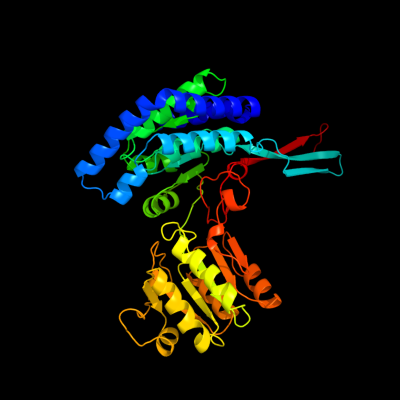
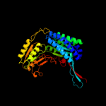
Fold:ALDH-like
Superfamily:ALDH-like
Family:ALDH-like |
| 2 | c2h5gA_
|
|
 |
100.0 |
36 |
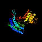
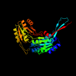
PDB header:oxidoreductase
Chain: A: PDB Molecule:delta 1-pyrroline-5-carboxylate synthetase;
PDBTitle: crystal structure of human pyrroline-5-carboxylate synthetase
|
| 3 | c3hazA_
|
|
 |
100.0 |
19 |
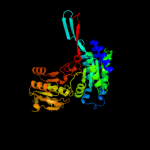
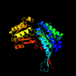
PDB header:oxidoreductase
Chain: A: PDB Molecule:proline dehydrogenase;
PDBTitle: crystal structure of bifunctional proline utilization a2 (puta) protein
|
| 4 | d1uzba_
|
|
 |
100.0 |
17 |
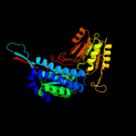
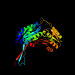
Fold:ALDH-like
Superfamily:ALDH-like
Family:ALDH-like |
| 5 | d1a4sa_
|
|
 |
100.0 |
17 |
Fold:ALDH-like
Superfamily:ALDH-like
Family:ALDH-like |
| 6 | d1vlua_
|
|
 |
100.0 |
36 |
Fold:ALDH-like
Superfamily:ALDH-like
Family:ALDH-like |
| 7 | c3qanB_
|
|
 |
100.0 |
17 |
PDB header:oxidoreductase
Chain: B: PDB Molecule:1-pyrroline-5-carboxylate dehydrogenase 1;
PDBTitle: crystal structure of 1-pyrroline-5-carboxylate dehydrogenase from2 bacillus halodurans
|
| 8 | c2jg7G_
|
|
 |
100.0 |
19 |
PDB header:oxidoreductase
Chain: G: PDB Molecule:antiquitin;
PDBTitle: crystal structure of seabream antiquitin and elucidation of2 its substrate specificity
|
| 9 | c3ed6B_
|
|
 |
100.0 |
18 |
PDB header:oxidoreductase
Chain: B: PDB Molecule:betaine aldehyde dehydrogenase;
PDBTitle: 1.7 angstrom resolution crystal structure of betaine aldehyde2 dehydrogenase (betb) from staphylococcus aureus
|
| 10 | d1ad3a_
|
|
 |
100.0 |
16 |
Fold:ALDH-like
Superfamily:ALDH-like
Family:ALDH-like |
| 11 | d1bxsa_
|
|
 |
100.0 |
20 |
Fold:ALDH-like
Superfamily:ALDH-like
Family:ALDH-like |
| 12 | d1o9ja_
|
|
 |
100.0 |
18 |
Fold:ALDH-like
Superfamily:ALDH-like
Family:ALDH-like |
| 13 | c2ve5H_
|
|
 |
100.0 |
17 |
PDB header:oxidoreductase
Chain: H: PDB Molecule:betaine aldehyde dehydrogenase;
PDBTitle: crystallographic structure of betaine aldehyde2 dehydrogenase from pseudomonas aeruginosa
|
| 14 | c2d4eB_
|
|
 |
100.0 |
17 |
PDB header:oxidoreductase
Chain: B: PDB Molecule:5-carboxymethyl-2-hydroxymuconate semialdehyde
PDBTitle: crystal structure of the hpcc from thermus thermophilus hb8
|
| 15 | d1ag8a_
|
|
 |
100.0 |
18 |
Fold:ALDH-like
Superfamily:ALDH-like
Family:ALDH-like |
| 16 | c2o2qA_
|
|
 |
100.0 |
16 |
PDB header:oxidoreductase
Chain: A: PDB Molecule:formyltetrahydrofolate dehydrogenase;
PDBTitle: crystal structure of the c-terminal domain of rat2 10'formyltetrahydrofolate dehydrogenase in complex with nadp
|
| 17 | d1euha_
|
|
 |
100.0 |
17 |
Fold:ALDH-like
Superfamily:ALDH-like
Family:ALDH-like |
| 18 | c3rh9A_
|
|
 |
100.0 |
17 |
PDB header:oxidoreductase
Chain: A: PDB Molecule:succinate-semialdehyde dehydrogenase (nad(p)(+));
PDBTitle: the crystal structure of oxidoreductase from marinobacter aquaeolei
|
| 19 | c3prlD_
|
|
 |
100.0 |
19 |
PDB header:oxidoreductase
Chain: D: PDB Molecule:nadp-dependent glyceraldehyde-3-phosphate dehydrogenase;
PDBTitle: crystal structure of nadp-dependent glyceraldehyde-3-phosphate2 dehydrogenase from bacillus halodurans c-125
|
| 20 | d1wnda_
|
|
 |
100.0 |
16 |
Fold:ALDH-like
Superfamily:ALDH-like
Family:ALDH-like |
| 21 | c2w8qA_ |
|
not modelled |
100.0 |
17 |
PDB header:oxidoreductase
Chain: A: PDB Molecule:succinate-semialdehyde dehydrogenase,
PDBTitle: the crystal structure of human ssadh in complex with ssa.
|
| 22 | d1o04a_ |
|
not modelled |
100.0 |
18 |
Fold:ALDH-like
Superfamily:ALDH-like
Family:ALDH-like |
| 23 | c3iwkB_ |
|
not modelled |
100.0 |
18 |
PDB header:oxidoreductase
Chain: B: PDB Molecule:aminoaldehyde dehydrogenase;
PDBTitle: crystal structure of aminoaldehyde dehydrogenase 1 from2 pisum sativum (psamadh1)
|
| 24 | d1ky8a_ |
|
not modelled |
100.0 |
20 |
Fold:ALDH-like
Superfamily:ALDH-like
Family:ALDH-like |
| 25 | c3ifgH_ |
|
not modelled |
100.0 |
16 |
PDB header:oxidoreductase
Chain: H: PDB Molecule:succinate-semialdehyde dehydrogenase (nadp+);
PDBTitle: crystal structure of succinate-semialdehyde dehydrogenase from2 burkholderia pseudomallei, part 1 of 2
|
| 26 | c3ek1C_ |
|
not modelled |
100.0 |
19 |
PDB header:oxidoreductase
Chain: C: PDB Molecule:aldehyde dehydrogenase;
PDBTitle: crystal structure of aldehyde dehydrogenase from brucella2 melitensis biovar abortus 2308
|
| 27 | c1t90B_ |
|
not modelled |
100.0 |
17 |
PDB header:oxidoreductase
Chain: B: PDB Molecule:probable methylmalonate-semialdehyde
PDBTitle: crystal structure of methylmalonate semialdehyde2 dehydrogenase from bacillus subtilis
|
| 28 | c3rosA_ |
|
not modelled |
100.0 |
16 |
PDB header:oxidoreductase
Chain: A: PDB Molecule:nad-dependent aldehyde dehydrogenase;
PDBTitle: crystal structure of nad-dependent aldehyde dehydrogenase from2 lactobacillus acidophilus
|
| 29 | c3r31A_ |
|
not modelled |
100.0 |
18 |
PDB header:oxidoreductase
Chain: A: PDB Molecule:betaine aldehyde dehydrogenase;
PDBTitle: crystal structure of betaine aldehyde dehydrogenase from agrobacterium2 tumefaciens
|
| 30 | c3k2wD_ |
|
not modelled |
100.0 |
19 |
PDB header:oxidoreductase
Chain: D: PDB Molecule:betaine-aldehyde dehydrogenase;
PDBTitle: crystal structure of betaine-aldehyde dehydrogenase from2 pseudoalteromonas atlantica t6c
|
| 31 | c3jz4C_ |
|
not modelled |
100.0 |
19 |
PDB header:oxidoreductase
Chain: C: PDB Molecule:succinate-semialdehyde dehydrogenase [nadp+];
PDBTitle: crystal structure of e. coli nadp dependent enzyme
|
| 32 | c3b4wA_ |
|
not modelled |
100.0 |
19 |
PDB header:oxidoreductase
Chain: A: PDB Molecule:aldehyde dehydrogenase;
PDBTitle: crystal structure of mycobacterium tuberculosis aldehyde dehydrogenase2 complexed with nad+
|
| 33 | c3i44A_ |
|
not modelled |
100.0 |
16 |
PDB header:oxidoreductase
Chain: A: PDB Molecule:aldehyde dehydrogenase;
PDBTitle: crystal structure of aldehyde dehydrogenase from bartonella2 henselae at 2.0a resolution
|
| 34 | c1vluB_
|
|
 |
100.0 |
38 |
PDB header:oxidoreductase
Chain: B: PDB Molecule:gamma-glutamyl phosphate reductase;
PDBTitle: crystal structure of gamma-glutamyl phosphate reductase (yor323c) from2 saccharomyces cerevisiae at 2.40 a resolution
|
| 35 | c3k9dD_ |
|
not modelled |
100.0 |
19 |
PDB header:oxidoreductase
Chain: D: PDB Molecule:aldehyde dehydrogenase;
PDBTitle: crystal structure of probable aldehyde dehydrogenase from listeria2 monocytogenes egd-e
|
| 36 | c2hg2A_ |
|
not modelled |
100.0 |
19 |
PDB header:oxidoreductase
Chain: A: PDB Molecule:aldehyde dehydrogenase a;
PDBTitle: structure of lactaldehyde dehydrogenase
|
| 37 | c3pqaA_ |
|
not modelled |
100.0 |
16 |
PDB header:oxidoreductase
Chain: A: PDB Molecule:lactaldehyde dehydrogenase;
PDBTitle: crystal structure of glyceraldehyde-3-phosphate dehydrogenase gapn2 from methanocaldococcus jannaschii dsm 2661
|
| 38 | c3v4cB_ |
|
not modelled |
100.0 |
18 |
PDB header:oxidoreductase
Chain: B: PDB Molecule:aldehyde dehydrogenase (nadp+);
PDBTitle: crystal structure of a semialdehyde dehydrogenase from sinorhizobium2 meliloti 1021
|
| 39 | c3efvC_ |
|
not modelled |
100.0 |
17 |
PDB header:oxidoreductase
Chain: C: PDB Molecule:putative succinate-semialdehyde dehydrogenase;
PDBTitle: crystal structure of a putative succinate-semialdehyde dehydrogenase2 from salmonella typhimurium lt2 with bound nad
|
| 40 | d1ez0a_ |
|
not modelled |
100.0 |
18 |
Fold:ALDH-like
Superfamily:ALDH-like
Family:ALDH-like |
| 41 | c3ju8B_ |
|
not modelled |
100.0 |
20 |
PDB header:oxidoreductase
Chain: B: PDB Molecule:succinylglutamic semialdehyde dehydrogenase;
PDBTitle: crystal structure of succinylglutamic semialdehyde dehydrogenase from2 pseudomonas aeruginosa.
|
| 42 | d1bi9a_ |
|
not modelled |
100.0 |
18 |
Fold:ALDH-like
Superfamily:ALDH-like
Family:ALDH-like |
| 43 | c2vroB_ |
|
not modelled |
100.0 |
20 |
PDB header:oxidoreductase
Chain: B: PDB Molecule:aldehyde dehydrogenase;
PDBTitle: crystal structure of aldehyde dehydrogenase from2 burkholderia xenovorans lb400
|
| 44 | c3lnsD_ |
|
not modelled |
100.0 |
18 |
PDB header:oxidoreductase
Chain: D: PDB Molecule:benzaldehyde dehydrogenase;
PDBTitle: benzaldehyde dehydrogenase, a class 3 aldehyde dehydrogenase, with2 bound nadp+ and benzoate adduct
|
| 45 | c3r64A_ |
|
not modelled |
100.0 |
17 |
PDB header:oxidoreductase
Chain: A: PDB Molecule:nad dependent benzaldehyde dehydrogenase;
PDBTitle: crystal structure of a nad-dependent benzaldehyde dehydrogenase from2 corynebacterium glutamicum
|
| 46 | c3my7A_ |
|
not modelled |
100.0 |
19 |
PDB header:oxidoreductase
Chain: A: PDB Molecule:alcohol dehydrogenase/acetaldehyde dehydrogenase;
PDBTitle: the crystal structure of the acdh domain of an alcohol dehydrogenase2 from vibrio parahaemolyticus to 2.25a
|
| 47 | d1k75a_ |
|
not modelled |
98.5 |
16 |
Fold:ALDH-like
Superfamily:ALDH-like
Family:L-histidinol dehydrogenase HisD |
| 48 | c3jtpB_ |
|
not modelled |
50.5 |
14 |
PDB header:protein binding
Chain: B: PDB Molecule:adapter protein meca 1;
PDBTitle: crystal structure of the c-terminal domain of meca
|
| 49 | c2yukA_ |
|
not modelled |
47.6 |
26 |
PDB header:transferase
Chain: A: PDB Molecule:myeloid/lymphoid or mixed-lineage leukemia
PDBTitle: solution structure of the hmg box of human myeloid/lymphoid2 or mixed-lineage leukemia protein 3 homolog
|
| 50 | d1k99a_ |
|
not modelled |
36.6 |
17 |
Fold:HMG-box
Superfamily:HMG-box
Family:HMG-box |
| 51 | d1s7ia_ |
|
not modelled |
35.7 |
11 |
Fold:Ferredoxin-like
Superfamily:Dimeric alpha+beta barrel
Family:DGPF domain (Pfam 04946) |
| 52 | d1g62a_ |
|
not modelled |
32.9 |
17 |
Fold:Pentein, beta/alpha-propeller
Superfamily:Pentein
Family:Ribosome anti-association factor eIF6 (aIF6) |
| 53 | c2crjA_ |
|
not modelled |
31.2 |
18 |
PDB header:gene regulation
Chain: A: PDB Molecule:swi/snf-related matrix-associated actin-
PDBTitle: solution structure of the hmg domain of mouse hmg domain2 protein hmgx2
|
| 54 | c3fghA_ |
|
not modelled |
30.2 |
13 |
PDB header:transcription
Chain: A: PDB Molecule:transcription factor a, mitochondrial;
PDBTitle: human mitochondrial transcription factor a box b
|
| 55 | c2ec4A_ |
|
not modelled |
29.1 |
8 |
PDB header:structural genomics, unknown function
Chain: A: PDB Molecule:fas-associated factor 1;
PDBTitle: solution structure of the uas domain from human fas-2 associated factor 1
|
| 56 | c2qguA_ |
|
not modelled |
27.7 |
9 |
PDB header:structural genomics, unknown function
Chain: A: PDB Molecule:probable signal peptide protein;
PDBTitle: three-dimensional structure of the phospholipid-binding protein from2 ralstonia solanacearum q8xv73_ralsq in complex with a phospholipid at3 the resolution 1.53 a. northeast structural genomics consortium4 target rsr89
|
| 57 | c3c8eB_ |
|
not modelled |
26.3 |
21 |
PDB header:transferase
Chain: B: PDB Molecule:yghu, glutathione s-transferase homologue;
PDBTitle: crystal structure analysis of yghu from e. coli
|
| 58 | c2eqzA_ |
|
not modelled |
26.3 |
15 |
PDB header:transcription
Chain: A: PDB Molecule:high mobility group protein b3;
PDBTitle: solution structure of the first hmg-box domain from high2 mobility group protein b3
|
| 59 | d1acoa2 |
|
not modelled |
25.8 |
7 |
Fold:Aconitase iron-sulfur domain
Superfamily:Aconitase iron-sulfur domain
Family:Aconitase iron-sulfur domain |
| 60 | d1j3xa_ |
|
not modelled |
25.6 |
13 |
Fold:HMG-box
Superfamily:HMG-box
Family:HMG-box |
| 61 | c1j3xA_ |
|
not modelled |
25.6 |
13 |
PDB header:dna binding protein
Chain: A: PDB Molecule:high mobility group protein 2;
PDBTitle: solution structure of the n-terminal domain of the hmgb2
|
| 62 | d1wo8a1 |
|
not modelled |
24.4 |
10 |
Fold:Methylglyoxal synthase-like
Superfamily:Methylglyoxal synthase-like
Family:Methylglyoxal synthase, MgsA |
| 63 | c3e5bB_ |
|
not modelled |
24.3 |
16 |
PDB header:lyase
Chain: B: PDB Molecule:isocitrate lyase;
PDBTitle: 2.4 a crystal structure of isocitrate lyase from brucella2 melitensis
|
| 64 | c2cs1A_ |
|
not modelled |
23.8 |
26 |
PDB header:dna binding protein
Chain: A: PDB Molecule:pms1 protein homolog 1;
PDBTitle: solution structure of the hmg domain of human dna mismatch2 repair protein
|
| 65 | d1y5ea1 |
|
not modelled |
23.6 |
14 |
Fold:Molybdenum cofactor biosynthesis proteins
Superfamily:Molybdenum cofactor biosynthesis proteins
Family:MogA-like |
| 66 | d1hsma_ |
|
not modelled |
21.1 |
19 |
Fold:HMG-box
Superfamily:HMG-box
Family:HMG-box |
| 67 | d2ftsa3 |
|
not modelled |
21.0 |
14 |
Fold:Molybdenum cofactor biosynthesis proteins
Superfamily:Molybdenum cofactor biosynthesis proteins
Family:MoeA central domain-like |
| 68 | c5acnA_ |
|
not modelled |
20.8 |
7 |
PDB header:lyase(carbon-oxygen)
Chain: A: PDB Molecule:aconitase;
PDBTitle: structure of activated aconitase. formation of the (4fe-4s)2 cluster in the crystal
|
| 69 | d1j46a_ |
|
not modelled |
20.5 |
20 |
Fold:HMG-box
Superfamily:HMG-box
Family:HMG-box |
| 70 | c2co9A_ |
|
not modelled |
20.4 |
15 |
PDB header:transcription
Chain: A: PDB Molecule:thymus high mobility group box protein tox;
PDBTitle: solution structure of the hmg_box domain of thymus high2 mobility group box protein tox from mouse
|
| 71 | d1lwma_ |
|
not modelled |
20.4 |
16 |
Fold:HMG-box
Superfamily:HMG-box
Family:HMG-box |
| 72 | d1vdha_ |
|
not modelled |
20.3 |
7 |
Fold:Ferredoxin-like
Superfamily:Dimeric alpha+beta barrel
Family:Chlorite dismutase-like |
| 73 | c3g5oC_ |
|
not modelled |
20.2 |
24 |
PDB header:toxin/antitoxin
Chain: C: PDB Molecule:uncharacterized protein rv2866;
PDBTitle: the crystal structure of the toxin-antitoxin complex relbe2 (rv2865-2 2866) from mycobacterium tuberculosis
|
| 74 | d1eucb2 |
|
not modelled |
20.0 |
4 |
Fold:ATP-grasp
Superfamily:Glutathione synthetase ATP-binding domain-like
Family:Succinyl-CoA synthetase, beta-chain, N-terminal domain |
| 75 | d1v64a_ |
|
not modelled |
19.4 |
18 |
Fold:HMG-box
Superfamily:HMG-box
Family:HMG-box |
| 76 | d1v63a_ |
|
not modelled |
19.2 |
5 |
Fold:HMG-box
Superfamily:HMG-box
Family:HMG-box |
| 77 | d1j3da_ |
|
not modelled |
19.0 |
21 |
Fold:HMG-box
Superfamily:HMG-box
Family:HMG-box |
| 78 | d1u2ca2 |
|
not modelled |
18.9 |
14 |
Fold:Dystroglycan, domain 2
Superfamily:Dystroglycan, domain 2
Family:Dystroglycan, domain 2 |
| 79 | c2yvqA_ |
|
not modelled |
18.7 |
23 |
PDB header:ligase
Chain: A: PDB Molecule:carbamoyl-phosphate synthase;
PDBTitle: crystal structure of mgs domain of carbamoyl-phosphate2 synthetase from homo sapiens
|
| 80 | d1hmfa_ |
|
not modelled |
18.5 |
19 |
Fold:HMG-box
Superfamily:HMG-box
Family:HMG-box |
| 81 | c1hmfA_ |
|
not modelled |
18.5 |
19 |
PDB header:dna-binding
Chain: A: PDB Molecule:high mobility group protein fragment-b;
PDBTitle: structure of the hmg box motif in the b-domain of hmg1
|
| 82 | d2lefa_ |
|
not modelled |
18.1 |
14 |
Fold:HMG-box
Superfamily:HMG-box
Family:HMG-box |
| 83 | d1t0tv_ |
|
not modelled |
17.9 |
7 |
Fold:Ferredoxin-like
Superfamily:Dimeric alpha+beta barrel
Family:Chlorite dismutase-like |
| 84 | d2b3ya2 |
|
not modelled |
17.5 |
14 |
Fold:Aconitase iron-sulfur domain
Superfamily:Aconitase iron-sulfur domain
Family:Aconitase iron-sulfur domain |
| 85 | d1ckta_ |
|
not modelled |
16.5 |
14 |
Fold:HMG-box
Superfamily:HMG-box
Family:HMG-box |
| 86 | c3labA_ |
|
not modelled |
16.0 |
13 |
PDB header:structural genomics, unknown function
Chain: A: PDB Molecule:putative kdpg (2-keto-3-deoxy-6-phosphogluconate)
PDBTitle: crystal structure of a putative kdpg (2-keto-3-deoxy-6-2 phosphogluconate) aldolase from oleispira antarctica
|
| 87 | c2b3yB_ |
|
not modelled |
15.4 |
14 |
PDB header:lyase
Chain: B: PDB Molecule:iron-responsive element binding protein 1;
PDBTitle: structure of a monoclinic crystal form of human cytosolic aconitase2 (irp1)
|
| 88 | d1gt0d_ |
|
not modelled |
15.2 |
14 |
Fold:HMG-box
Superfamily:HMG-box
Family:HMG-box |
| 89 | c1wx2A_ |
|
not modelled |
15.1 |
16 |
PDB header:oxidoreductase/metal transport
Chain: A: PDB Molecule:tyrosinase;
PDBTitle: crystal structure of the oxy-form of the copper-bound streptomyces2 castaneoglobisporus tyrosinase complexed with a caddie protein3 prepared by the addition of hydrogenperoxide
|
| 90 | d1wmia1 |
|
not modelled |
14.9 |
25 |
Fold:RelE-like
Superfamily:RelE-like
Family:RelE-like |
| 91 | c2vxhF_ |
|
not modelled |
14.5 |
12 |
PDB header:oxidoreductase
Chain: F: PDB Molecule:chlorite dismutase;
PDBTitle: the crystal structure of chlorite dismutase: a detox enzyme2 producing molecular oxygen
|
| 92 | c1v8dC_ |
|
not modelled |
14.2 |
21 |
PDB header:structural genomics, unknown function
Chain: C: PDB Molecule:hypothetical protein (tt1679);
PDBTitle: crystal structure of the conserved hypothetical protein2 tt1679 from thermus thermophilus
|
| 93 | d2dlxa1 |
|
not modelled |
14.0 |
8 |
Fold:Thioredoxin fold
Superfamily:Thioredoxin-like
Family:UAS domain |
| 94 | c1aabA_ |
|
not modelled |
13.5 |
15 |
PDB header:dna-binding
Chain: A: PDB Molecule:high mobility group protein;
PDBTitle: nmr structure of rat hmg1 hmga fragment
|
| 95 | d1aaba_ |
|
not modelled |
13.5 |
15 |
Fold:HMG-box
Superfamily:HMG-box
Family:HMG-box |
| 96 | c1usdA_ |
|
not modelled |
13.5 |
28 |
PDB header:signaling protein
Chain: A: PDB Molecule:vasodilator-stimulated phosphoprotein;
PDBTitle: human vasp tetramerisation domain l352m
|
| 97 | d2nu7b2 |
|
not modelled |
13.2 |
9 |
Fold:ATP-grasp
Superfamily:Glutathione synthetase ATP-binding domain-like
Family:Succinyl-CoA synthetase, beta-chain, N-terminal domain |
| 98 | c2lhjA_ |
|
not modelled |
12.8 |
12 |
PDB header:structural genomics, unknown function
Chain: A: PDB Molecule:high mobility group protein homolog nhp1;
PDBTitle: nmr structure of the high mobility group protein-like protein nhp12 from babesia bovis t2bo (baboa.00841.a)
|
| 99 | c3bpqD_ |
|
not modelled |
12.7 |
14 |
PDB header:toxin
Chain: D: PDB Molecule:toxin rele3;
PDBTitle: crystal structure of relb-rele antitoxin-toxin complex from2 methanococcus jannaschii
|