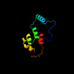
1 d1qgia_
47.0
14
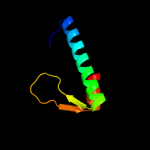
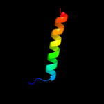
Fold: Lysozyme-likeSuperfamily: Lysozyme-likeFamily: Chitosanase2 d1eysh2
38.3
38
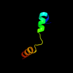
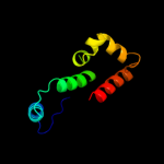
Fold: Single transmembrane helixSuperfamily: Photosystem II reaction centre subunit H, transmembrane regionFamily: Photosystem II reaction centre subunit H, transmembrane region3 d3ckca1
31.3
17
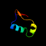
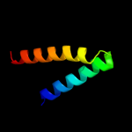
Fold: alpha-alpha superhelixSuperfamily: TPR-likeFamily: SusD-like4 c3pr3B_
26.7
33
PDB header: isomeraseChain: B: PDB Molecule: glucose-6-phosphate isomerase;PDBTitle: crystal structure of plasmodium falciparum glucose-6-phosphate2 isomerase (pf14_0341) in complex with fructose-6-phosphate
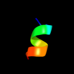
5 c1sqwA_
16.4
21
PDB header: unknown functionChain: A: PDB Molecule: saccharomyces cerevisiae nip7p homolog;PDBTitle: crystal structure of kd93, a novel protein expressed in the2 human pro
6 d2ajta1
16.0
67
Fold: Reductase/isomerase/elongation factor common domainSuperfamily: FucI/AraA C-terminal domain-likeFamily: AraA C-terminal domain-like7 c1w8jD_
15.8
18
PDB header: motor proteinChain: D: PDB Molecule: myosin va;PDBTitle: crystal structure of myosin v motor domain -2 nucleotide-free
8 d1oeda_
15.4
14
Fold: Neurotransmitter-gated ion-channel transmembrane poreSuperfamily: Neurotransmitter-gated ion-channel transmembrane poreFamily: Neurotransmitter-gated ion-channel transmembrane pore9 d1lkxa_
13.2
21
Fold: P-loop containing nucleoside triphosphate hydrolasesSuperfamily: P-loop containing nucleoside triphosphate hydrolasesFamily: Motor proteins10 d2h8pc1
12.8
15
Fold: Voltage-gated potassium channelsSuperfamily: Voltage-gated potassium channelsFamily: Voltage-gated potassium channels11 d1mnda2
12.6
20
Fold: P-loop containing nucleoside triphosphate hydrolasesSuperfamily: P-loop containing nucleoside triphosphate hydrolasesFamily: Motor proteins12 c1eysH_
12.0
38
PDB header: electron transportChain: H: PDB Molecule: photosynthetic reaction center;PDBTitle: crystal structure of photosynthetic reaction center from a2 thermophilic bacterium, thermochromatium tepidum
13 c1br2C_
10.8
13
PDB header: muscle proteinChain: C: PDB Molecule: myosin;PDBTitle: smooth muscle myosin motor domain complexed with mgadp.alf4
14 d2b0ja1
10.5
15
Fold: 6-phosphogluconate dehydrogenase C-terminal domain-likeSuperfamily: 6-phosphogluconate dehydrogenase C-terminal domain-likeFamily: HMD dimerization domain-like15 d1kx5b_
9.8
36
Fold: Histone-foldSuperfamily: Histone-foldFamily: Nucleosome core histones16 d1nh1a_
9.3
29
Fold: Antivirulence factorSuperfamily: Antivirulence factorFamily: Antivirulence factor17 c1nh1A_
9.3
29
PDB header: avirulence proteinChain: A: PDB Molecule: avirulence b protein;PDBTitle: crystal structure of the type iii effector avrb from2 pseudomonas syringae.
18 c3ujhB_
8.7
31
PDB header: isomeraseChain: B: PDB Molecule: glucose-6-phosphate isomerase;PDBTitle: crystal structure of substrate-bound glucose-6-phosphate isomerase2 from toxoplasma gondii
19 c2zw3B_
8.4
20
PDB header: cell adhesionChain: B: PDB Molecule: gap junction beta-2 protein;PDBTitle: structure of the connexin-26 gap junction channel at 3.52 angstrom resolution
20 c2rndA_
7.8
22
PDB header: endocytosisChain: A: PDB Molecule: myc box-dependent-interacting protein 1;PDBTitle: structure of the n-terminal barpeptide in dpc micelles
21 c2erpA_
not modelled
7.7
22
PDB header: toxinChain: A: PDB Molecule: vascular apoptosis-inducing protein 1;PDBTitle: crystal structure of vascular apoptosis-inducing protein-1(inhibitor-2 bound form)
22 c3lhnB_
not modelled
7.4
83
PDB header: lipid binding proteinChain: B: PDB Molecule: lipoprotein;PDBTitle: crystal structure of putative lipoprotein (np_718719.1) from2 shewanella oneidensis at 1.42 a resolution
23 d2foka1
not modelled
7.4
31
Fold: DNA/RNA-binding 3-helical bundleSuperfamily: "Winged helix" DNA-binding domainFamily: Restriction endonuclease FokI, N-terminal (recognition) domain24 d1u1ia2
not modelled
7.2
25
Fold: FwdE/GAPDH domain-likeSuperfamily: Glyceraldehyde-3-phosphate dehydrogenase-like, C-terminal domainFamily: Dihydrodipicolinate reductase-like25 c2kjyA_
not modelled
7.1
45
PDB header: signaling proteinChain: A: PDB Molecule: protein phosphatase 1 regulatory subunit 12a;PDBTitle: mypt1(658-714)
26 c2fcgF_
not modelled
6.9
50
PDB header: antimicrobial proteinChain: F: PDB Molecule: antibacterial protein fall-39, core peptide;PDBTitle: solution structure of the c-terminal fragment of human ll-37
27 d1khda1
not modelled
6.3
12
Fold: Methionine synthase domain-likeSuperfamily: Nucleoside phosphorylase/phosphoribosyltransferase N-terminal domainFamily: Nucleoside phosphorylase/phosphoribosyltransferase N-terminal domain28 c1w9iA_
not modelled
6.2
21
PDB header: myosinChain: A: PDB Molecule: myosin ii heavy chain;PDBTitle: myosin ii dictyostelium discoideum motor domain s456y bound2 with mgadp-befx
29 c1k6nH_
not modelled
6.2
47
PDB header: photosynthesisChain: H: PDB Molecule: photosynthetic reaction center h subunit;PDBTitle: e(l212)a,d(l213)a double mutant structure of photosynthetic reaction2 center from rhodobacter sphaeroides
30 c1g8xB_
not modelled
6.0
18
PDB header: structural proteinChain: B: PDB Molecule: myosin ii heavy chain fused to alpha-actinin 3;PDBTitle: structure of a genetically engineered molecular motor
31 d1cfea_
not modelled
5.9
38
Fold: PR-1-likeSuperfamily: PR-1-likeFamily: PR-1-like32 d1d0xa2
not modelled
5.8
18
Fold: P-loop containing nucleoside triphosphate hydrolasesSuperfamily: P-loop containing nucleoside triphosphate hydrolasesFamily: Motor proteins33 d1vjpa2
not modelled
5.7
40
Fold: FwdE/GAPDH domain-likeSuperfamily: Glyceraldehyde-3-phosphate dehydrogenase-like, C-terminal domainFamily: Dihydrodipicolinate reductase-like34 d1rzhh2
not modelled
5.7
41
Fold: Single transmembrane helixSuperfamily: Photosystem II reaction centre subunit H, transmembrane regionFamily: Photosystem II reaction centre subunit H, transmembrane region35 c2vb6A_
not modelled
5.3
24
PDB header: motor proteinChain: A: PDB Molecule: myosin vi;PDBTitle: myosin vi (md-insert2-cam, delta insert1) post-rigor state (2 crystal form 2)