Phyre Home Retrieve Phyre Job Id
Emaill.a.kelley@imperial.ac.uk
DescriptionP76001
DateWed Jan 25 15:21:05 GMT 2012
Unique Job ID5b22f41c30a74fff

Summary 

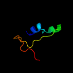
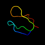
Top model
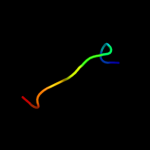
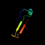
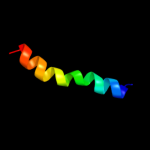
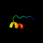
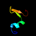
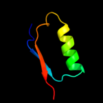
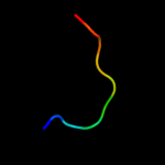
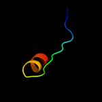
Image coloured by rainbow N → C terminus
Model (left) based on template d1ig4a_
Top template information
Fold:DNA-binding domain
Superfamily:DNA-binding domain
Family:Methyl-CpG-binding domain, MBD
Confidence and coverage
Confidence: 50.9% Coverage: 25%
31 residues ( 25% of your sequence) have been modelled with 50.9% confidence by the single highest scoring template.
You may wish to submit your sequence to Phyrealarm. This will automatically scan your sequence every week for new potential templates as they appear in the Phyre2 library.
Please note: You must be registered and logged in to use Phyrealarm.
3D viewing
Interactive 3D view in Jmol

Sequence analysis 

Secondary structure and disorder prediction 

   1........10.........20.........30.........40.........50.........60
Sequence  MNMMRIFYIGLSGVGMMFSSMASGNDAGGLQSPACGVVCDPYICVNSDGISPELTRKYLG
Secondary structure 


SS confidence 


Disorder  ??


??????????

?


Disorder confidence 


   .........70.........80.........90.........100.........110.........120
Sequence  EKAAENLQSLQGYDPSEFTFANGVFCDVKEKLCRDDRYFGVDGKRSGKINQTTTKMLFMC
Secondary structure 


SS confidence 


Disorder 


?????
??


?
???????????


??
Disorder confidence 


   ..
Sequence  RE
Secondary structure 

SS confidence 

Disorder  ??
Disorder confidence 


Confidence Key
High(9)                    Low (0)
?Disordered
Alpha helix
Beta strand

Domain analysis 

Hover over an aligned region to see model and summary info

Please note, only up to the top 20 hits are modelled to reduce computer load

RankAligned region

PDB 1ig4 chain A

3D model

Region: 51 - 91
Aligned: 31
Modelled: 41
Confidence: 50.9%
Identity: 32%
Fold: DNA-binding domain
Superfamily: DNA-binding domain
Family: Methyl-CpG-binding domain, MBD

Phyre2

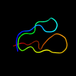
PDB 2a2b chain A

3D model

Region: 79 - 97
Aligned: 17
Modelled: 19
Confidence: 49.6%
Identity: 41%
PDB header:antibiotic
Chain: A: PDB Molecule:bacteriocin curvacin a;
PDBTitle: curvacin a

Phyre2

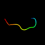
PDB 2q3e chain H

3D model

Region: 1 - 21
Aligned: 21
Modelled: 21
Confidence: 16.6%
Identity: 29%
PDB header:oxidoreductase
Chain: H: PDB Molecule:udp-glucose 6-dehydrogenase;
PDBTitle: structure of human udp-glucose dehydrogenase complexed with nadh and2 udp-glucose

Phyre2

PDB 1flc chain A domain 1

3D model

Region: 39 - 49
Aligned: 11
Modelled: 11
Confidence: 11.7%
Identity: 45%
Fold: Viral protein domain
Superfamily: Viral protein domain
Family: Hemagglutinin domain of haemagglutinin-esterase-fusion glycoprotein HEF1

Phyre2

PDB 1ua7 chain A domain 1

3D model

Region: 79 - 88
Aligned: 10
Modelled: 10
Confidence: 11.0%
Identity: 10%
Fold: Glycosyl hydrolase domain
Superfamily: Glycosyl hydrolase domain
Family: alpha-Amylases, C-terminal beta-sheet domain

Phyre2

PDB 3ebk chain A

3D model

Region: 78 - 113
Aligned: 32
Modelled: 36
Confidence: 9.9%
Identity: 22%
PDB header:allergen
Chain: A: PDB Molecule:allergen bla g 4;
PDBTitle: crystal structure of major allergens, bla g 4 from2 cockroaches

Phyre2

PDB 2yqp chain A

3D model

Region: 27 - 44
Aligned: 18
Modelled: 18
Confidence: 8.5%
Identity: 44%
PDB header:gene regulation, hydrolase
Chain: A: PDB Molecule:probable atp-dependent rna helicase ddx59;
PDBTitle: solution structure of the zf-hit domain in dead (asp-glu-2 ala-asp) box polypeptide 59

Phyre2

PDB 1q90 chain R

3D model

Region: 1 - 24
Aligned: 24
Modelled: 24
Confidence: 8.5%
Identity: 33%
Fold: Single transmembrane helix
Superfamily: ISP transmembrane anchor
Family: ISP transmembrane anchor

Phyre2

PDB 1q90 chain R

3D model

Region: 1 - 24
Aligned: 24
Modelled: 24
Confidence: 8.5%
Identity: 33%
PDB header:photosynthesis
Chain: R: PDB Molecule:cytochrome b6-f complex iron-sulfur subunit;
PDBTitle: structure of the cytochrome b6f (plastohydroquinone : plastocyanin2 oxidoreductase) from chlamydomonas reinhardtii

Phyre2

PDB 3pln chain A

3D model

Region: 2 - 21
Aligned: 20
Modelled: 20
Confidence: 8.1%
Identity: 25%
PDB header:oxidoreductase
Chain: A: PDB Molecule:udp-glucose 6-dehydrogenase;
PDBTitle: crystal structure of klebsiella pneumoniae udp-glucose 6-dehydrogenase2 complexed with udp-glucose

Phyre2

PDB 1qki chain A domain 2

3D model

Region: 58 - 85
Aligned: 20
Modelled: 28
Confidence: 7.6%
Identity: 45%
Fold: FwdE/GAPDH domain-like
Superfamily: Glyceraldehyde-3-phosphate dehydrogenase-like, C-terminal domain
Family: Glucose 6-phosphate dehydrogenase-like

Phyre2

PDB 3ggp chain A

3D model

Region: 1 - 41
Aligned: 36
Modelled: 38
Confidence: 7.5%
Identity: 19%
PDB header:oxidoreductase
Chain: A: PDB Molecule:prephenate dehydrogenase;
PDBTitle: crystal structure of prephenate dehydrogenase from a. aeolicus in2 complex with hydroxyphenyl propionate and nad+

Phyre2

PDB 1hx0 chain A domain 1

3D model

Region: 79 - 88
Aligned: 10
Modelled: 10
Confidence: 7.1%
Identity: 40%
Fold: Glycosyl hydrolase domain
Superfamily: Glycosyl hydrolase domain
Family: alpha-Amylases, C-terminal beta-sheet domain

Phyre2

PDB 3prj chain B

3D model

Region: 1 - 21
Aligned: 21
Modelled: 21
Confidence: 7.0%
Identity: 29%
PDB header:oxidoreductase
Chain: B: PDB Molecule:udp-glucose 6-dehydrogenase;
PDBTitle: role of packing defects in the evolution of allostery and induced fit2 in human udp-glucose dehydrogenase.

Phyre2

PDB 1g94 chain A domain 1

3D model

Region: 79 - 88
Aligned: 10
Modelled: 10
Confidence: 6.6%
Identity: 40%
Fold: Glycosyl hydrolase domain
Superfamily: Glycosyl hydrolase domain
Family: alpha-Amylases, C-terminal beta-sheet domain

Phyre2

PDB 1jae chain A domain 1

3D model

Region: 79 - 88
Aligned: 10
Modelled: 10
Confidence: 6.6%
Identity: 40%
Fold: Glycosyl hydrolase domain
Superfamily: Glycosyl hydrolase domain
Family: alpha-Amylases, C-terminal beta-sheet domain

Phyre2

PDB 2bhl chain B

3D model

Region: 58 - 85
Aligned: 20
Modelled: 28
Confidence: 6.6%
Identity: 45%
PDB header:oxidoreductase (choh(d)-nadp)
Chain: B: PDB Molecule:glucose-6-phosphate 1-dehydrogenase;
PDBTitle: x-ray structure of human glucose-6-phosphate dehydrogenase2 (deletion variant) complexed with glucose-6-phosphate

Phyre2

PDB 1e0f chain I

3D model

Region: 83 - 110
Aligned: 27
Modelled: 28
Confidence: 6.5%
Identity: 37%
PDB header:coagulation/crystal structure/heparin-b
Chain: I: PDB Molecule:haemadin;
PDBTitle: crystal structure of the human alpha-thrombin-haemadin2 complex: an exosite ii-binding inhibitor

Phyre2

PDB 1e0f chain I

3D model

Region: 83 - 110
Aligned: 27
Modelled: 28
Confidence: 6.5%
Identity: 37%
Fold: Knottins (small inhibitors, toxins, lectins)
Superfamily: Leech antihemostatic proteins
Family: Hirudin-like

Phyre2

PDB 3dhp chain A domain 1

3D model

Region: 79 - 88
Aligned: 10
Modelled: 10
Confidence: 6.3%
Identity: 40%
Fold: Glycosyl hydrolase domain
Superfamily: Glycosyl hydrolase domain
Family: alpha-Amylases, C-terminal beta-sheet domain

Phyre2
1

d1ig4a_
2

c2a2bA_
3

c2q3eH_
4

d1flca1
5

d1ua7a1
6

c3ebkA_
7

c2yqpA_
8

d1q90r_
9

c1q90R_
10

c3plnA_
11

d1qkia2
12

c3ggpA_
13

d1hx0a1
14

c3prjB_
15

d1g94a1
16

d1jaea1
17

c2bhlB_
18

c1e0fI_
19

d1e0fi_
20

d3dhpa1
21


22


23


24


25


26


27


28


29


Detailed template information 

#
Template Alignment Coverage3D Model Confidence
% i.d. Template Information
1d1ig4a_


50.9 32 Fold:DNA-binding domain
Superfamily:DNA-binding domain
Family:Methyl-CpG-binding domain, MBD
2c2a2bA_


49.6 41 PDB header:antibiotic
Chain: A: PDB Molecule:bacteriocin curvacin a;
PDBTitle: curvacin a
3c2q3eH_


16.6 29 PDB header:oxidoreductase
Chain: H: PDB Molecule:udp-glucose 6-dehydrogenase;
PDBTitle: structure of human udp-glucose dehydrogenase complexed with nadh and2 udp-glucose
4d1flca1


11.7 45 Fold:Viral protein domain
Superfamily:Viral protein domain
Family:Hemagglutinin domain of haemagglutinin-esterase-fusion glycoprotein HEF1
5d1ua7a1


11.0 10 Fold:Glycosyl hydrolase domain
Superfamily:Glycosyl hydrolase domain
Family:alpha-Amylases, C-terminal beta-sheet domain
6c3ebkA_


9.9 22 PDB header:allergen
Chain: A: PDB Molecule:allergen bla g 4;
PDBTitle: crystal structure of major allergens, bla g 4 from2 cockroaches
7c2yqpA_


8.5 44 PDB header:gene regulation, hydrolase
Chain: A: PDB Molecule:probable atp-dependent rna helicase ddx59;
PDBTitle: solution structure of the zf-hit domain in dead (asp-glu-2 ala-asp) box polypeptide 59
8d1q90r_


8.5 33 Fold:Single transmembrane helix
Superfamily:ISP transmembrane anchor
Family:ISP transmembrane anchor
9c1q90R_


8.5 33 PDB header:photosynthesis
Chain: R: PDB Molecule:cytochrome b6-f complex iron-sulfur subunit;
PDBTitle: structure of the cytochrome b6f (plastohydroquinone : plastocyanin2 oxidoreductase) from chlamydomonas reinhardtii
10c3plnA_


8.1 25 PDB header:oxidoreductase
Chain: A: PDB Molecule:udp-glucose 6-dehydrogenase;
PDBTitle: crystal structure of klebsiella pneumoniae udp-glucose 6-dehydrogenase2 complexed with udp-glucose
11d1qkia2


7.6 45 Fold:FwdE/GAPDH domain-like
Superfamily:Glyceraldehyde-3-phosphate dehydrogenase-like, C-terminal domain
Family:Glucose 6-phosphate dehydrogenase-like
12c3ggpA_


7.5 19 PDB header:oxidoreductase
Chain: A: PDB Molecule:prephenate dehydrogenase;
PDBTitle: crystal structure of prephenate dehydrogenase from a. aeolicus in2 complex with hydroxyphenyl propionate and nad+
13d1hx0a1


7.1 40 Fold:Glycosyl hydrolase domain
Superfamily:Glycosyl hydrolase domain
Family:alpha-Amylases, C-terminal beta-sheet domain
14c3prjB_


7.0 29 PDB header:oxidoreductase
Chain: B: PDB Molecule:udp-glucose 6-dehydrogenase;
PDBTitle: role of packing defects in the evolution of allostery and induced fit2 in human udp-glucose dehydrogenase.
15d1g94a1


6.6 40 Fold:Glycosyl hydrolase domain
Superfamily:Glycosyl hydrolase domain
Family:alpha-Amylases, C-terminal beta-sheet domain
16d1jaea1


6.6 40 Fold:Glycosyl hydrolase domain
Superfamily:Glycosyl hydrolase domain
Family:alpha-Amylases, C-terminal beta-sheet domain
17c2bhlB_


6.6 45 PDB header:oxidoreductase (choh(d)-nadp)
Chain: B: PDB Molecule:glucose-6-phosphate 1-dehydrogenase;
PDBTitle: x-ray structure of human glucose-6-phosphate dehydrogenase2 (deletion variant) complexed with glucose-6-phosphate
18c1e0fI_


6.5 37 PDB header:coagulation/crystal structure/heparin-b
Chain: I: PDB Molecule:haemadin;
PDBTitle: crystal structure of the human alpha-thrombin-haemadin2 complex: an exosite ii-binding inhibitor
19d1e0fi_


6.5 37 Fold:Knottins (small inhibitors, toxins, lectins)
Superfamily:Leech antihemostatic proteins
Family:Hirudin-like
20d3dhpa1


6.3 40 Fold:Glycosyl hydrolase domain
Superfamily:Glycosyl hydrolase domain
Family:alpha-Amylases, C-terminal beta-sheet domain
21d1t27a_


not modelled 6.2 16 Fold:TBP-like
Superfamily:Bet v1-like
Family:Phoshatidylinositol transfer protein, PITP
22d1r0va3


not modelled 6.1 29 Fold:MutS N-terminal domain-like
Superfamily:tRNA-intron endonuclease N-terminal domain-like
Family:tRNA-intron endonuclease N-terminal domain-like
23d1rmka_


not modelled 6.0 45 Fold:Knottins (small inhibitors, toxins, lectins)
Superfamily:omega toxin-like
Family:Conotoxin
24d1h9aa1


not modelled 6.0 33 Fold:NAD(P)-binding Rossmann-fold domains
Superfamily:NAD(P)-binding Rossmann-fold domains
Family:Glyceraldehyde-3-phosphate dehydrogenase-like, N-terminal domain
25c2l3bA_


not modelled 5.9 29 PDB header:structural genomics, unknown function
Chain: A: PDB Molecule:conserved protein found in conjugate transposon;
PDBTitle: solution nmr structure of the bt_0084 lipoprotein from bacteroides2 thetaiotaomicron, northeast structural genomics consortium target3 btr376
26d2a1la1


not modelled 5.9 20 Fold:TBP-like
Superfamily:Bet v1-like
Family:Phoshatidylinositol transfer protein, PITP
27c2l7qA_


not modelled 5.9 33 PDB header:structural genomics, unknown function
Chain: A: PDB Molecule:conserved protein found in conjugate transposon;
PDBTitle: solution nmr structure of conjugate transposon protein bvu_1572(27-2 141) from bacteroides vulgatus, northeast structural genomics3 consortium target bvr155
28c1e0fJ_


not modelled 5.8 37 PDB header:coagulation/crystal structure/heparin-b
Chain: J: PDB Molecule:haemadin;
PDBTitle: crystal structure of the human alpha-thrombin-haemadin2 complex: an exosite ii-binding inhibitor
29c1qkiE_


not modelled 5.5 45 PDB header:oxidoreductase
Chain: E: PDB Molecule:glucose-6-phosphate 1-dehydrogenase;
PDBTitle: x-ray structure of human glucose 6-phosphate dehydrogenase2 (variant canton r459l) complexed with structural nadp+

Binding site prediction 

Due to computational demand, binding site predictions are not run for batch jobs

If you want to predict binding sites, please manually submit your model of choice to 3DLigandSite


Phyre is for academic use only

Please cite: Protein structure prediction on the web: a case study using the Phyre server
Kelley LA and Sternberg MJE. Nature Protocols 4, 363 - 371 (2009) [pdf] [Import into BibTeX]
 
If you use the binding site predictions from 3DLigandSite, please also cite:
3DLigandSite: predicting ligand-binding sites using similar structures.
Wass MN, Kelley LA and Sternberg MJ Nucleic Acids Research 38, W469-73 (2010) [PubMed]
 
© Structural Bioinformatics Group
Imperial College London
Lawrence Kelley, Benjamin Jefferys
Disclaimer
Terms and Conditions
Component software
Template detection: HHpred 1.51
Secondary structure prediction: Psi-pred 2.5
Disorder prediction: Disopred 2.4
Transmembrane prediction: Memsat_SVM
Multi-template modelling and ab initio: Poing 1.0