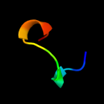
Phyre Home Retrieve Phyre Job Id
Emaill.a.kelley@imperial.ac.uk
DescriptionP76547
DateThu Jan 5 12:24:24 GMT 2012
Unique Job ID579a475f46b22738

Summary 

Top model
Image coloured by rainbow N → C terminus
Model (left) based on template c4a1oB_
Top template information
PDB header:transferase-hydrolase
Chain: B: PDB Molecule:bifunctional purine biosynthesis protein purh;
PDBTitle: crystal structure of mycobacterium tuberculosis purh complexed with2 aicar and a novel nucleotide cfair, at 2.48 a resolution.
Confidence and coverage
Confidence: 26.1% Coverage: 42%
83 residues ( 42% of your sequence) have been modelled with 26.1% confidence by the single highest scoring template.
You may wish to submit your sequence to Phyrealarm. This will automatically scan your sequence every week for new potential templates as they appear in the Phyre2 library.
Please note: You must be registered and logged in to use Phyrealarm.
3D viewing
Interactive 3D view in Jmol

Sequence analysis 

Secondary structure and disorder prediction 

   1........10.........20.........30.........40.........50.........60
Sequence  MSLIRTETRDTKRAADPLHDLRSKPFSEWGEDEIRRFNLIDALLEFVYTDTSSPFGIGMT
Secondary structure 


SS confidence 


Disorder  ???????????????
??
?


?
??


Disorder confidence 


   .........70.........80.........90.........100.........110.........120
Sequence  FDYTECWEIGVRDDCLVMTRVKPVHPEYAKHWNMKGVMNDKTRFHADKWVGYSKVLAWVS
Secondary structure 


SS confidence 


Disorder 


Disorder confidence 


   .........130.........140.........150.........160.........170.........180
Sequence  LSHKDTFTGAKRFQYFQTMYDMERQINANLPVGGLPNVDTERTGKLFQRDDFSEDSHAND
Secondary structure 


SS confidence 


Disorder 


??


??
?????????????
Disorder confidence 


   .........190.......
Sequence  PKLVGDDYVPQAPEQIN
Secondary structure 


SS confidence 


Disorder  ?


???????
Disorder confidence 


Confidence Key
High(9)                    Low (0)
?Disordered
Alpha helix
Beta strand

Domain analysis 

Hover over an aligned region to see model and summary info

Please note, only up to the top 20 hits are modelled to reduce computer load

RankAligned region

PDB 4a1o chain B

3D model

Region: 89 - 177
Aligned: 83
Modelled: 89
Confidence: 26.1%
Identity: 19%
PDB header:transferase-hydrolase
Chain: B: PDB Molecule:bifunctional purine biosynthesis protein purh;
PDBTitle: crystal structure of mycobacterium tuberculosis purh complexed with2 aicar and a novel nucleotide cfair, at 2.48 a resolution.

Phyre2

PDB 2g39 chain A domain 1

3D model

Region: 145 - 196
Aligned: 43
Modelled: 43
Confidence: 24.3%
Identity: 21%
Fold: NagB/RpiA/CoA transferase-like
Superfamily: NagB/RpiA/CoA transferase-like
Family: CoA transferase alpha subunit-like

Phyre2

PDB 2no8 chain A

3D model

Region: 21 - 37
Aligned: 16
Modelled: 17
Confidence: 13.6%
Identity: 31%
PDB header:immune system
Chain: A: PDB Molecule:colicin-e2 immunity protein;
PDBTitle: nmr structure analysis of the colicin immuntiy protein im2

Phyre2

PDB 2l95 chain A

3D model

Region: 25 - 37
Aligned: 13
Modelled: 13
Confidence: 13.5%
Identity: 8%
PDB header:hydrolase
Chain: A: PDB Molecule:crammer;
PDBTitle: solution structure of cytotoxic t-lymphocyte antigent-2(ctla protein),2 crammer at ph 6.0

Phyre2

PDB 3bq7 chain A

3D model

Region: 24 - 37
Aligned: 14
Modelled: 14
Confidence: 12.3%
Identity: 29%
PDB header:transferase
Chain: A: PDB Molecule:diacylglycerol kinase delta;
PDBTitle: sam domain of diacylglycerol kinase delta1 (e35g)

Phyre2

PDB 1iv8 chain A domain 2

3D model

Region: 2 - 16
Aligned: 15
Modelled: 15
Confidence: 12.3%
Identity: 47%
Fold: TIM beta/alpha-barrel
Superfamily: (Trans)glycosidases
Family: Amylase, catalytic domain

Phyre2

PDB 1zo0 chain A domain 1

3D model

Region: 55 - 115
Aligned: 41
Modelled: 46
Confidence: 11.9%
Identity: 7%
Fold: Acyl-CoA N-acyltransferases (Nat)
Superfamily: Acyl-CoA N-acyltransferases (Nat)
Family: Ornithine decarboxylase antizyme-like

Phyre2

PDB 2csb chain A domain 4

3D model

Region: 86 - 115
Aligned: 30
Modelled: 30
Confidence: 11.8%
Identity: 33%
Fold: SAM domain-like
Superfamily: RuvA domain 2-like
Family: Topoisomerase V repeat domain

Phyre2

PDB 2xqo chain A

3D model

Region: 178 - 190
Aligned: 13
Modelled: 13
Confidence: 11.6%
Identity: 38%
PDB header:hydrolase
Chain: A: PDB Molecule:cellulosome enzyme, dockerin type i;
PDBTitle: ctcel124: a cellulase from clostridium thermocellum

Phyre2

PDB 3hje chain A

3D model

Region: 2 - 16
Aligned: 15
Modelled: 15
Confidence: 11.1%
Identity: 47%
PDB header:transferase
Chain: A: PDB Molecule:704aa long hypothetical glycosyltransferase;
PDBTitle: crystal structure of sulfolobus tokodaii hypothetical2 maltooligosyl trehalose synthase

Phyre2

PDB 1v85 chain A

3D model

Region: 19 - 73
Aligned: 55
Modelled: 55
Confidence: 10.3%
Identity: 11%
PDB header:apoptosis
Chain: A: PDB Molecule:similar to ring finger protein 36;
PDBTitle: sterile alpha motif (sam) domain of mouse bifunctional2 apoptosis regulator

Phyre2

PDB 3es5 chain A

3D model

Region: 127 - 150
Aligned: 22
Modelled: 24
Confidence: 9.1%
Identity: 45%
PDB header:virus
Chain: A: PDB Molecule:putative capsid protein;
PDBTitle: crystal structure of partitivirus (psv-f)

Phyre2

PDB 1gxh chain A

3D model

Region: 25 - 37
Aligned: 13
Modelled: 13
Confidence: 8.9%
Identity: 23%
Fold: Acyl carrier protein-like
Superfamily: Colicin E immunity proteins
Family: Colicin E immunity proteins

Phyre2

PDB 3op0 chain B

3D model

Region: 38 - 97
Aligned: 58
Modelled: 60
Confidence: 8.7%
Identity: 21%
PDB header:signaling protein/signaling protein regu
Chain: B: PDB Molecule:signal transduction protein cbl-c;
PDBTitle: crystal structure of cbl-c (cbl-3) tkb domain in complex with egfr2 py1069 peptide

Phyre2

PDB 2nog chain A

3D model

Region: 19 - 37
Aligned: 19
Modelled: 19
Confidence: 8.4%
Identity: 21%
PDB header:dna binding protein
Chain: A: PDB Molecule:iswi protein;
PDBTitle: sant domain structure of xenopus remodeling factor iswi

Phyre2

PDB 2dld chain A domain 2

3D model

Region: 154 - 178
Aligned: 25
Modelled: 25
Confidence: 8.3%
Identity: 24%
Fold: Flavodoxin-like
Superfamily: Formate/glycerate dehydrogenase catalytic domain-like
Family: Formate/glycerate dehydrogenases, substrate-binding domain

Phyre2

PDB 3bs7 chain A

3D model

Region: 24 - 34
Aligned: 11
Modelled: 11
Confidence: 8.1%
Identity: 18%
PDB header:signaling protein
Chain: A: PDB Molecule:protein aveugle;
PDBTitle: crystal structure of the sterile alpha motif (sam) domain2 of hyphen/aveugle

Phyre2

PDB 1l6l chain K

3D model

Region: 134 - 157
Aligned: 24
Modelled: 24
Confidence: 8.0%
Identity: 29%
PDB header:lipid transport
Chain: K: PDB Molecule:apolipoprotein a-ii;
PDBTitle: structures of apolipoprotein a-ii and a lipid surrogate2 complex provide insights into apolipoprotein-lipid3 interactions

Phyre2

PDB 2vlq chain A domain 1

3D model

Region: 25 - 37
Aligned: 13
Modelled: 13
Confidence: 7.4%
Identity: 23%
Fold: Acyl carrier protein-like
Superfamily: Colicin E immunity proteins
Family: Colicin E immunity proteins

Phyre2

PDB 1j4a chain A domain 2

3D model

Region: 154 - 166
Aligned: 13
Modelled: 13
Confidence: 7.4%
Identity: 31%
Fold: Flavodoxin-like
Superfamily: Formate/glycerate dehydrogenase catalytic domain-like
Family: Formate/glycerate dehydrogenases, substrate-binding domain

Phyre2
1

c4a1oB_
2

d2g39a1
3

c2no8A_
4

c2l95A_
5

c3bq7A_
6

d1iv8a2
7

d1zo0a1
8

d2csba4
9

c2xqoA_
10

c3hjeA_
11

c1v85A_
12

c3es5A_
13

d1gxha_
14

c3op0B_
15

c2nogA_
16

d2dlda2
17

c3bs7A_
18

c1l6lK_
19

d2vlqa1
20

d1j4aa2
21


22


23


24


25


26


27


28


29


30


31


32


33


34


35


36


Detailed template information 

#
Template Alignment Coverage3D Model Confidence
% i.d. Template Information
1c4a1oB_


26.1 19 PDB header:transferase-hydrolase
Chain: B: PDB Molecule:bifunctional purine biosynthesis protein purh;
PDBTitle: crystal structure of mycobacterium tuberculosis purh complexed with2 aicar and a novel nucleotide cfair, at 2.48 a resolution.
2d2g39a1


24.3 21 Fold:NagB/RpiA/CoA transferase-like
Superfamily:NagB/RpiA/CoA transferase-like
Family:CoA transferase alpha subunit-like
3c2no8A_


13.6 31 PDB header:immune system
Chain: A: PDB Molecule:colicin-e2 immunity protein;
PDBTitle: nmr structure analysis of the colicin immuntiy protein im2
4c2l95A_


13.5 8 PDB header:hydrolase
Chain: A: PDB Molecule:crammer;
PDBTitle: solution structure of cytotoxic t-lymphocyte antigent-2(ctla protein),2 crammer at ph 6.0
5c3bq7A_


12.3 29 PDB header:transferase
Chain: A: PDB Molecule:diacylglycerol kinase delta;
PDBTitle: sam domain of diacylglycerol kinase delta1 (e35g)
6d1iv8a2


12.3 47 Fold:TIM beta/alpha-barrel
Superfamily:(Trans)glycosidases
Family:Amylase, catalytic domain
7d1zo0a1


11.9 7 Fold:Acyl-CoA N-acyltransferases (Nat)
Superfamily:Acyl-CoA N-acyltransferases (Nat)
Family:Ornithine decarboxylase antizyme-like
8d2csba4


11.8 33 Fold:SAM domain-like
Superfamily:RuvA domain 2-like
Family:Topoisomerase V repeat domain
9c2xqoA_


11.6 38 PDB header:hydrolase
Chain: A: PDB Molecule:cellulosome enzyme, dockerin type i;
PDBTitle: ctcel124: a cellulase from clostridium thermocellum
10c3hjeA_


11.1 47 PDB header:transferase
Chain: A: PDB Molecule:704aa long hypothetical glycosyltransferase;
PDBTitle: crystal structure of sulfolobus tokodaii hypothetical2 maltooligosyl trehalose synthase
11c1v85A_


10.3 11 PDB header:apoptosis
Chain: A: PDB Molecule:similar to ring finger protein 36;
PDBTitle: sterile alpha motif (sam) domain of mouse bifunctional2 apoptosis regulator
12c3es5A_


9.1 45 PDB header:virus
Chain: A: PDB Molecule:putative capsid protein;
PDBTitle: crystal structure of partitivirus (psv-f)
13d1gxha_


8.9 23 Fold:Acyl carrier protein-like
Superfamily:Colicin E immunity proteins
Family:Colicin E immunity proteins
14c3op0B_


8.7 21 PDB header:signaling protein/signaling protein regu
Chain: B: PDB Molecule:signal transduction protein cbl-c;
PDBTitle: crystal structure of cbl-c (cbl-3) tkb domain in complex with egfr2 py1069 peptide
15c2nogA_


8.4 21 PDB header:dna binding protein
Chain: A: PDB Molecule:iswi protein;
PDBTitle: sant domain structure of xenopus remodeling factor iswi
16d2dlda2


8.3 24 Fold:Flavodoxin-like
Superfamily:Formate/glycerate dehydrogenase catalytic domain-like
Family:Formate/glycerate dehydrogenases, substrate-binding domain
17c3bs7A_


8.1 18 PDB header:signaling protein
Chain: A: PDB Molecule:protein aveugle;
PDBTitle: crystal structure of the sterile alpha motif (sam) domain2 of hyphen/aveugle
18c1l6lK_


8.0 29 PDB header:lipid transport
Chain: K: PDB Molecule:apolipoprotein a-ii;
PDBTitle: structures of apolipoprotein a-ii and a lipid surrogate2 complex provide insights into apolipoprotein-lipid3 interactions
19d2vlqa1


7.4 23 Fold:Acyl carrier protein-like
Superfamily:Colicin E immunity proteins
Family:Colicin E immunity proteins
20d1j4aa2


7.4 31 Fold:Flavodoxin-like
Superfamily:Formate/glycerate dehydrogenase catalytic domain-like
Family:Formate/glycerate dehydrogenases, substrate-binding domain
21c1ofcX_


not modelled 7.4 29 PDB header:nuclear protein
Chain: X: PDB Molecule:iswi protein;
PDBTitle: nucleosome recognition module of iswi atpase
22d2cqqa1


not modelled 7.3 27 Fold:DNA/RNA-binding 3-helical bundle
Superfamily:Homeodomain-like
Family:Myb/SANT domain
23d1f0la2


not modelled 7.3 29 Fold:ADP-ribosylation
Superfamily:ADP-ribosylation
Family:ADP-ribosylating toxins
24d2hqya2


not modelled 6.9 19 Fold:Acyl-CoA N-acyltransferases (Nat)
Superfamily:Acyl-CoA N-acyltransferases (Nat)
Family:FemXAB nonribosomal peptidyltransferases
25c2wsfG_


not modelled 6.7 47 PDB header:photosynthesis
Chain: G: PDB Molecule:photosystem i reaction center subunit v,
PDBTitle: improved model of plant photosystem i
26c3f75P_


not modelled 6.7 38 PDB header:hydrolase
Chain: P: PDB Molecule:cathepsin l propeptide;
PDBTitle: activated toxoplasma gondii cathepsin l (tgcpl) in complex with its2 propeptide
27c2wscK_


not modelled 6.5 26 PDB header:photosynthesis
Chain: K: PDB Molecule:photosystem i reaction center subunit psak,
PDBTitle: improved model of plant photosystem i
28d2fu5a1


not modelled 6.5 25 Fold:Mss4-like
Superfamily:Mss4-like
Family:RabGEF Mss4
29d3buxb1


not modelled 6.2 24 Fold:EF Hand-like
Superfamily:EF-hand
Family:EF-hand modules in multidomain proteins
30d1ayia_


not modelled 6.2 23 Fold:Acyl carrier protein-like
Superfamily:Colicin E immunity proteins
Family:Colicin E immunity proteins
31d1wwva1


not modelled 6.1 14 Fold:SAM domain-like
Superfamily:SAM/Pointed domain
Family:SAM (sterile alpha motif) domain
32c3qodB_


not modelled 6.1 14 PDB header:dna binding protein
Chain: B: PDB Molecule:heterocyst differentiation protein;
PDBTitle: crystal structure of heterocyst differentiation protein, hetr from2 fischerella mv11
33d1dpsa_


not modelled 5.7 71 Fold:Ferritin-like
Superfamily:Ferritin-like
Family:Ferritin
34d1t92a_


not modelled 5.7 18 Fold:Pili subunits
Superfamily:Pili subunits
Family:Pseudopilin
35c3cecA_


not modelled 5.5 57 PDB header:transcription
Chain: A: PDB Molecule:putative antidote protein of plasmid maintenance system;
PDBTitle: crystal structure of a putative antidote protein of plasmid2 maintenance system (npun_f2943) from nostoc punctiforme pcc 73102 at3 1.60 a resolution
36c2o01G_


not modelled 5.3 47 PDB header:photosynthesis
Chain: G: PDB Molecule:photosystem i reaction center subunit v,
PDBTitle: the structure of a plant photosystem i supercomplex at 3.42 angstrom resolution

Binding site prediction 

Due to computational demand, binding site predictions are not run for batch jobs

If you want to predict binding sites, please manually submit your model of choice to 3DLigandSite


Phyre is for academic use only

Please cite: Protein structure prediction on the web: a case study using the Phyre server
Kelley LA and Sternberg MJE. Nature Protocols 4, 363 - 371 (2009) [pdf] [Import into BibTeX]
 
If you use the binding site predictions from 3DLigandSite, please also cite:
3DLigandSite: predicting ligand-binding sites using similar structures.
Wass MN, Kelley LA and Sternberg MJ Nucleic Acids Research 38, W469-73 (2010) [PubMed]
 
© Structural Bioinformatics Group
Imperial College London
Lawrence Kelley, Benjamin Jefferys
Disclaimer
Terms and Conditions
Component software
Template detection: HHpred 1.51
Secondary structure prediction: Psi-pred 2.5
Disorder prediction: Disopred 2.4
Transmembrane prediction: Memsat_SVM
Multi-template modelling and ab initio: Poing 1.0