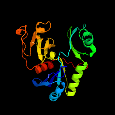
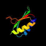
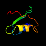
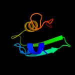
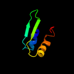
1 c1yf5L_
100.0
22
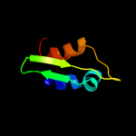
PDB header: transport proteinChain: L: PDB Molecule: general secretion pathway protein l;PDBTitle: cyto-epsl: the cytoplasmic domain of epsl, an inner membrane component2 of the type ii secretion system of vibrio cholerae
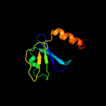
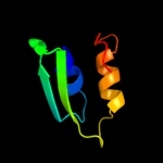
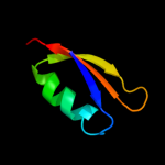
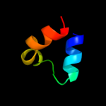
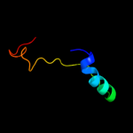
2 d2bh1a2
100.0
24
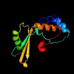
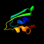
Fold: Ribonuclease H-like motifSuperfamily: Actin-like ATPase domainFamily: Cyto-EpsL domain3 c2w7vB_
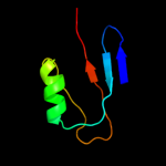
99.8
11
PDB header: transport proteinChain: B: PDB Molecule: general secretion pathway protein l;PDBTitle: periplasmic domain of epsl from vibrio parahaemolyticus
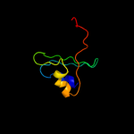
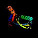
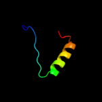
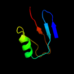
4 d2bh1a1
99.7
18
Fold: Ribonuclease H-like motifSuperfamily: Actin-like ATPase domainFamily: Cyto-EpsL domain5 c2ychA_
98.0
16
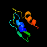
PDB header: cell cycleChain: A: PDB Molecule: competence protein pilm;PDBTitle: pilm-piln type iv pilus biogenesis complex
6 d1p6ta2
27.1
11
Fold: Ferredoxin-likeSuperfamily: HMA, heavy metal-associated domainFamily: HMA, heavy metal-associated domain7 d1s6ua_
25.7
5
Fold: Ferredoxin-likeSuperfamily: HMA, heavy metal-associated domainFamily: HMA, heavy metal-associated domain8 c2l3mA_
21.0
8
PDB header: metal binding proteinChain: A: PDB Molecule: copper-ion-binding protein;PDBTitle: solution structure of the putative copper-ion-binding protein from2 bacillus anthracis str. ames
9 d1e4ft1
19.7
6
Fold: Ribonuclease H-like motifSuperfamily: Actin-like ATPase domainFamily: Actin/HSP7010 d2qifa1
16.9
10
Fold: Ferredoxin-likeSuperfamily: HMA, heavy metal-associated domainFamily: HMA, heavy metal-associated domain11 d2aw0a_
16.7
4
Fold: Ferredoxin-likeSuperfamily: HMA, heavy metal-associated domainFamily: HMA, heavy metal-associated domain12 d2dyna_
12.9
16
Fold: PH domain-like barrelSuperfamily: PH domain-likeFamily: Pleckstrin-homology domain (PH domain)13 d1dcja_
12.9
19
Fold: IF3-likeSuperfamily: SirA-likeFamily: SirA-like14 d1cpza_
10.8
4
Fold: Ferredoxin-likeSuperfamily: HMA, heavy metal-associated domainFamily: HMA, heavy metal-associated domain15 d1sb6a_
10.4
6
Fold: Ferredoxin-likeSuperfamily: HMA, heavy metal-associated domainFamily: HMA, heavy metal-associated domain16 c2ky4A_
9.4
3
PDB header: photosynthesisChain: A: PDB Molecule: phycobilisome linker polypeptide;PDBTitle: solution nmr structure of the pbs linker domain of phycobilisome2 linker polypeptide from anabaena sp. northeast structural genomics3 consortium target nsr123e
17 d1uv7a_
8.8
6
Fold: RRF/tRNA synthetase additional domain-likeSuperfamily: General secretion pathway protein M, EpsMFamily: General secretion pathway protein M, EpsM18 c1uv7A_
8.8
6
PDB header: transportChain: A: PDB Molecule: general secretion pathway protein m;PDBTitle: periplasmic domain of epsm from vibrio cholerae
19 d1p6ta1
8.6
12
Fold: Ferredoxin-likeSuperfamily: HMA, heavy metal-associated domainFamily: HMA, heavy metal-associated domain20 c3p14C_
7.8
16
PDB header: isomeraseChain: C: PDB Molecule: l-rhamnose isomerase;PDBTitle: crystal structure of l-rhamnose isomerase with a novel high thermo-2 stability from bacillus halodurans
21 c2x29A_
not modelled
7.7
30
PDB header: cell adhesionChain: A: PDB Molecule: tumor necrosis factor ligand superfamily memberPDBTitle: crystal structure of human4-1bb ligand ectodomain
22 c3dxsX_
not modelled
7.7
11
PDB header: hydrolaseChain: X: PDB Molecule: copper-transporting atpase ran1;PDBTitle: crystal structure of a copper binding domain from hma7, a p-2 type atpase
23 d1d8wa_
not modelled
7.3
24
Fold: TIM beta/alpha-barrelSuperfamily: Xylose isomerase-likeFamily: L-rhamnose isomerase24 c2jpiA_
not modelled
7.2
6
PDB header: structural genomicsChain: A: PDB Molecule: hypothetical protein;PDBTitle: chemical shift assignments of pa4090 from pseudomonas2 aeruginosa
25 c2yciX_
not modelled
7.1
19
PDB header: transferaseChain: X: PDB Molecule: 5-methyltetrahydrofolate corrinoid/iron sulfur proteinPDBTitle: methyltransferase native
26 d1f44a1
not modelled
7.1
4
Fold: SAM domain-likeSuperfamily: lambda integrase-like, N-terminal domainFamily: lambda integrase-like, N-terminal domain27 c2k2pA_
not modelled
7.0
11
PDB header: structural genomics, unknown functionChain: A: PDB Molecule: uncharacterized protein atu1203;PDBTitle: solution nmr structure of protein atu1203 from agrobacterium2 tumefaciens. northeast structural genomics consortium (nesg) target3 att10, ontario center for structural proteomics target atc1183
28 d1q8la_
not modelled
7.0
5
Fold: Ferredoxin-likeSuperfamily: HMA, heavy metal-associated domainFamily: HMA, heavy metal-associated domain29 c2dhiA_
not modelled
7.0
9
PDB header: signaling proteinChain: A: PDB Molecule: pleckstrin homology domain-containing family bPDBTitle: solution structure of the ph domain of evectin-2 from mouse
30 c2kkhA_
not modelled
6.6
10
PDB header: metal transportChain: A: PDB Molecule: putative heavy metal transporter;PDBTitle: structure of the zinc binding domain of the atpase hma4
31 c1y3kA_
not modelled
5.9
5
PDB header: hydrolaseChain: A: PDB Molecule: copper-transporting atpase 1;PDBTitle: solution structure of the apo form of the fifth domain of2 menkes protein
32 d1afia_
not modelled
5.6
7
Fold: Ferredoxin-likeSuperfamily: HMA, heavy metal-associated domainFamily: HMA, heavy metal-associated domain33 c2rloA_
not modelled
5.6
6
PDB header: signaling proteinChain: A: PDB Molecule: centaurin-gamma 1;PDBTitle: split ph domain of pi3-kinase enhancer
34 c3lstB_
not modelled
5.4
26
PDB header: transferaseChain: B: PDB Molecule: calo1 methyltransferase;PDBTitle: crystal structure of calo1, methyltransferase in calicheamicin2 biosynthesis, sah bound form
35 d1f6ya_
not modelled
5.3
18
Fold: TIM beta/alpha-barrelSuperfamily: Dihydropteroate synthetase-likeFamily: Methyltetrahydrofolate-utiluzing methyltransferases