Phyre Home Retrieve Phyre Job Id
Emaill.a.kelley@imperial.ac.uk
DescriptionP64481
DateThu Jan 5 12:08:48 GMT 2012
Unique Job ID51f3c11526153b95

Summary 

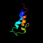
Top model
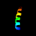
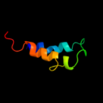
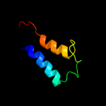
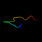
Image coloured by rainbow N → C terminus
Model (left) based on template d1ca1a1
Top template information
Fold:Phospholipase C/P1 nuclease
Superfamily:Phospholipase C/P1 nuclease
Family:Phospholipase C
Confidence and coverage
Confidence: 40.5% Coverage: 10%
19 residues ( 10% of your sequence) have been modelled with 40.5% confidence by the single highest scoring template.
You may wish to submit your sequence to Phyrealarm. This will automatically scan your sequence every week for new potential templates as they appear in the Phyre2 library.
Please note: You must be registered and logged in to use Phyrealarm.
3D viewing
Interactive 3D view in Jmol

Sequence analysis 

Secondary structure and disorder prediction 

   1........10.........20.........30.........40.........50.........60
Sequence  MTAEGHLLFSIACAVFAKNAELTPVLAQGDWWHIVPSAILTCLLPDIDHPKSFLGQRLKW
Secondary structure 


SS confidence 


Disorder  ???


??????


Disorder confidence 


   .........70.........80.........90.........100.........110.........120
Sequence  ISKPIARAFGHRGFTHSLLAVFALLATFYLKVPEGWFIPADALQGMVLGYLSHILADMLT
Secondary structure 


SS confidence 


Disorder 


??????


Disorder confidence 


   .........130.........140.........150.........160.........170.........180
Sequence  PAGVPLLWPCRWRFRLPILVPQKGNQLERFICMALFVWSVWMPHSLPENSAVRWSSQMIN
Secondary structure 


SS confidence 


Disorder 


?


?
???


Disorder confidence 


   .........190......
Sequence  TLQIQFHRLIKHQVEY
Secondary structure 

SS confidence 


Disorder 


???
Disorder confidence 


Confidence Key
High(9)                    Low (0)
?Disordered
Alpha helix
Beta strand

Domain analysis 

Hover over an aligned region to see model and summary info

Please note, only up to the top 20 hits are modelled to reduce computer load

RankAligned region

PDB 1ca1 chain A domain 1

3D model

Region: 104 - 122
Aligned: 19
Modelled: 19
Confidence: 40.5%
Identity: 32%
Fold: Phospholipase C/P1 nuclease
Superfamily: Phospholipase C/P1 nuclease
Family: Phospholipase C

Phyre2

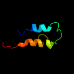
PDB 1olp chain A domain 1

3D model

Region: 104 - 121
Aligned: 18
Modelled: 18
Confidence: 31.2%
Identity: 33%
Fold: Phospholipase C/P1 nuclease
Superfamily: Phospholipase C/P1 nuclease
Family: Phospholipase C

Phyre2

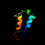
PDB 1gyg chain A

3D model

Region: 106 - 122
Aligned: 17
Modelled: 17
Confidence: 26.9%
Identity: 35%
PDB header:hydrolase
Chain: A: PDB Molecule:phospholipase c;
PDBTitle: r32 closed form of alpha-toxin from clostridium perfringens2 strain cer89l43

Phyre2

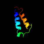
PDB 1kho chain A domain 1

3D model

Region: 104 - 121
Aligned: 18
Modelled: 18
Confidence: 26.7%
Identity: 33%
Fold: Phospholipase C/P1 nuclease
Superfamily: Phospholipase C/P1 nuclease
Family: Phospholipase C

Phyre2

PDB 1y2k chain A domain 1

3D model

Region: 5 - 51
Aligned: 43
Modelled: 47
Confidence: 18.8%
Identity: 23%
Fold: HD-domain/PDEase-like
Superfamily: HD-domain/PDEase-like
Family: PDEase

Phyre2

PDB 1xot chain B

3D model

Region: 5 - 51
Aligned: 43
Modelled: 47
Confidence: 18.0%
Identity: 23%
PDB header:hydrolase
Chain: B: PDB Molecule:camp-specific 3',5'-cyclic phosphodiesterase 4b;
PDBTitle: catalytic domain of human phosphodiesterase 4b in complex with2 vardenafil

Phyre2

PDB 2r8q chain A

3D model

Region: 5 - 51
Aligned: 43
Modelled: 47
Confidence: 13.8%
Identity: 16%
PDB header:hydrolase
Chain: A: PDB Molecule:class i phosphodiesterase pdeb1;
PDBTitle: structure of lmjpdeb1 in complex with ibmx

Phyre2

PDB 3ecm chain A

3D model

Region: 5 - 51
Aligned: 43
Modelled: 47
Confidence: 12.9%
Identity: 26%
PDB header:hydrolase
Chain: A: PDB Molecule:high affinity camp-specific and ibmx-insensitive
PDBTitle: crystal structure of the unliganded pde8a catalytic domain

Phyre2

PDB 1z1l chain A

3D model

Region: 5 - 51
Aligned: 43
Modelled: 47
Confidence: 12.8%
Identity: 30%
PDB header:hydrolase
Chain: A: PDB Molecule:cgmp-dependent 3',5'-cyclic phosphodiesterase;
PDBTitle: the crystal structure of the phosphodiesterase 2a catalytic2 domain

Phyre2

PDB 3ec2 chain A

3D model

Region: 2 - 12
Aligned: 11
Modelled: 11
Confidence: 11.9%
Identity: 18%
PDB header:replication
Chain: A: PDB Molecule:dna replication protein dnac;
PDBTitle: crystal structure of the dnac helicase loader

Phyre2

PDB 3dy8 chain A domain 1

3D model

Region: 5 - 51
Aligned: 43
Modelled: 47
Confidence: 11.5%
Identity: 19%
Fold: HD-domain/PDEase-like
Superfamily: HD-domain/PDEase-like
Family: PDEase

Phyre2

PDB 1ah7 chain A

3D model

Region: 104 - 122
Aligned: 19
Modelled: 19
Confidence: 11.2%
Identity: 32%
Fold: Phospholipase C/P1 nuclease
Superfamily: Phospholipase C/P1 nuclease
Family: Phospholipase C

Phyre2

PDB 1f0j chain A

3D model

Region: 5 - 51
Aligned: 43
Modelled: 47
Confidence: 11.1%
Identity: 23%
Fold: HD-domain/PDEase-like
Superfamily: HD-domain/PDEase-like
Family: PDEase

Phyre2

PDB 3bjc chain A

3D model

Region: 12 - 51
Aligned: 36
Modelled: 40
Confidence: 11.0%
Identity: 17%
PDB header:hydrolase
Chain: A: PDB Molecule:cgmp-specific 3',5'-cyclic phosphodiesterase;
PDBTitle: crystal structure of the pde5a catalytic domain in complex2 with a novel inhibitor

Phyre2

PDB 2w58 chain B

3D model

Region: 2 - 12
Aligned: 11
Modelled: 11
Confidence: 10.4%
Identity: 36%
PDB header:hydrolase
Chain: B: PDB Molecule:primosome component (helicase loader);
PDBTitle: crystal structure of the dnai

Phyre2

PDB 2qgz chain A

3D model

Region: 2 - 12
Aligned: 11
Modelled: 11
Confidence: 10.4%
Identity: 27%
PDB header:hydrolase
Chain: A: PDB Molecule:putative primosome component;
PDBTitle: crystal structure of a putative primosome component from2 streptococcus pyogenes serotype m3. northeast structural3 genomics target dr58

Phyre2

PDB 3cvg chain C

3D model

Region: 115 - 133
Aligned: 19
Modelled: 19
Confidence: 9.5%
Identity: 21%
PDB header:metal binding protein
Chain: C: PDB Molecule:putative metal binding protein;
PDBTitle: crystal structure of a periplasmic putative metal binding protein

Phyre2

PDB 1p3q chain Q

3D model

Region: 40 - 48
Aligned: 9
Modelled: 4
Confidence: 9.0%
Identity: 44%
Fold: RuvA C-terminal domain-like
Superfamily: UBA-like
Family: CUE domain

Phyre2

PDB 1tbf chain A

3D model

Region: 5 - 51
Aligned: 43
Modelled: 47
Confidence: 8.9%
Identity: 19%
Fold: HD-domain/PDEase-like
Superfamily: HD-domain/PDEase-like
Family: PDEase

Phyre2

PDB 1xoz chain A

3D model

Region: 5 - 51
Aligned: 43
Modelled: 47
Confidence: 8.9%
Identity: 19%
PDB header:hydrolase
Chain: A: PDB Molecule:cgmp-specific 3',5'-cyclic phosphodiesterase;
PDBTitle: catalytic domain of human phosphodiesterase 5a in complex2 with tadalafil

Phyre2
1

d1ca1a1
2

d1olpa1
3

c1gygA_
4

d1khoa1
5

d1y2ka1
6

c1xotB_
7

c2r8qA_
8

c3ecmA_
9

c1z1lA_
10

c3ec2A_
11

d3dy8a1
12

d1ah7a_
13

d1f0ja_
14

c3bjcA_
15

c2w58B_
16

c2qgzA_
17

c3cvgC_
18

d1p3qq_
19

d1tbfa_
20

c1xozA_
21


22


23


24


25


26


27


28


29


30


31


32


33


Detailed template information 

#
Template Alignment Coverage3D Model Confidence
% i.d. Template Information
1d1ca1a1


40.5 32 Fold:Phospholipase C/P1 nuclease
Superfamily:Phospholipase C/P1 nuclease
Family:Phospholipase C
2d1olpa1


31.2 33 Fold:Phospholipase C/P1 nuclease
Superfamily:Phospholipase C/P1 nuclease
Family:Phospholipase C
3c1gygA_


26.9 35 PDB header:hydrolase
Chain: A: PDB Molecule:phospholipase c;
PDBTitle: r32 closed form of alpha-toxin from clostridium perfringens2 strain cer89l43
4d1khoa1


26.7 33 Fold:Phospholipase C/P1 nuclease
Superfamily:Phospholipase C/P1 nuclease
Family:Phospholipase C
5d1y2ka1


18.8 23 Fold:HD-domain/PDEase-like
Superfamily:HD-domain/PDEase-like
Family:PDEase
6c1xotB_


18.0 23 PDB header:hydrolase
Chain: B: PDB Molecule:camp-specific 3',5'-cyclic phosphodiesterase 4b;
PDBTitle: catalytic domain of human phosphodiesterase 4b in complex with2 vardenafil
7c2r8qA_


13.8 16 PDB header:hydrolase
Chain: A: PDB Molecule:class i phosphodiesterase pdeb1;
PDBTitle: structure of lmjpdeb1 in complex with ibmx
8c3ecmA_


12.9 26 PDB header:hydrolase
Chain: A: PDB Molecule:high affinity camp-specific and ibmx-insensitive
PDBTitle: crystal structure of the unliganded pde8a catalytic domain
9c1z1lA_


12.8 30 PDB header:hydrolase
Chain: A: PDB Molecule:cgmp-dependent 3',5'-cyclic phosphodiesterase;
PDBTitle: the crystal structure of the phosphodiesterase 2a catalytic2 domain
10c3ec2A_


11.9 18 PDB header:replication
Chain: A: PDB Molecule:dna replication protein dnac;
PDBTitle: crystal structure of the dnac helicase loader
11d3dy8a1


11.5 19 Fold:HD-domain/PDEase-like
Superfamily:HD-domain/PDEase-like
Family:PDEase
12d1ah7a_


11.2 32 Fold:Phospholipase C/P1 nuclease
Superfamily:Phospholipase C/P1 nuclease
Family:Phospholipase C
13d1f0ja_


11.1 23 Fold:HD-domain/PDEase-like
Superfamily:HD-domain/PDEase-like
Family:PDEase
14c3bjcA_


11.0 17 PDB header:hydrolase
Chain: A: PDB Molecule:cgmp-specific 3',5'-cyclic phosphodiesterase;
PDBTitle: crystal structure of the pde5a catalytic domain in complex2 with a novel inhibitor
15c2w58B_


10.4 36 PDB header:hydrolase
Chain: B: PDB Molecule:primosome component (helicase loader);
PDBTitle: crystal structure of the dnai
16c2qgzA_


10.4 27 PDB header:hydrolase
Chain: A: PDB Molecule:putative primosome component;
PDBTitle: crystal structure of a putative primosome component from2 streptococcus pyogenes serotype m3. northeast structural3 genomics target dr58
17c3cvgC_


9.5 21 PDB header:metal binding protein
Chain: C: PDB Molecule:putative metal binding protein;
PDBTitle: crystal structure of a periplasmic putative metal binding protein
18d1p3qq_


9.0 44 Fold:RuvA C-terminal domain-like
Superfamily:UBA-like
Family:CUE domain
19d1tbfa_


8.9 19 Fold:HD-domain/PDEase-like
Superfamily:HD-domain/PDEase-like
Family:PDEase
20c1xozA_


8.9 19 PDB header:hydrolase
Chain: A: PDB Molecule:cgmp-specific 3',5'-cyclic phosphodiesterase;
PDBTitle: catalytic domain of human phosphodiesterase 5a in complex2 with tadalafil
21c1olpB_


not modelled 8.4 32 PDB header:hydrolase
Chain: B: PDB Molecule:alpha-toxin;
PDBTitle: alpha toxin from clostridium absonum
22c3qi4A_


not modelled 8.4 19 PDB header:hydrolase
Chain: A: PDB Molecule:high affinity cgmp-specific 3',5'-cyclic phosphodiesterase
PDBTitle: crystal structure of pde9a(q453e) in complex with ibmx
23c1wqdA_


not modelled 8.3 56 PDB header:toxin
Chain: A: PDB Molecule:omtx2;
PDBTitle: an unusual fold for potassium channel blockers: nmr2 structure of three toxins from the scorpion opisthacanthus3 madagascariensis
24c1zklA_


not modelled 8.1 21 PDB header:hydrolase
Chain: A: PDB Molecule:high-affinity camp-specific 3',5'-cyclic
PDBTitle: multiple determinants for inhibitor selectivity of cyclic2 nucleotide phosphodiesterases
25d2h44a1


not modelled 7.7 16 Fold:HD-domain/PDEase-like
Superfamily:HD-domain/PDEase-like
Family:PDEase
26c2kjqA_


not modelled 7.6 27 PDB header:replication
Chain: A: PDB Molecule:dnaa-related protein;
PDBTitle: solution structure of protein nmb1076 from neisseria meningitidis.2 northeast structural genomics consortium target mr101b.
27c3g3nA_


not modelled 7.4 21 PDB header:hydrolase
Chain: A: PDB Molecule:high affinity camp-specific 3',5'-cyclic
PDBTitle: pde7a catalytic domain in complex with 3-(2,6-2 difluorophenyl)-2-(methylthio)quinazolin-4(3h)-one
28d1so2a_


not modelled 7.3 24 Fold:HD-domain/PDEase-like
Superfamily:HD-domain/PDEase-like
Family:PDEase
29d1p3qr_


not modelled 7.0 43 Fold:RuvA C-terminal domain-like
Superfamily:UBA-like
Family:CUE domain
30c2lf6A_


not modelled 6.8 23 PDB header:signaling protein
Chain: A: PDB Molecule:effector protein hopab1;
PDBTitle: solution nmr structure of hopabpph1448_220_320 from pseudomonas2 syringae pv. phaseolicola str. 1448a, midwest center for structural3 genomics target apc40132.4 and northeast structural genomics4 consortium target pst3a
31d1mn3a_


not modelled 6.1 44 Fold:RuvA C-terminal domain-like
Superfamily:UBA-like
Family:CUE domain
32c3ibjB_


not modelled 5.9 30 PDB header:hydrolase
Chain: B: PDB Molecule:cgmp-dependent 3',5'-cyclic phosphodiesterase;
PDBTitle: x-ray structure of pde2a
33c2o8hA_


not modelled 5.4 18 PDB header:hydrolase
Chain: A: PDB Molecule:phosphodiesterase-10a;
PDBTitle: crystal structure of the catalytic domain of rat2 phosphodiesterase 10a

Binding site prediction 

Due to computational demand, binding site predictions are not run for batch jobs

If you want to predict binding sites, please manually submit your model of choice to 3DLigandSite


Phyre is for academic use only

Please cite: Protein structure prediction on the web: a case study using the Phyre server
Kelley LA and Sternberg MJE. Nature Protocols 4, 363 - 371 (2009) [pdf] [Import into BibTeX]
 
If you use the binding site predictions from 3DLigandSite, please also cite:
3DLigandSite: predicting ligand-binding sites using similar structures.
Wass MN, Kelley LA and Sternberg MJ Nucleic Acids Research 38, W469-73 (2010) [PubMed]
 
© Structural Bioinformatics Group
Imperial College London
Lawrence Kelley, Benjamin Jefferys
Disclaimer
Terms and Conditions
Component software
Template detection: HHpred 1.51
Secondary structure prediction: Psi-pred 2.5
Disorder prediction: Disopred 2.4
Transmembrane prediction: Memsat_SVM
Multi-template modelling and ab initio: Poing 1.0