Phyre Home Retrieve Phyre Job Id
Emaill.a.kelley@imperial.ac.uk
DescriptionP75818
DateThu Jan 5 12:14:31 GMT 2012
Unique Job ID51d2266632419c90

Summary 

Top model
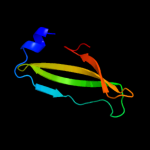
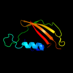
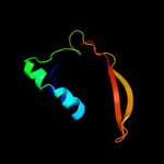
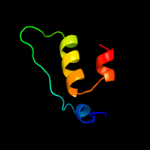
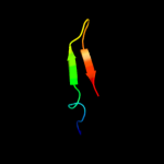
Image coloured by rainbow N → C terminus
Model (left) based on template c2qguA_
Top template information
PDB header:structural genomics, unknown function
Chain: A: PDB Molecule:probable signal peptide protein;
PDBTitle: three-dimensional structure of the phospholipid-binding protein from2 ralstonia solanacearum q8xv73_ralsq in complex with a phospholipid at3 the resolution 1.53 a. northeast structural genomics consortium4 target rsr89
Confidence and coverage
Confidence: 84.4% Coverage: 37%
64 residues ( 37% of your sequence) have been modelled with 84.4% confidence by the single highest scoring template.
You may wish to submit your sequence to Phyrealarm. This will automatically scan your sequence every week for new potential templates as they appear in the Phyre2 library.
Please note: You must be registered and logged in to use Phyrealarm.
3D viewing
Interactive 3D view in Jmol

Sequence analysis 

Secondary structure and disorder prediction 

   1........10.........20.........30.........40.........50.........60
Sequence  MRYSKLTMLIPCALLLSACTTVTPAYKDNGTRSGPCVEGGPDNVAQQFYDYRILHRSNDI
Secondary structure 


SS confidence 


Disorder  ????
?


?


?

??????????????


Disorder confidence 


   .........70.........80.........90.........100.........110.........120
Sequence  TALRPYLSDKLATLLSDASRDNNHRELLTNDPFSSRTTLPDSAHVASASTIPNRDARNIP
Secondary structure 


SS confidence 


Disorder 


?????????
?


??


??

???
??????


Disorder confidence 


   .........130.........140.........150.........160.........170.
Sequence  LRVDLKQGDQGWQDEVLMIQEGQCWVIDDVRYLGGSVHATAGTLRQSIENR
Secondary structure 


SS confidence 


Disorder 


?
????


????????


????
Disorder confidence 


Confidence Key
High(9)                    Low (0)
?Disordered
Alpha helix
Beta strand

Domain analysis 

Hover over an aligned region to see model and summary info

Please note, only up to the top 20 hits are modelled to reduce computer load

RankAligned region

PDB 2qgu chain A

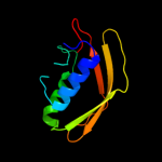
3D model

Region: 93 - 156
Aligned: 64
Modelled: 64
Confidence: 84.4%
Identity: 11%
PDB header:structural genomics, unknown function
Chain: A: PDB Molecule:probable signal peptide protein;
PDBTitle: three-dimensional structure of the phospholipid-binding protein from2 ralstonia solanacearum q8xv73_ralsq in complex with a phospholipid at3 the resolution 1.53 a. northeast structural genomics consortium4 target rsr89

Phyre2

PDB 3duk chain D

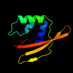
3D model

Region: 104 - 153
Aligned: 47
Modelled: 50
Confidence: 78.3%
Identity: 9%
PDB header:unknown function
Chain: D: PDB Molecule:ntf2-like protein of unknown function;
PDBTitle: crystal structure of a ntf2-like protein of unknown function2 (mfla_0564) from methylobacillus flagellatus kt at 2.200 a resolution

Phyre2

PDB 2cw9 chain A domain 1

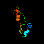
3D model

Region: 57 - 154
Aligned: 94
Modelled: 98
Confidence: 77.9%
Identity: 16%
Fold: Cystatin-like
Superfamily: NTF2-like
Family: TIM44-like

Phyre2

PDB 1vqq chain A domain 1

3D model

Region: 92 - 148
Aligned: 56
Modelled: 57
Confidence: 76.0%
Identity: 11%
Fold: Cystatin-like
Superfamily: NTF2-like
Family: Penicillin binding protein 2a (PBP2A), N-terminal domain

Phyre2

PDB 3qk9 chain B

3D model

Region: 57 - 154
Aligned: 94
Modelled: 98
Confidence: 75.0%
Identity: 11%
PDB header:protein transport
Chain: B: PDB Molecule:mitochondrial import inner membrane translocase subunit
PDBTitle: yeast tim44 c-terminal domain complexed with cymal-3

Phyre2

PDB 3fka chain D

3D model

Region: 98 - 153
Aligned: 52
Modelled: 56
Confidence: 71.4%
Identity: 17%
PDB header:unknown function
Chain: D: PDB Molecule:uncharacterized ntf-2 like protein;
PDBTitle: crystal structure of a ntf-2 like protein of unknown function2 (spo1084) from silicibacter pomeroyi dss-3 at 1.69 a resolution

Phyre2

PDB 3blz chain A domain 1

3D model

Region: 104 - 153
Aligned: 47
Modelled: 50
Confidence: 64.6%
Identity: 11%
Fold: Cystatin-like
Superfamily: NTF2-like
Family: Sbal0622-like

Phyre2

PDB 1w8o chain A domain 3

3D model

Region: 84 - 145
Aligned: 61
Modelled: 62
Confidence: 50.6%
Identity: 10%
Fold: 6-bladed beta-propeller
Superfamily: Sialidases
Family: Sialidases (neuraminidases)

Phyre2

PDB 2fxt chain A domain 1

3D model

Region: 57 - 127
Aligned: 66
Modelled: 71
Confidence: 35.3%
Identity: 9%
Fold: Cystatin-like
Superfamily: NTF2-like
Family: TIM44-like

Phyre2

PDB 1dmu chain A

3D model

Region: 106 - 145
Aligned: 39
Modelled: 40
Confidence: 32.1%
Identity: 10%
Fold: Restriction endonuclease-like
Superfamily: Restriction endonuclease-like
Family: Restriction endonuclease BglI

Phyre2

PDB 3f7s chain A

3D model

Region: 93 - 164
Aligned: 72
Modelled: 72
Confidence: 31.5%
Identity: 17%
PDB header:unknown function
Chain: A: PDB Molecule:uncharacterized ntf2-like protein;
PDBTitle: crystal structure of a ntf2-like protein of unknown function (pp_4556)2 from pseudomonas putida kt2440 at 2.11 a resolution

Phyre2

PDB 2ber chain A

3D model

Region: 84 - 145
Aligned: 60
Modelled: 62
Confidence: 22.7%
Identity: 8%
PDB header:hydrolase
Chain: A: PDB Molecule:bacterial sialidase;
PDBTitle: y370g active site mutant of the sialidase from2 micromonospora viridifaciens in complex with beta-neu5ac3 (sialic acid).

Phyre2

PDB 1m98 chain A domain 2

3D model

Region: 22 - 63
Aligned: 40
Modelled: 42
Confidence: 21.6%
Identity: 20%
Fold: Cystatin-like
Superfamily: NTF2-like
Family: Orange carotenoid protein, C-terminal domain

Phyre2

PDB 3gwr chain A

3D model

Region: 37 - 147
Aligned: 96
Modelled: 93
Confidence: 20.3%
Identity: 18%
PDB header:protein binding
Chain: A: PDB Molecule:putative calcium/calmodulin-dependent protein kinase type
PDBTitle: crystal structure of putative calcium/calmodulin-dependent protein2 kinase type ii association domain (yp_315894.1) from thiobacillus3 denitrificans atcc 25259 at 2.00 a resolution

Phyre2

PDB 3k7c chain C

3D model

Region: 55 - 147
Aligned: 90
Modelled: 93
Confidence: 16.1%
Identity: 11%
PDB header:protein binding
Chain: C: PDB Molecule:putative ntf2-like transpeptidase;
PDBTitle: crystal structure of putative ntf2-like transpeptidase (np_281412.1)2 from campylobacter jejuni at 2.00 a resolution

Phyre2

PDB 1kke chain A domain 2

3D model

Region: 85 - 160
Aligned: 55
Modelled: 76
Confidence: 14.9%
Identity: 35%
Fold: Virus attachment protein globular domain
Superfamily: Virus attachment protein globular domain
Family: Reovirus attachment protein sigma 1 head domain

Phyre2

PDB 2j2z chain A domain 2

3D model

Region: 129 - 149
Aligned: 21
Modelled: 21
Confidence: 13.2%
Identity: 19%
Fold: C2 domain-like
Superfamily: Periplasmic chaperone C-domain
Family: Periplasmic chaperone C-domain

Phyre2

PDB 1ub1 chain A

3D model

Region: 130 - 155
Aligned: 26
Modelled: 26
Confidence: 12.7%
Identity: 19%
Fold: DNA-binding domain
Superfamily: DNA-binding domain
Family: Methyl-CpG-binding domain, MBD

Phyre2

PDB 1kke chain A

3D model

Region: 85 - 160
Aligned: 55
Modelled: 76
Confidence: 12.5%
Identity: 35%
PDB header:viral protein
Chain: A: PDB Molecule:sigma 1 protein;
PDBTitle: crystal structure of reovirus attachment protein sigma12 trimer

Phyre2

PDB 3cnx chain A domain 1

3D model

Region: 102 - 147
Aligned: 46
Modelled: 46
Confidence: 10.7%
Identity: 9%
Fold: Cystatin-like
Superfamily: NTF2-like
Family: SAV4671-like

Phyre2
1

c2qguA_
2

c3dukD_
3

d2cw9a1
4

d1vqqa1
5

c3qk9B_
6

c3fkaD_
7

d3blza1
8

d1w8oa3
9

d2fxta1
10

d1dmua_
11

c3f7sA_
12

c2berA_
13

d1m98a2
14

c3gwrA_
15

c3k7cC_
16

d1kkea2
17

d2j2za2
18

d1ub1a_
19

c1kkeA_
20

d3cnxa1
21


22


23


24


25


26


27


28


29


30


31


32


33


34


35


36


37


38


39


40


41


42


43


44


Detailed template information 

#
Template Alignment Coverage3D Model Confidence
% i.d. Template Information
1c2qguA_


84.4 11 PDB header:structural genomics, unknown function
Chain: A: PDB Molecule:probable signal peptide protein;
PDBTitle: three-dimensional structure of the phospholipid-binding protein from2 ralstonia solanacearum q8xv73_ralsq in complex with a phospholipid at3 the resolution 1.53 a. northeast structural genomics consortium4 target rsr89
2c3dukD_


78.3 9 PDB header:unknown function
Chain: D: PDB Molecule:ntf2-like protein of unknown function;
PDBTitle: crystal structure of a ntf2-like protein of unknown function2 (mfla_0564) from methylobacillus flagellatus kt at 2.200 a resolution
3d2cw9a1


77.9 16 Fold:Cystatin-like
Superfamily:NTF2-like
Family:TIM44-like
4d1vqqa1


76.0 11 Fold:Cystatin-like
Superfamily:NTF2-like
Family:Penicillin binding protein 2a (PBP2A), N-terminal domain
5c3qk9B_


75.0 11 PDB header:protein transport
Chain: B: PDB Molecule:mitochondrial import inner membrane translocase subunit
PDBTitle: yeast tim44 c-terminal domain complexed with cymal-3
6c3fkaD_


71.4 17 PDB header:unknown function
Chain: D: PDB Molecule:uncharacterized ntf-2 like protein;
PDBTitle: crystal structure of a ntf-2 like protein of unknown function2 (spo1084) from silicibacter pomeroyi dss-3 at 1.69 a resolution
7d3blza1


64.6 11 Fold:Cystatin-like
Superfamily:NTF2-like
Family:Sbal0622-like
8d1w8oa3


50.6 10 Fold:6-bladed beta-propeller
Superfamily:Sialidases
Family:Sialidases (neuraminidases)
9d2fxta1


35.3 9 Fold:Cystatin-like
Superfamily:NTF2-like
Family:TIM44-like
10d1dmua_


32.1 10 Fold:Restriction endonuclease-like
Superfamily:Restriction endonuclease-like
Family:Restriction endonuclease BglI
11c3f7sA_


31.5 17 PDB header:unknown function
Chain: A: PDB Molecule:uncharacterized ntf2-like protein;
PDBTitle: crystal structure of a ntf2-like protein of unknown function (pp_4556)2 from pseudomonas putida kt2440 at 2.11 a resolution
12c2berA_


22.7 8 PDB header:hydrolase
Chain: A: PDB Molecule:bacterial sialidase;
PDBTitle: y370g active site mutant of the sialidase from2 micromonospora viridifaciens in complex with beta-neu5ac3 (sialic acid).
13d1m98a2


21.6 20 Fold:Cystatin-like
Superfamily:NTF2-like
Family:Orange carotenoid protein, C-terminal domain
14c3gwrA_


20.3 18 PDB header:protein binding
Chain: A: PDB Molecule:putative calcium/calmodulin-dependent protein kinase type
PDBTitle: crystal structure of putative calcium/calmodulin-dependent protein2 kinase type ii association domain (yp_315894.1) from thiobacillus3 denitrificans atcc 25259 at 2.00 a resolution
15c3k7cC_


16.1 11 PDB header:protein binding
Chain: C: PDB Molecule:putative ntf2-like transpeptidase;
PDBTitle: crystal structure of putative ntf2-like transpeptidase (np_281412.1)2 from campylobacter jejuni at 2.00 a resolution
16d1kkea2


14.9 35 Fold:Virus attachment protein globular domain
Superfamily:Virus attachment protein globular domain
Family:Reovirus attachment protein sigma 1 head domain
17d2j2za2


13.2 19 Fold:C2 domain-like
Superfamily:Periplasmic chaperone C-domain
Family:Periplasmic chaperone C-domain
18d1ub1a_


12.7 19 Fold:DNA-binding domain
Superfamily:DNA-binding domain
Family:Methyl-CpG-binding domain, MBD
19c1kkeA_


12.5 35 PDB header:viral protein
Chain: A: PDB Molecule:sigma 1 protein;
PDBTitle: crystal structure of reovirus attachment protein sigma12 trimer
20d3cnxa1


10.7 9 Fold:Cystatin-like
Superfamily:NTF2-like
Family:SAV4671-like
21c1mwuA_


not modelled 10.1 11 PDB header:biosynthetic protein
Chain: A: PDB Molecule:penicillin-binding protein 2a;
PDBTitle: structure of methicillin acyl-penicillin binding protein 2a2 from methicillin resistant staphylococcus aureus strain3 27r at 2.60 a resolution.
22d2duya1


not modelled 9.9 27 Fold:SAM domain-like
Superfamily:RuvA domain 2-like
Family:ComEA-like
23d2r4ia1


not modelled 9.7 9 Fold:Cystatin-like
Superfamily:NTF2-like
Family:CHU142-like
24d1g2qa_


not modelled 9.6 15 Fold:PRTase-like
Superfamily:PRTase-like
Family:Phosphoribosyltransferases (PRTases)
25c2zxkB_


not modelled 9.4 24 PDB header:oxidoreductase
Chain: B: PDB Molecule:red chlorophyll catabolite reductase,
PDBTitle: crystal structure of semet-red chlorophyll catabolite2 reductase
26c3lo2A_


not modelled 9.2 35 PDB header:antimicrobial protein
Chain: A: PDB Molecule:neutrophil defensin 1;
PDBTitle: crystal structure of human alpha-defensin 1 (y21a mutant)
27c3lo2B_


not modelled 9.2 35 PDB header:antimicrobial protein
Chain: B: PDB Molecule:neutrophil defensin 1;
PDBTitle: crystal structure of human alpha-defensin 1 (y21a mutant)
28c3gzrA_


not modelled 9.2 16 PDB header:unknown function
Chain: A: PDB Molecule:uncharacterized protein with a ntf2-like fold;
PDBTitle: crystal structure of an uncharacterized protein with a cystatin-like2 fold (cc_2572) from caulobacter vibrioides at 1.40 a resolution
29c2veeC_


not modelled 9.1 12 PDB header:transport protein
Chain: C: PDB Molecule:protoglobin;
PDBTitle: structure of protoglobin from methanosarcina acetivorans2 c2a
30d1ea9c2


not modelled 8.5 23 Fold:Glycosyl hydrolase domain
Superfamily:Glycosyl hydrolase domain
Family:alpha-Amylases, C-terminal beta-sheet domain
31c2e26A_


not modelled 7.8 13 PDB header:signaling protein
Chain: A: PDB Molecule:reelin;
PDBTitle: crystal structure of two repeat fragment of reelin
32c3s6xA_


not modelled 7.8 35 PDB header:viral protein
Chain: A: PDB Molecule:outer capsid protein sigma-1;
PDBTitle: structure of reovirus attachment protein sigma1 in complex with alpha-2 2,3-sialyllactose
33d1sf9a_


not modelled 7.2 11 Fold:SH3-like barrel
Superfamily:Hypothetical protein YfhH
Family:Hypothetical protein YfhH
34c2v6yA_


not modelled 7.1 30 PDB header:hydrolase
Chain: A: PDB Molecule:aaa family atpase, p60 katanin;
PDBTitle: structure of the mit domain from a s. solfataricus vps4-2 like atpase
35d1kt0a2


not modelled 6.8 19 Fold:FKBP-like
Superfamily:FKBP-like
Family:FKBP immunophilin/proline isomerase
36c3ke7A_


not modelled 6.7 11 PDB header:isomerase
Chain: A: PDB Molecule:putative ketosteroid isomerase;
PDBTitle: crystal structure of putative ketosteroid isomerase (yp_001303366.1)2 from parabacteroides distasonis atcc 8503 at 1.45 a resolution
37d2gxfa1


not modelled 6.7 8 Fold:Cystatin-like
Superfamily:NTF2-like
Family:YybH-like
38c3lo1A_


not modelled 6.6 35 PDB header:antimicrobial protein
Chain: A: PDB Molecule:neutrophil defensin 1;
PDBTitle: crystal structure of human alpha-defensin 1 (y16a mutant)
39d3bb9a1


not modelled 6.6 14 Fold:Cystatin-like
Superfamily:NTF2-like
Family:SO0125-like
40c3ie4A_


not modelled 6.3 15 PDB header:immune system
Chain: A: PDB Molecule:gram-negative binding protein 3;
PDBTitle: b-glucan binding domain of drosophila gnbp3 defines a novel2 family of pattern recognition receptor
41c2ky8A_


not modelled 5.6 27 PDB header:transcription/dna
Chain: A: PDB Molecule:methyl-cpg-binding domain protein 2;
PDBTitle: solution structure and dynamic analysis of chicken mbd2 methyl binding2 domain bound to a target methylated dna sequence
42d2dloa2


not modelled 5.5 43 Fold:Glucocorticoid receptor-like (DNA-binding domain)
Superfamily:Glucocorticoid receptor-like (DNA-binding domain)
Family:LIM domain
43d3b7ca1


not modelled 5.5 16 Fold:Cystatin-like
Superfamily:NTF2-like
Family:SO0125-like
44c2vtwF_


not modelled 5.3 19 PDB header:viral protein
Chain: F: PDB Molecule:fiber protein 2;
PDBTitle: structure of the c-terminal head domain of the fowl2 adenovirus type 1 short fibre

Binding site prediction 

Due to computational demand, binding site predictions are not run for batch jobs

If you want to predict binding sites, please manually submit your model of choice to 3DLigandSite


Phyre is for academic use only

Please cite: Protein structure prediction on the web: a case study using the Phyre server
Kelley LA and Sternberg MJE. Nature Protocols 4, 363 - 371 (2009) [pdf] [Import into BibTeX]
 
If you use the binding site predictions from 3DLigandSite, please also cite:
3DLigandSite: predicting ligand-binding sites using similar structures.
Wass MN, Kelley LA and Sternberg MJ Nucleic Acids Research 38, W469-73 (2010) [PubMed]
 
© Structural Bioinformatics Group
Imperial College London
Lawrence Kelley, Benjamin Jefferys
Disclaimer
Terms and Conditions
Component software
Template detection: HHpred 1.51
Secondary structure prediction: Psi-pred 2.5
Disorder prediction: Disopred 2.4
Transmembrane prediction: Memsat_SVM
Multi-template modelling and ab initio: Poing 1.0