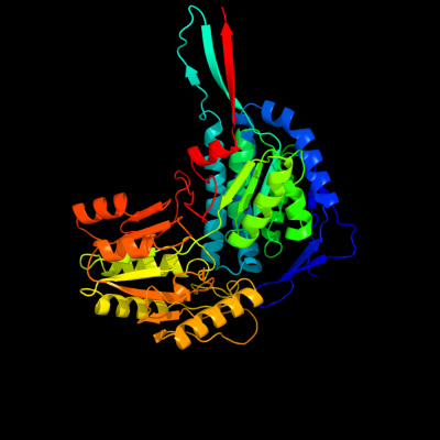
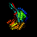
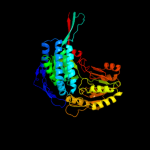
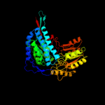
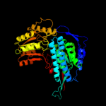
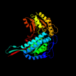
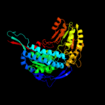
| 1 | c2hg2A_
|
|
 |
100.0 |
100 |
PDB header:oxidoreductase
Chain: A: PDB Molecule:aldehyde dehydrogenase a;
PDBTitle: structure of lactaldehyde dehydrogenase
|
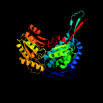
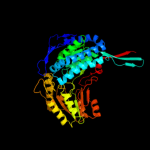
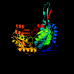
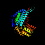
| 2 | c3ed6B_
|
|
 |
100.0 |
36 |
PDB header:oxidoreductase
Chain: B: PDB Molecule:betaine aldehyde dehydrogenase;
PDBTitle: 1.7 angstrom resolution crystal structure of betaine aldehyde2 dehydrogenase (betb) from staphylococcus aureus
|
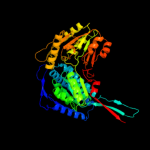
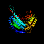
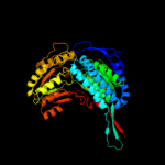
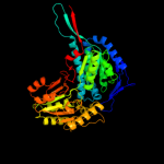
| 3 | c2o2qA_
|
|
 |
100.0 |
33 |
PDB header:oxidoreductase
Chain: A: PDB Molecule:formyltetrahydrofolate dehydrogenase;
PDBTitle: crystal structure of the c-terminal domain of rat2 10'formyltetrahydrofolate dehydrogenase in complex with nadp
|
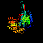
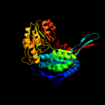
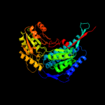
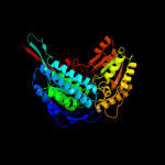
| 4 | d1a4sa_
|
|
 |
100.0 |
35 |
Fold:ALDH-like
Superfamily:ALDH-like
Family:ALDH-like |
| 5 | c3ifgH_
|
|
 |
100.0 |
34 |
PDB header:oxidoreductase
Chain: H: PDB Molecule:succinate-semialdehyde dehydrogenase (nadp+);
PDBTitle: crystal structure of succinate-semialdehyde dehydrogenase from2 burkholderia pseudomallei, part 1 of 2
|
| 6 | d1bxsa_
|
|
 |
100.0 |
33 |
Fold:ALDH-like
Superfamily:ALDH-like
Family:ALDH-like |
| 7 | d1o9ja_
|
|
 |
100.0 |
33 |
Fold:ALDH-like
Superfamily:ALDH-like
Family:ALDH-like |
| 8 | c3rh9A_
|
|
 |
100.0 |
35 |
PDB header:oxidoreductase
Chain: A: PDB Molecule:succinate-semialdehyde dehydrogenase (nad(p)(+));
PDBTitle: the crystal structure of oxidoreductase from marinobacter aquaeolei
|
| 9 | c2d4eB_
|
|
 |
100.0 |
32 |
PDB header:oxidoreductase
Chain: B: PDB Molecule:5-carboxymethyl-2-hydroxymuconate semialdehyde
PDBTitle: crystal structure of the hpcc from thermus thermophilus hb8
|
| 10 | c2jg7G_
|
|
 |
100.0 |
28 |
PDB header:oxidoreductase
Chain: G: PDB Molecule:antiquitin;
PDBTitle: crystal structure of seabream antiquitin and elucidation of2 its substrate specificity
|
| 11 | c3jz4C_
|
|
 |
100.0 |
39 |
PDB header:oxidoreductase
Chain: C: PDB Molecule:succinate-semialdehyde dehydrogenase [nadp+];
PDBTitle: crystal structure of e. coli nadp dependent enzyme
|
| 12 | c3ek1C_
|
|
 |
100.0 |
36 |
PDB header:oxidoreductase
Chain: C: PDB Molecule:aldehyde dehydrogenase;
PDBTitle: crystal structure of aldehyde dehydrogenase from brucella2 melitensis biovar abortus 2308
|
| 13 | c3k2wD_
|
|
 |
100.0 |
43 |
PDB header:oxidoreductase
Chain: D: PDB Molecule:betaine-aldehyde dehydrogenase;
PDBTitle: crystal structure of betaine-aldehyde dehydrogenase from2 pseudoalteromonas atlantica t6c
|
| 14 | c2w8qA_
|
|
 |
100.0 |
36 |
PDB header:oxidoreductase
Chain: A: PDB Molecule:succinate-semialdehyde dehydrogenase,
PDBTitle: the crystal structure of human ssadh in complex with ssa.
|
| 15 | c3iwkB_
|
|
 |
100.0 |
35 |
PDB header:oxidoreductase
Chain: B: PDB Molecule:aminoaldehyde dehydrogenase;
PDBTitle: crystal structure of aminoaldehyde dehydrogenase 1 from2 pisum sativum (psamadh1)
|
| 16 | d1wnda_
|
|
 |
100.0 |
38 |
Fold:ALDH-like
Superfamily:ALDH-like
Family:ALDH-like |
| 17 | c2ve5H_
|
|
 |
100.0 |
35 |
PDB header:oxidoreductase
Chain: H: PDB Molecule:betaine aldehyde dehydrogenase;
PDBTitle: crystallographic structure of betaine aldehyde2 dehydrogenase from pseudomonas aeruginosa
|
| 18 | c3qanB_
|
|
 |
100.0 |
32 |
PDB header:oxidoreductase
Chain: B: PDB Molecule:1-pyrroline-5-carboxylate dehydrogenase 1;
PDBTitle: crystal structure of 1-pyrroline-5-carboxylate dehydrogenase from2 bacillus halodurans
|
| 19 | d1o04a_
|
|
 |
100.0 |
32 |
Fold:ALDH-like
Superfamily:ALDH-like
Family:ALDH-like |
| 20 | c3r31A_
|
|
 |
100.0 |
34 |
PDB header:oxidoreductase
Chain: A: PDB Molecule:betaine aldehyde dehydrogenase;
PDBTitle: crystal structure of betaine aldehyde dehydrogenase from agrobacterium2 tumefaciens
|
| 21 | d1ag8a_ |
|
not modelled |
100.0 |
32 |
Fold:ALDH-like
Superfamily:ALDH-like
Family:ALDH-like |
| 22 | d1uzba_ |
|
not modelled |
100.0 |
31 |
Fold:ALDH-like
Superfamily:ALDH-like
Family:ALDH-like |
| 23 | c1t90B_ |
|
not modelled |
100.0 |
30 |
PDB header:oxidoreductase
Chain: B: PDB Molecule:probable methylmalonate-semialdehyde
PDBTitle: crystal structure of methylmalonate semialdehyde2 dehydrogenase from bacillus subtilis
|
| 24 | c3i44A_ |
|
not modelled |
100.0 |
33 |
PDB header:oxidoreductase
Chain: A: PDB Molecule:aldehyde dehydrogenase;
PDBTitle: crystal structure of aldehyde dehydrogenase from bartonella2 henselae at 2.0a resolution
|
| 25 | c3b4wA_ |
|
not modelled |
100.0 |
34 |
PDB header:oxidoreductase
Chain: A: PDB Molecule:aldehyde dehydrogenase;
PDBTitle: crystal structure of mycobacterium tuberculosis aldehyde dehydrogenase2 complexed with nad+
|
| 26 | d1euha_ |
|
not modelled |
100.0 |
33 |
Fold:ALDH-like
Superfamily:ALDH-like
Family:ALDH-like |
| 27 | c3prlD_ |
|
not modelled |
100.0 |
35 |
PDB header:oxidoreductase
Chain: D: PDB Molecule:nadp-dependent glyceraldehyde-3-phosphate dehydrogenase;
PDBTitle: crystal structure of nadp-dependent glyceraldehyde-3-phosphate2 dehydrogenase from bacillus halodurans c-125
|
| 28 | d1ky8a_ |
|
not modelled |
100.0 |
27 |
Fold:ALDH-like
Superfamily:ALDH-like
Family:ALDH-like |
| 29 | c3ju8B_ |
|
not modelled |
100.0 |
28 |
PDB header:oxidoreductase
Chain: B: PDB Molecule:succinylglutamic semialdehyde dehydrogenase;
PDBTitle: crystal structure of succinylglutamic semialdehyde dehydrogenase from2 pseudomonas aeruginosa.
|
| 30 | d1bi9a_ |
|
not modelled |
100.0 |
32 |
Fold:ALDH-like
Superfamily:ALDH-like
Family:ALDH-like |
| 31 | c3rosA_ |
|
not modelled |
100.0 |
28 |
PDB header:oxidoreductase
Chain: A: PDB Molecule:nad-dependent aldehyde dehydrogenase;
PDBTitle: crystal structure of nad-dependent aldehyde dehydrogenase from2 lactobacillus acidophilus
|
| 32 | c3hazA_ |
|
not modelled |
100.0 |
29 |
PDB header:oxidoreductase
Chain: A: PDB Molecule:proline dehydrogenase;
PDBTitle: crystal structure of bifunctional proline utilization a2 (puta) protein
|
| 33 | c3efvC_ |
|
not modelled |
100.0 |
34 |
PDB header:oxidoreductase
Chain: C: PDB Molecule:putative succinate-semialdehyde dehydrogenase;
PDBTitle: crystal structure of a putative succinate-semialdehyde dehydrogenase2 from salmonella typhimurium lt2 with bound nad
|
| 34 | c3r64A_ |
|
not modelled |
100.0 |
31 |
PDB header:oxidoreductase
Chain: A: PDB Molecule:nad dependent benzaldehyde dehydrogenase;
PDBTitle: crystal structure of a nad-dependent benzaldehyde dehydrogenase from2 corynebacterium glutamicum
|
| 35 | c2vroB_ |
|
not modelled |
100.0 |
23 |
PDB header:oxidoreductase
Chain: B: PDB Molecule:aldehyde dehydrogenase;
PDBTitle: crystal structure of aldehyde dehydrogenase from2 burkholderia xenovorans lb400
|
| 36 | c3pqaA_ |
|
not modelled |
100.0 |
35 |
PDB header:oxidoreductase
Chain: A: PDB Molecule:lactaldehyde dehydrogenase;
PDBTitle: crystal structure of glyceraldehyde-3-phosphate dehydrogenase gapn2 from methanocaldococcus jannaschii dsm 2661
|
| 37 | d1ad3a_ |
|
not modelled |
100.0 |
26 |
Fold:ALDH-like
Superfamily:ALDH-like
Family:ALDH-like |
| 38 | c3v4cB_ |
|
not modelled |
100.0 |
23 |
PDB header:oxidoreductase
Chain: B: PDB Molecule:aldehyde dehydrogenase (nadp+);
PDBTitle: crystal structure of a semialdehyde dehydrogenase from sinorhizobium2 meliloti 1021
|
| 39 | d1ez0a_ |
|
not modelled |
100.0 |
22 |
Fold:ALDH-like
Superfamily:ALDH-like
Family:ALDH-like |
| 40 | c3lnsD_ |
|
not modelled |
100.0 |
25 |
PDB header:oxidoreductase
Chain: D: PDB Molecule:benzaldehyde dehydrogenase;
PDBTitle: benzaldehyde dehydrogenase, a class 3 aldehyde dehydrogenase, with2 bound nadp+ and benzoate adduct
|
| 41 | c3k9dD_ |
|
not modelled |
100.0 |
18 |
PDB header:oxidoreductase
Chain: D: PDB Molecule:aldehyde dehydrogenase;
PDBTitle: crystal structure of probable aldehyde dehydrogenase from listeria2 monocytogenes egd-e
|
| 42 | d1o20a_ |
|
not modelled |
100.0 |
19 |
Fold:ALDH-like
Superfamily:ALDH-like
Family:ALDH-like |
| 43 | c3my7A_ |
|
not modelled |
100.0 |
21 |
PDB header:oxidoreductase
Chain: A: PDB Molecule:alcohol dehydrogenase/acetaldehyde dehydrogenase;
PDBTitle: the crystal structure of the acdh domain of an alcohol dehydrogenase2 from vibrio parahaemolyticus to 2.25a
|
| 44 | c2h5gA_ |
|
not modelled |
100.0 |
19 |
PDB header:oxidoreductase
Chain: A: PDB Molecule:delta 1-pyrroline-5-carboxylate synthetase;
PDBTitle: crystal structure of human pyrroline-5-carboxylate synthetase
|
| 45 | d1vlua_ |
|
not modelled |
100.0 |
22 |
Fold:ALDH-like
Superfamily:ALDH-like
Family:ALDH-like |
| 46 | c1vluB_ |
|
not modelled |
100.0 |
22 |
PDB header:oxidoreductase
Chain: B: PDB Molecule:gamma-glutamyl phosphate reductase;
PDBTitle: crystal structure of gamma-glutamyl phosphate reductase (yor323c) from2 saccharomyces cerevisiae at 2.40 a resolution
|
| 47 | d1k75a_ |
|
not modelled |
98.5 |
17 |
Fold:ALDH-like
Superfamily:ALDH-like
Family:L-histidinol dehydrogenase HisD |
| 48 | c3fa4D_ |
|
not modelled |
62.5 |
16 |
PDB header:lyase
Chain: D: PDB Molecule:2,3-dimethylmalate lyase;
PDBTitle: crystal structure of 2,3-dimethylmalate lyase, a pep mutase/isocitrate2 lyase superfamily member, triclinic crystal form
|
| 49 | c2yvqA_ |
|
not modelled |
54.1 |
12 |
PDB header:ligase
Chain: A: PDB Molecule:carbamoyl-phosphate synthase;
PDBTitle: crystal structure of mgs domain of carbamoyl-phosphate2 synthetase from homo sapiens
|
| 50 | c3e5bB_ |
|
not modelled |
50.9 |
14 |
PDB header:lyase
Chain: B: PDB Molecule:isocitrate lyase;
PDBTitle: 2.4 a crystal structure of isocitrate lyase from brucella2 melitensis
|
| 51 | d1a9xa2 |
|
not modelled |
47.6 |
17 |
Fold:Methylglyoxal synthase-like
Superfamily:Methylglyoxal synthase-like
Family:Carbamoyl phosphate synthetase, large subunit allosteric, C-terminal domain |
| 52 | d1y5ea1 |
|
not modelled |
46.6 |
15 |
Fold:Molybdenum cofactor biosynthesis proteins
Superfamily:Molybdenum cofactor biosynthesis proteins
Family:MogA-like |
| 53 | d1s7ia_ |
|
not modelled |
42.6 |
17 |
Fold:Ferredoxin-like
Superfamily:Dimeric alpha+beta barrel
Family:DGPF domain (Pfam 04946) |
| 54 | d1wo8a1 |
|
not modelled |
42.3 |
13 |
Fold:Methylglyoxal synthase-like
Superfamily:Methylglyoxal synthase-like
Family:Methylglyoxal synthase, MgsA |
| 55 | c2yvkA_ |
|
not modelled |
42.2 |
19 |
PDB header:isomerase
Chain: A: PDB Molecule:methylthioribose-1-phosphate isomerase;
PDBTitle: crystal structure of 5-methylthioribose 1-phosphate2 isomerase product complex from bacillus subtilis
|
| 56 | d2bona1 |
|
not modelled |
41.3 |
15 |
Fold:NAD kinase/diacylglycerol kinase-like
Superfamily:NAD kinase/diacylglycerol kinase-like
Family:Diacylglycerol kinase-like |
| 57 | d2ftsa3 |
|
not modelled |
38.4 |
21 |
Fold:Molybdenum cofactor biosynthesis proteins
Superfamily:Molybdenum cofactor biosynthesis proteins
Family:MoeA central domain-like |
| 58 | d1uz5a3 |
|
not modelled |
36.9 |
16 |
Fold:Molybdenum cofactor biosynthesis proteins
Superfamily:Molybdenum cofactor biosynthesis proteins
Family:MoeA central domain-like |
| 59 | c1uz5A_ |
|
not modelled |
34.6 |
16 |
PDB header:molybdopterin biosynthesis
Chain: A: PDB Molecule:402aa long hypothetical molybdopterin
PDBTitle: the crystal structure of molybdopterin biosynthesis moea2 protein from pyrococcus horikosii
|
| 60 | c2ixaA_ |
|
not modelled |
33.3 |
15 |
PDB header:hydrolase
Chain: A: PDB Molecule:alpha-n-acetylgalactosaminidase;
PDBTitle: a-zyme, n-acetylgalactosaminidase
|
| 61 | c2l69A_ |
|
not modelled |
28.6 |
21 |
PDB header:de novo protein
Chain: A: PDB Molecule:rossmann 2x3 fold protein;
PDBTitle: solution nmr structure of de novo designed protein, p-loop ntpase2 fold, northeast structural genomics consortium target or28
|
| 62 | d2g2ca1 |
|
not modelled |
26.8 |
10 |
Fold:Molybdenum cofactor biosynthesis proteins
Superfamily:Molybdenum cofactor biosynthesis proteins
Family:MogA-like |
| 63 | d1k99a_ |
|
not modelled |
23.5 |
11 |
Fold:HMG-box
Superfamily:HMG-box
Family:HMG-box |
| 64 | c2ec4A_ |
|
not modelled |
23.3 |
16 |
PDB header:structural genomics, unknown function
Chain: A: PDB Molecule:fas-associated factor 1;
PDBTitle: solution structure of the uas domain from human fas-2 associated factor 1
|
| 65 | d1xxaa_ |
|
not modelled |
22.7 |
21 |
Fold:DCoH-like
Superfamily:C-terminal domain of arginine repressor
Family:C-terminal domain of arginine repressor |
| 66 | c3fghA_ |
|
not modelled |
22.7 |
11 |
PDB header:transcription
Chain: A: PDB Molecule:transcription factor a, mitochondrial;
PDBTitle: human mitochondrial transcription factor a box b
|
| 67 | c2crjA_ |
|
not modelled |
21.8 |
18 |
PDB header:gene regulation
Chain: A: PDB Molecule:swi/snf-related matrix-associated actin-
PDBTitle: solution structure of the hmg domain of mouse hmg domain2 protein hmgx2
|
| 68 | c2is8A_ |
|
not modelled |
21.3 |
10 |
PDB header:structural protein
Chain: A: PDB Molecule:molybdopterin biosynthesis enzyme, moab;
PDBTitle: crystal structure of the molybdopterin biosynthesis enzyme moab2 (ttha0341) from thermus theromophilus hb8
|
| 69 | d1u0ta_ |
|
not modelled |
21.3 |
15 |
Fold:NAD kinase/diacylglycerol kinase-like
Superfamily:NAD kinase/diacylglycerol kinase-like
Family:NAD kinase-like |
| 70 | c3jtpB_ |
|
not modelled |
19.6 |
27 |
PDB header:protein binding
Chain: B: PDB Molecule:adapter protein meca 1;
PDBTitle: crystal structure of the c-terminal domain of meca
|
| 71 | d1wu2a3 |
|
not modelled |
19.5 |
13 |
Fold:Molybdenum cofactor biosynthesis proteins
Superfamily:Molybdenum cofactor biosynthesis proteins
Family:MoeA central domain-like |
| 72 | c2eqzA_ |
|
not modelled |
19.3 |
11 |
PDB header:transcription
Chain: A: PDB Molecule:high mobility group protein b3;
PDBTitle: solution structure of the first hmg-box domain from high2 mobility group protein b3
|
| 73 | d1j3xa_ |
|
not modelled |
18.8 |
11 |
Fold:HMG-box
Superfamily:HMG-box
Family:HMG-box |
| 74 | c1j3xA_ |
|
not modelled |
18.8 |
11 |
PDB header:dna binding protein
Chain: A: PDB Molecule:high mobility group protein 2;
PDBTitle: solution structure of the n-terminal domain of the hmgb2
|
| 75 | c3b8iF_ |
|
not modelled |
17.3 |
18 |
PDB header:lyase
Chain: F: PDB Molecule:pa4872 oxaloacetate decarboxylase;
PDBTitle: crystal structure of oxaloacetate decarboxylase from pseudomonas2 aeruginosa (pa4872) in complex with oxalate and mg2+.
|
| 76 | c3v4gA_ |
|
not modelled |
17.2 |
21 |
PDB header:dna binding protein
Chain: A: PDB Molecule:arginine repressor;
PDBTitle: 1.60 angstrom resolution crystal structure of an arginine repressor2 from vibrio vulnificus cmcp6
|
| 77 | c3rfuC_ |
|
not modelled |
16.9 |
12 |
PDB header:hydrolase, membrane protein
Chain: C: PDB Molecule:copper efflux atpase;
PDBTitle: crystal structure of a copper-transporting pib-type atpase
|
| 78 | c2nqqA_ |
|
not modelled |
16.1 |
14 |
PDB header:biosynthetic protein
Chain: A: PDB Molecule:molybdopterin biosynthesis protein moea;
PDBTitle: moea r137q
|
| 79 | d2ioja1 |
|
not modelled |
16.1 |
17 |
Fold:MurF and HprK N-domain-like
Superfamily:HprK N-terminal domain-like
Family:DRTGG domain |
| 80 | c3f4lF_ |
|
not modelled |
16.1 |
16 |
PDB header:oxidoreductase
Chain: F: PDB Molecule:putative oxidoreductase yhhx;
PDBTitle: crystal structure of a probable oxidoreductase yhhx in2 triclinic form. northeast structural genomics target er647
|
| 81 | c2pjkA_ |
|
not modelled |
15.9 |
15 |
PDB header:structural genomics, unknown function
Chain: A: PDB Molecule:178aa long hypothetical molybdenum cofactor
PDBTitle: structure of hypothetical molybdenum cofactor biosynthesis2 protein b from sulfolobus tokodaii
|
| 82 | d1mkza_ |
|
not modelled |
15.7 |
15 |
Fold:Molybdenum cofactor biosynthesis proteins
Superfamily:Molybdenum cofactor biosynthesis proteins
Family:MogA-like |
| 83 | d2lefa_ |
|
not modelled |
15.3 |
14 |
Fold:HMG-box
Superfamily:HMG-box
Family:HMG-box |
| 84 | d1hsma_ |
|
not modelled |
14.8 |
18 |
Fold:HMG-box
Superfamily:HMG-box
Family:HMG-box |
| 85 | c2cs1A_ |
|
not modelled |
14.7 |
18 |
PDB header:dna binding protein
Chain: A: PDB Molecule:pms1 protein homolog 1;
PDBTitle: solution structure of the hmg domain of human dna mismatch2 repair protein
|
| 86 | c2co9A_ |
|
not modelled |
14.6 |
15 |
PDB header:transcription
Chain: A: PDB Molecule:thymus high mobility group box protein tox;
PDBTitle: solution structure of the hmg_box domain of thymus high2 mobility group box protein tox from mouse
|
| 87 | c3dtyA_ |
|
not modelled |
14.5 |
16 |
PDB header:oxidoreductase
Chain: A: PDB Molecule:oxidoreductase, gfo/idh/moca family;
PDBTitle: crystal structure of an oxidoreductase from pseudomonas2 syringae
|
| 88 | d1j46a_ |
|
not modelled |
14.2 |
8 |
Fold:HMG-box
Superfamily:HMG-box
Family:HMG-box |
| 89 | c2bonB_ |
|
not modelled |
13.9 |
9 |
PDB header:transferase
Chain: B: PDB Molecule:lipid kinase;
PDBTitle: structure of an escherichia coli lipid kinase (yegs)
|
| 90 | c3ereD_ |
|
not modelled |
13.6 |
19 |
PDB header:dna binding protein/dna
Chain: D: PDB Molecule:arginine repressor;
PDBTitle: crystal structure of the arginine repressor protein from mycobacterium2 tuberculosis in complex with the dna operator
|
| 91 | c2qiwA_ |
|
not modelled |
13.6 |
15 |
PDB header:transferase
Chain: A: PDB Molecule:pep phosphonomutase;
PDBTitle: crystal structure of a putative phosphoenolpyruvate phosphonomutase2 (ncgl1015, cgl1060) from corynebacterium glutamicum atcc 13032 at3 1.80 a resolution
|
| 92 | d1lwma_ |
|
not modelled |
13.0 |
21 |
Fold:HMG-box
Superfamily:HMG-box
Family:HMG-box |
| 93 | d1v64a_ |
|
not modelled |
12.8 |
13 |
Fold:HMG-box
Superfamily:HMG-box
Family:HMG-box |
| 94 | c1jr2A_ |
|
not modelled |
12.2 |
13 |
PDB header:lyase
Chain: A: PDB Molecule:uroporphyrinogen-iii synthase;
PDBTitle: structure of uroporphyrinogen iii synthase
|
| 95 | d1jr2a_ |
|
not modelled |
12.2 |
13 |
Fold:HemD-like
Superfamily:HemD-like
Family:HemD-like |
| 96 | c3peiA_ |
|
not modelled |
12.0 |
14 |
PDB header:hydrolase
Chain: A: PDB Molecule:cytosol aminopeptidase;
PDBTitle: crystal structure of cytosol aminopeptidase from francisella2 tularensis
|
| 97 | d1v63a_ |
|
not modelled |
11.9 |
5 |
Fold:HMG-box
Superfamily:HMG-box
Family:HMG-box |
| 98 | c3gfgB_ |
|
not modelled |
11.9 |
12 |
PDB header:oxidoreductase
Chain: B: PDB Molecule:uncharacterized oxidoreductase yvaa;
PDBTitle: structure of putative oxidoreductase yvaa from bacillus subtilis in2 triclinic form
|
| 99 | c1h6dL_ |
|
not modelled |
11.8 |
18 |
PDB header:protein translocation
Chain: L: PDB Molecule:precursor form of glucose-fructose
PDBTitle: oxidized precursor form of glucose-fructose oxidoreductase2 from zymomonas mobilis complexed with glycerol
|