1 c2oxlA_
58.9
20
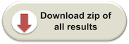
PDB header: gene regulationChain: A: PDB Molecule: hypothetical protein ymgb;PDBTitle: structure and function of the e. coli protein ymgb: a protein critical2 for biofilm formation and acid resistance
2 c2dt7A_
39.7
33
PDB header: rna binding proteinChain: A: PDB Molecule: splicing factor 3a subunit 3;PDBTitle: solution structure of the second surp domain of human2 splicing factor sf3a120 in complex with a fragment of3 human splicing factor sf3a60
3 c3cl3D_
32.2
39
PDB header: viral protein/signaling proteinChain: D: PDB Molecule: nf-kappa-b essential modulator;PDBTitle: crystal structure of a vflip-ikkgamma complex: insights2 into viral activation of the ikk signalosome
4 c2k6oA_
16.8
32
PDB header: antimicrobial proteinChain: A: PDB Molecule: cathelicidin antimicrobial peptide;PDBTitle: human ll-37 structure
5 c2fcgF_
15.2
38
PDB header: antimicrobial proteinChain: F: PDB Molecule: antibacterial protein fall-39, core peptide;PDBTitle: solution structure of the c-terminal fragment of human ll-37
6 c1ij2C_
13.4
40
PDB header: transcriptionChain: C: PDB Molecule: general control protein gcn4;PDBTitle: gcn4-pvtl coiled-coil trimer with threonine at the a(16)2 position
7 c1ztaA_
13.3
47
PDB header: dna-binding motifChain: A: PDB Molecule: leucine zipper monomer;PDBTitle: the solution structure of a leucine-zipper motif peptide
8 c1ij2B_
12.5
40
PDB header: transcriptionChain: B: PDB Molecule: general control protein gcn4;PDBTitle: gcn4-pvtl coiled-coil trimer with threonine at the a(16)2 position
9 c1ij3B_
12.1
40
PDB header: transcriptionChain: B: PDB Molecule: general control protein gcn4;PDBTitle: gcn4-pvsl coiled-coil trimer with serine at the a(16)2 position
10 c1ij3C_
12.1
40
PDB header: transcriptionChain: C: PDB Molecule: general control protein gcn4;PDBTitle: gcn4-pvsl coiled-coil trimer with serine at the a(16)2 position
11 c1rb6C_
12.0
40
PDB header: dna binding proteinChain: C: PDB Molecule: general control protein gcn4;PDBTitle: antiparallel trimer of gcn4-leucine zipper core mutant as2 n16a tetragonal form
12 c3k7zB_
12.0
40
PDB header: dna binding proteinChain: B: PDB Molecule: general control protein gcn4;PDBTitle: gcn4-leucine zipper core mutant as n16a trigonal automatic2 solution
13 c3k7zA_
12.0
40
PDB header: dna binding proteinChain: A: PDB Molecule: general control protein gcn4;PDBTitle: gcn4-leucine zipper core mutant as n16a trigonal automatic2 solution
14 c1rb1A_
12.0
40
PDB header: dna binding proteinChain: A: PDB Molecule: general control protein gcn4;PDBTitle: gcn4-leucine zipper core mutant as n16a trigonal automatic2 solution
15 c1rb1B_
12.0
40
PDB header: dna binding proteinChain: B: PDB Molecule: general control protein gcn4;PDBTitle: gcn4-leucine zipper core mutant as n16a trigonal automatic2 solution
16 c1swiA_
11.4
40
PDB header: leucine zipperChain: A: PDB Molecule: gcn4p1;PDBTitle: gcn4-leucine zipper core mutant as n16a complexed with2 benzene
17 c2l5gA_
11.4
31
PDB header: transcription regulatorChain: A: PDB Molecule: g protein pathway suppressor 2;PDBTitle: co-ordinates and 1h, 13c and 15n chemical shift assignments for the2 complex of gps2 53-90 and smrt 167-207
18 d1gh6a_
9.7
20
Fold: Long alpha-hairpinSuperfamily: Chaperone J-domainFamily: Chaperone J-domain19 c2l6lA_
9.5
27
PDB header: chaperoneChain: A: PDB Molecule: dnaj homolog subfamily c member 24;PDBTitle: solution structure of human j-protein co-chaperone, dph4
20 c1ce0B_
9.5
33
PDB header: hiv-1 envelope proteinChain: B: PDB Molecule: protein (leucine zipper model h38-p1);PDBTitle: trimerization specificity in hiv-1 gp41: analysis with a2 gcn4 leucine zipper model
21 c2ovcA_
not modelled
9.4
64
PDB header: transport proteinChain: A: PDB Molecule: potassium voltage-gated channel subfamily kqt member 4;PDBTitle: crystal structure of a coiled-coil tetramerization domain from kv7.42 channels
22 d2bcgg2
not modelled
8.9
21
Fold: FAD/NAD(P)-binding domainSuperfamily: FAD/NAD(P)-binding domainFamily: GDI-like N domain23 c2khfA_
not modelled
8.7
20
PDB header: antimicrobial proteinChain: A: PDB Molecule: plnj;PDBTitle: plantaricin j in dpc-micelles
24 c2w0cR_
not modelled
8.4
71
PDB header: virusChain: R: PDB Molecule: protein p3;PDBTitle: x-ray structure of the entire lipid-containing2 bacteriophage pm2
25 c2voiB_
not modelled
8.2
33
PDB header: apoptosisChain: B: PDB Molecule: bh3-interacting domain death agonist p13;PDBTitle: structure of mouse a1 bound to the bid bh3-domain
26 d1nz6a_
not modelled
8.2
45
Fold: Long alpha-hairpinSuperfamily: Chaperone J-domainFamily: Chaperone J-domain27 c2o37A_
not modelled
8.1
27
PDB header: chaperoneChain: A: PDB Molecule: protein sis1;PDBTitle: j-domain of sis1 protein, hsp40 co-chaperone from2 saccharomyces cerevisiae.
28 c2g7rA_
not modelled
7.7
29
PDB header: hydrolaseChain: A: PDB Molecule: mucosa-associated lymphoid tissue lymphoma translocationPDBTitle: x-ray structure of the death domain of the human mucosa associated2 lymphoid tissue lymphoma translocation protein 1
29 c3ag7A_
not modelled
7.2
11
PDB header: plant proteinChain: A: PDB Molecule: putative uncharacterized protein f9e10.5;PDBTitle: an auxilin-like j-domain containing protein, jac1 j-domain
30 c2ys8A_
not modelled
7.2
27
PDB header: protein bindingChain: A: PDB Molecule: rab-related gtp-binding protein rabj;PDBTitle: solution structure of the dnaj-like domain from human ras-2 associated protein rap1
31 c2l06A_
not modelled
7.0
86
PDB header: protein bindingChain: A: PDB Molecule: phycobilisome lcm core-membrane linker polypeptide;PDBTitle: solution nmr structure of the pbs linker polypeptide domain (fragment2 254-400) of phycobilisome linker protein apce from synechocystis sp.3 pcc 6803. northeast structural genomics consortium target sgr209c
32 c2guzO_
not modelled
6.8
36
PDB header: chaperone, protein transportChain: O: PDB Molecule: mitochondrial import inner membrane translocasePDBTitle: structure of the tim14-tim16 complex of the mitochondrial2 protein import motor
33 c2ochA_
not modelled
6.8
36
PDB header: chaperoneChain: A: PDB Molecule: hypothetical protein dnj-12;PDBTitle: j-domain of dnj-12 from caenorhabditis elegans
34 c2ctrA_
not modelled
6.6
36
PDB header: chaperoneChain: A: PDB Molecule: dnaj homolog subfamily b member 9;PDBTitle: solution structure of j-domain from human dnaj subfamily b2 menber 9
35 c2ky4A_
not modelled
6.4
56
PDB header: photosynthesisChain: A: PDB Molecule: phycobilisome linker polypeptide;PDBTitle: solution nmr structure of the pbs linker domain of phycobilisome2 linker polypeptide from anabaena sp. northeast structural genomics3 consortium target nsr123e
36 d1fd3a_
not modelled
6.4
67
Fold: Defensin-likeSuperfamily: Defensin-likeFamily: Defensin37 c2xzrA_
not modelled
6.3
24
PDB header: cell adhesionChain: A: PDB Molecule: immunoglobulin-binding protein eibd;PDBTitle: escherichia coli immunoglobulin-binding protein eibd 391-438 fused2 to gcn4 adaptors
38 c2dn9A_
not modelled
6.3
55
PDB header: apoptosis, chaperoneChain: A: PDB Molecule: dnaj homolog subfamily a member 3;PDBTitle: solution structure of j-domain from the dnaj homolog, human2 tid1 protein
39 c2khgA_
not modelled
6.3
20
PDB header: antimicrobial proteinChain: A: PDB Molecule: plnj;PDBTitle: plantaricin j in tfe
40 d1xbla_
not modelled
6.1
27
Fold: Long alpha-hairpinSuperfamily: Chaperone J-domainFamily: Chaperone J-domain41 c1bq0A_
not modelled
6.1
27
PDB header: chaperoneChain: A: PDB Molecule: dnaj;PDBTitle: j-domain (residues 1-77) of the escherichia coli n-terminal2 fragment (residues 1-104) of the molecular chaperone dnaj,3 nmr, 20 structures
42 c2d96A_
not modelled
6.0
29
PDB header: transcriptionChain: A: PDB Molecule: nuclear factor nf-kappa-b p100 subunit;PDBTitle: solution structure of the death domain of nuclear factor nf-2 kappa-b p100
43 d1fafa_
not modelled
6.0
36
Fold: Long alpha-hairpinSuperfamily: Chaperone J-domainFamily: Chaperone J-domain44 d1ni7a_
not modelled
5.8
20
Fold: SufE/NifUSuperfamily: SufE/NifUFamily: SufE-like45 d1qmga2
not modelled
5.8
28
Fold: NAD(P)-binding Rossmann-fold domainsSuperfamily: NAD(P)-binding Rossmann-fold domainsFamily: 6-phosphogluconate dehydrogenase-like, N-terminal domain46 c2ctqA_
not modelled
5.7
27
PDB header: chaperoneChain: A: PDB Molecule: dnaj homolog subfamily c member 12;PDBTitle: solution structure of j-domain from human dnaj subfamily c2 menber 12
47 c2pjwV_
not modelled
5.7
22
PDB header: endocytosis/exocytosisChain: V: PDB Molecule: vacuolar protein sorting-associated protein 27;PDBTitle: the vps27/hse1 complex is a gat domain-based scaffold for2 ubiquitin-dependent sorting
48 d1e3ha6
not modelled
5.6
17
Fold: Ribonuclease PH domain 2-likeSuperfamily: Ribonuclease PH domain 2-likeFamily: Ribonuclease PH domain 2-like49 c2a3dA_
not modelled
5.4
35
PDB header: three-helix bundleChain: A: PDB Molecule: protein (de novo three-helix bundle);PDBTitle: solution structure of a de novo designed single chain three-2 helix bundle (a3d)
50 c2o7hF_
not modelled
5.4
33
PDB header: transcriptionChain: F: PDB Molecule: general control protein gcn4;PDBTitle: crystal structure of trimeric coiled coil gcn4 leucine zipper
51 c2cugA_
not modelled
5.3
36
PDB header: chaperoneChain: A: PDB Molecule: mkiaa0962 protein;PDBTitle: solution structure of the j domain of the pseudo dnaj2 protein, mouse hypothetical mkiaa0962