| 1 |
|
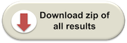
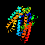
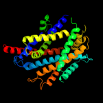
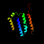
PDB 3lrc chain C
Region: 6 - 435
Aligned: 410
Modelled: 410
Confidence: 100.0%
Identity: 100%
PDB header:transport protein
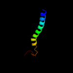
Chain: C: PDB Molecule:arginine/agmatine antiporter;
PDBTitle: structure of e. coli adic (p1)
Phyre2
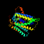
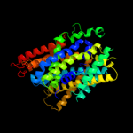
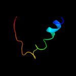
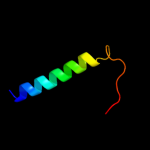
| 2 |
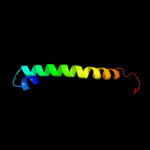
|
PDB 3gia chain A
Region: 6 - 443
Aligned: 427
Modelled: 427
Confidence: 100.0%
Identity: 18%
PDB header:transport protein
Chain: A: PDB Molecule:uncharacterized protein mj0609;
PDBTitle: crystal structure of apct transporter
Phyre2
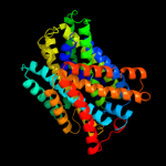
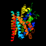
| 3 |
|
PDB 2jln chain A
Region: 3 - 445
Aligned: 430
Modelled: 430
Confidence: 100.0%
Identity: 13%
PDB header:membrane protein
Chain: A: PDB Molecule:mhp1;
PDBTitle: structure of mhp1, a nucleobase-cation-symport-1 family2 transporter
Phyre2
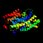
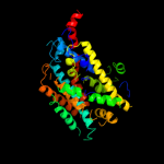
| 4 |
|
PDB 2xq2 chain A
Region: 8 - 444
Aligned: 429
Modelled: 429
Confidence: 99.6%
Identity: 12%
PDB header:transport protein
Chain: A: PDB Molecule:sodium/glucose cotransporter;
PDBTitle: structure of the k294a mutant of vsglt
Phyre2
| 5 |
|
PDB 3dh4 chain A
Region: 8 - 445
Aligned: 434
Modelled: 434
Confidence: 99.5%
Identity: 11%
PDB header:transport protein
Chain: A: PDB Molecule:sodium/glucose cotransporter;
PDBTitle: crystal structure of sodium/sugar symporter with bound galactose from2 vibrio parahaemolyticus
Phyre2
| 6 |
|
PDB 2a65 chain A domain 1
Region: 11 - 434
Aligned: 414
Modelled: 414
Confidence: 97.6%
Identity: 14%
Fold: SNF-like
Superfamily: SNF-like
Family: SNF-like
Phyre2
| 7 |
|
PDB 2w8a chain C
Region: 3 - 387
Aligned: 375
Modelled: 358
Confidence: 97.5%
Identity: 13%
PDB header:membrane protein
Chain: C: PDB Molecule:glycine betaine transporter betp;
PDBTitle: crystal structure of the sodium-coupled glycine betaine2 symporter betp from corynebacterium glutamicum with bound3 substrate
Phyre2
| 8 |
|
PDB 3hfx chain A
Region: 32 - 382
Aligned: 342
Modelled: 342
Confidence: 85.1%
Identity: 11%
PDB header:transport protein
Chain: A: PDB Molecule:l-carnitine/gamma-butyrobetaine antiporter;
PDBTitle: crystal structure of carnitine transporter
Phyre2
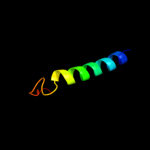
| 9 |
|
PDB 2knc chain A
Region: 403 - 445
Aligned: 43
Modelled: 43
Confidence: 55.1%
Identity: 7%
PDB header:cell adhesion
Chain: A: PDB Molecule:integrin alpha-iib;
PDBTitle: platelet integrin alfaiib-beta3 transmembrane-cytoplasmic2 heterocomplex
Phyre2
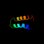
| 10 |
|
PDB 1fft chain B domain 2
Region: 417 - 444
Aligned: 28
Modelled: 28
Confidence: 23.3%
Identity: 11%
Fold: Transmembrane helix hairpin
Superfamily: Cytochrome c oxidase subunit II-like, transmembrane region
Family: Cytochrome c oxidase subunit II-like, transmembrane region
Phyre2
| 11 |
|
PDB 3qnq chain D
Region: 395 - 445
Aligned: 51
Modelled: 51
Confidence: 19.0%
Identity: 12%
PDB header:membrane protein, transport protein
Chain: D: PDB Molecule:pts system, cellobiose-specific iic component;
PDBTitle: crystal structure of the transporter chbc, the iic component from the2 n,n'-diacetylchitobiose-specific phosphotransferase system
Phyre2
| 12 |
|
PDB 3rko chain F
Region: 320 - 436
Aligned: 117
Modelled: 117
Confidence: 13.4%
Identity: 14%
PDB header:oxidoreductase
Chain: F: PDB Molecule:nadh-quinone oxidoreductase subunit j;
PDBTitle: crystal structure of the membrane domain of respiratory complex i from2 e. coli at 3.0 angstrom resolution
Phyre2
| 13 |
|
PDB 3dtu chain B domain 2
Region: 409 - 441
Aligned: 33
Modelled: 33
Confidence: 12.5%
Identity: 12%
Fold: Transmembrane helix hairpin
Superfamily: Cytochrome c oxidase subunit II-like, transmembrane region
Family: Cytochrome c oxidase subunit II-like, transmembrane region
Phyre2
| 14 |
|
PDB 1m57 chain H
Region: 409 - 441
Aligned: 33
Modelled: 33
Confidence: 8.2%
Identity: 12%
PDB header:oxidoreductase
Chain: H: PDB Molecule:cytochrome c oxidase;
PDBTitle: structure of cytochrome c oxidase from rhodobacter2 sphaeroides (eq(i-286) mutant))
Phyre2
| 15 |
|
PDB 2iub chain A domain 2
Region: 388 - 431
Aligned: 44
Modelled: 44
Confidence: 6.8%
Identity: 11%
Fold: Transmembrane helix hairpin
Superfamily: Magnesium transport protein CorA, transmembrane region
Family: Magnesium transport protein CorA, transmembrane region
Phyre2
| 16 |
|
PDB 1ar1 chain B
Region: 409 - 444
Aligned: 36
Modelled: 36
Confidence: 6.3%
Identity: 8%
PDB header:complex (oxidoreductase/antibody)
Chain: B: PDB Molecule:cytochrome c oxidase;
PDBTitle: structure at 2.7 angstrom resolution of the paracoccus2 denitrificans two-subunit cytochrome c oxidase complexed3 with an antibody fv fragment
Phyre2