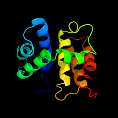
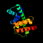
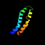
| 1 | d1ey1a_
|
|
 |
100.0 |
100 |
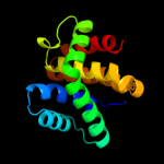
Fold:NusB-like
Superfamily:NusB-like
Family:Antitermination factor NusB |
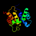
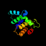
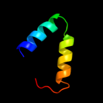
| 2 | d1tzva_
|
|
 |
100.0 |
35 |
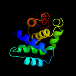
Fold:NusB-like
Superfamily:NusB-like
Family:Antitermination factor NusB |
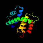
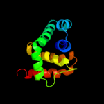
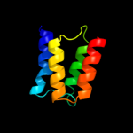
| 3 | c2jr0A_
|
|
 |
100.0 |
24 |
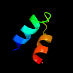
PDB header:transcription
Chain: A: PDB Molecule:n utilization substance protein b homolog;
PDBTitle: solution structure of nusb from aquifex aeolicus
|
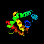
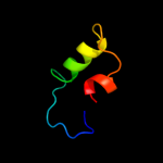
| 4 | d1eyva_
|
|
 |
100.0 |
34 |
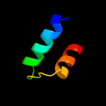
Fold:NusB-like
Superfamily:NusB-like
Family:Antitermination factor NusB |
| 5 | c1eyvA_
|
|
 |
100.0 |
34 |
PDB header:transcription
Chain: A: PDB Molecule:n-utilizing substance protein b homolog;
PDBTitle: the crystal structure of nusb from mycobacterium2 tuberculosis
|
| 6 | d1sqga1
|
|
 |
100.0 |
23 |
Fold:NusB-like
Superfamily:NusB-like
Family:RmsB N-terminal domain-like |
| 7 | c1sqgA_
|
|
 |
100.0 |
23 |
PDB header:transferase
Chain: A: PDB Molecule:sun protein;
PDBTitle: the crystal structure of the e. coli fmu apoenzyme at 1.652 a resolution
|
| 8 | c2yxlA_
|
|
 |
99.9 |
14 |
PDB header:transferase
Chain: A: PDB Molecule:450aa long hypothetical fmu protein;
PDBTitle: crystal structure of ph0851
|
| 9 | d1q8ca_
|
|
 |
81.4 |
18 |
Fold:NusB-like
Superfamily:NusB-like
Family:Hypothetical protein MG027 |
| 10 | c2zc2A_
|
|
 |
42.8 |
15 |
PDB header:replication
Chain: A: PDB Molecule:dnad-like replication protein;
PDBTitle: crystal structure of dnad-like replication protein from2 streptococcus mutans ua159, gi 24377835, residues 127-199
|
| 11 | d2i5ua1
|
|
 |
35.7 |
24 |
Fold:DnaD domain-like
Superfamily:DnaD domain-like
Family:DnaD domain |
| 12 | c3ls1A_
|
|
 |
32.8 |
8 |
PDB header:photosynthesis
Chain: A: PDB Molecule:sll1638 protein;
PDBTitle: crystal structure of cyanobacterial psbq from synechocystis2 sp. pcc 6803 complexed with zn2+
|
| 13 | c3pubA_
|
|
 |
30.8 |
21 |
PDB header:unknown function
Chain: A: PDB Molecule:30kda protein;
PDBTitle: crystal structure of the bombyx mori low molecular weight lipoprotein2 7 (bmlp7)
|
| 14 | d1v54e_
|
|
 |
24.5 |
18 |
Fold:alpha-alpha superhelix
Superfamily:Cytochrome c oxidase subunit E
Family:Cytochrome c oxidase subunit E |
| 15 | c2kebA_
|
|
 |
21.6 |
19 |
PDB header:dna binding protein
Chain: A: PDB Molecule:dna polymerase subunit alpha b;
PDBTitle: nmr solution structure of the n-terminal domain of the dna polymerase2 alpha p68 subunit
|
| 16 | c2y69R_
|
|
 |
20.9 |
19 |
PDB header:electron transport
Chain: R: PDB Molecule:cytochrome c oxidase subunit 5a;
PDBTitle: bovine heart cytochrome c oxidase re-refined with molecular2 oxygen
|
| 17 | d3c8ga1
|
|
 |
12.1 |
14 |
Fold:MtlR-like
Superfamily:MtlR-like
Family:MtlR-like |
| 18 | c2kz5A_
|
|
 |
11.8 |
16 |
PDB header:transcription
Chain: A: PDB Molecule:transcription factor nf-e2 45 kda subunit;
PDBTitle: solution nmr structure of transcription factor nf-e2 subunit's dna2 binding domain from homo sapiens, northeast structural genomics3 consortium target hr4653b
|
| 19 | c2hv8D_
|
|
 |
11.0 |
4 |
PDB header:protein transport
Chain: D: PDB Molecule:rab11 family-interacting protein 3;
PDBTitle: crystal structure of gtp-bound rab11 in complex with fip3
|
| 20 | d2ga1a1
|
|
 |
10.9 |
11 |
Fold:DNA/RNA-binding 3-helical bundle
Superfamily:Homeodomain-like
Family:Alr1493-like |
| 21 | c2z1dA_ |
|
not modelled |
9.3 |
15 |
PDB header:metal binding protein
Chain: A: PDB Molecule:hydrogenase expression/formation protein hypd;
PDBTitle: crystal structure of [nife] hydrogenase maturation protein, hypd from2 thermococcus kodakaraensis
|
| 22 | c3kaeC_ |
|
not modelled |
7.6 |
21 |
PDB header:protein binding
Chain: C: PDB Molecule:possible protein of nuclear scaffold;
PDBTitle: cdc27 n-terminus
|
| 23 | d1xmka1 |
|
not modelled |
6.9 |
27 |
Fold:DNA/RNA-binding 3-helical bundle
Superfamily:"Winged helix" DNA-binding domain
Family:Z-DNA binding domain |
| 24 | c2vckC_ |
|
not modelled |
6.7 |
39 |
PDB header:oxidoreductase
Chain: C: PDB Molecule:cyanobacterial phycoerythrobilin;
PDBTitle: structure of phycoerythrobilin synthase pebs from the2 cyanophage p-ssm2 in complex with the bound substrate3 biliverdin ixa
|
| 25 | c3c9pA_ |
|
not modelled |
6.1 |
11 |
PDB header:structural genomics, unknown function
Chain: A: PDB Molecule:uncharacterized protein sp1917;
PDBTitle: crystal structure of uncharacterized protein sp1917
|
| 26 | c1dxzA_ |
|
not modelled |
5.7 |
15 |
PDB header:transmembrane protein
Chain: A: PDB Molecule:acetylcholine receptor protein, alpha chain;
PDBTitle: m2 transmembrane segment of alpha-subunit of nicotinic2 acetylcholine receptor from torpedo californica, nmr, 203 structures
|
| 27 | d1rkta2 |
|
not modelled |
5.6 |
9 |
Fold:Tetracyclin repressor-like, C-terminal domain
Superfamily:Tetracyclin repressor-like, C-terminal domain
Family:Tetracyclin repressor-like, C-terminal domain |
| 28 | d1cmia_ |
|
not modelled |
5.6 |
5 |
Fold:DLC
Superfamily:DLC
Family:DLC |
| 29 | d1x4pa1 |
|
not modelled |
5.3 |
25 |
Fold:Surp module (SWAP domain)
Superfamily:Surp module (SWAP domain)
Family:Surp module (SWAP domain) |