1 d3bzka3
52.2
16
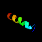
Fold: Tex N-terminal region-likeSuperfamily: Tex N-terminal region-likeFamily: Tex N-terminal region-like2 c1aq5C_
48.3
24
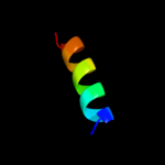
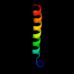
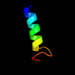
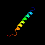
PDB header: coiled-coilChain: C: PDB Molecule: cartilage matrix protein;PDBTitle: high-resolution solution nmr structure of the trimeric coiled-coil2 domain of chicken cartilage matrix protein, 20 structures
3 c2xdjF_
40.9
31
PDB header: unknown functionChain: F: PDB Molecule: uncharacterized protein ybgf;PDBTitle: crystal structure of the n-terminal domain of e.coli ybgf
4 d1e52a_
40.6
22
Fold: Long alpha-hairpinSuperfamily: C-terminal UvrC-binding domain of UvrBFamily: C-terminal UvrC-binding domain of UvrB5 c2oceA_
36.3
16
PDB header: structural genomics, unknown functionChain: A: PDB Molecule: hypothetical protein pa5201;PDBTitle: crystal structure of tex family protein pa5201 from2 pseudomonas aeruginosa
6 c2pnvA_
32.1
23
PDB header: membrane proteinChain: A: PDB Molecule: small conductance calcium-activated potassiumPDBTitle: crystal structure of the leucine zipper domain of small-2 conductance ca2+-activated k+ (skca) channel from rattus3 norvegicus
7 d1nkzb_
24.9
37
Fold: Light-harvesting complex subunitsSuperfamily: Light-harvesting complex subunitsFamily: Light-harvesting complex subunits8 d1jb0i_
24.4
18
Fold: Single transmembrane helixSuperfamily: Subunit VIII of photosystem I reaction centre, PsaIFamily: Subunit VIII of photosystem I reaction centre, PsaI9 c2oqqB_
23.1
24
PDB header: transcriptionChain: B: PDB Molecule: transcription factor hy5;PDBTitle: crystal structure of hy5 leucine zipper homodimer from2 arabidopsis thaliana
10 c3okqA_
23.0
19
PDB header: protein bindingChain: A: PDB Molecule: bud site selection protein 6;PDBTitle: crystal structure of a core domain of yeast actin nucleation cofactor2 bud6
11 d1wrda1
22.6
17
Fold: Spectrin repeat-likeSuperfamily: GAT-like domainFamily: GAT domain12 c1b9uA_
20.8
32
PDB header: hydrolaseChain: A: PDB Molecule: protein (atp synthase);PDBTitle: membrane domain of the subunit b of the e.coli atp synthase
13 d1pfta_
20.7
50
Fold: Rubredoxin-likeSuperfamily: Zinc beta-ribbonFamily: Transcriptional factor domain14 c3mk7F_
20.2
21
PDB header: oxidoreductaseChain: F: PDB Molecule: cytochrome c oxidase, cbb3-type, subunit p;PDBTitle: the structure of cbb3 cytochrome oxidase
15 c3ixzB_
20.2
28
PDB header: hydrolaseChain: B: PDB Molecule: potassium-transporting atpase subunit beta;PDBTitle: pig gastric h+/k+-atpase complexed with aluminium fluoride
16 d1dl6a_
20.1
25
Fold: Rubredoxin-likeSuperfamily: Zinc beta-ribbonFamily: Transcriptional factor domain17 d1wr6a1
19.4
17
Fold: Spectrin repeat-likeSuperfamily: GAT-like domainFamily: GAT domain18 c3bz1y_
18.8
47
PDB header: electron transportChain: Y: PDB Molecule: photosystem ii protein y;PDBTitle: crystal structure of cyanobacterial photosystem ii (part 12 of 2). this file contains first monomer of psii dimer
19 c2d96A_
18.6
32
PDB header: transcriptionChain: A: PDB Molecule: nuclear factor nf-kappa-b p100 subunit;PDBTitle: solution structure of the death domain of nuclear factor nf-2 kappa-b p100
20 c3arcy_
18.1
47
PDB header: electron transport, photosynthesisChain: Y: PDB Molecule: protein ycf12;PDBTitle: crystal structure of oxygen-evolving photosystem ii at 1.9 angstrom2 resolution
21 c3arcY_
not modelled
18.1
47
PDB header: electron transport, photosynthesisChain: Y: PDB Molecule: protein ycf12;PDBTitle: crystal structure of oxygen-evolving photosystem ii at 1.9 angstrom2 resolution
22 d1nu9c2
not modelled
17.8
40
Fold: immunoglobulin/albumin-binding domain-likeSuperfamily: StaphylocoagulaseFamily: Staphylocoagulase23 c1a92B_
not modelled
17.6
24
PDB header: leucine zipperChain: B: PDB Molecule: delta antigen;PDBTitle: oligomerization domain of hepatitis delta antigen
24 c3a0hy_
not modelled
17.1
47
PDB header: electron transportChain: Y: PDB Molecule: photosystem ii reaction center protein ycf12;PDBTitle: crystal structure of i-substituted photosystem ii complex
25 c3a0hY_
not modelled
17.1
47
PDB header: electron transportChain: Y: PDB Molecule: photosystem ii reaction center protein ycf12;PDBTitle: crystal structure of i-substituted photosystem ii complex
26 c3m06F_
not modelled
16.9
14
PDB header: protein bindingChain: F: PDB Molecule: tnf receptor-associated factor 2;PDBTitle: crystal structure of traf2
27 c3ls1A_
not modelled
14.8
12
PDB header: photosynthesisChain: A: PDB Molecule: sll1638 protein;PDBTitle: crystal structure of cyanobacterial psbq from synechocystis2 sp. pcc 6803 complexed with zn2+
28 d1g7oa1
not modelled
14.5
14
Fold: GST C-terminal domain-likeSuperfamily: GST C-terminal domain-likeFamily: Glutathione S-transferase (GST), C-terminal domain29 d1o3xa_
not modelled
14.3
24
Fold: Spectrin repeat-likeSuperfamily: GAT-like domainFamily: GAT domain30 d1bf5a1
not modelled
14.2
17
Fold: STAT-likeSuperfamily: STATFamily: STAT31 c2jgoB_
not modelled
14.2
30
PDB header: de novo proteinChain: B: PDB Molecule: coil ser l9c;PDBTitle: stucture of the arsenated de novo designed peptide coil ser2 l9c
32 c3ljmC_
not modelled
14.2
30
PDB header: de novo proteinChain: C: PDB Molecule: coil ser l9c;PDBTitle: structure of de novo designed apo peptide coil ser l9c
33 c3ljmB_
not modelled
14.2
30
PDB header: de novo proteinChain: B: PDB Molecule: coil ser l9c;PDBTitle: structure of de novo designed apo peptide coil ser l9c
34 c2jgoC_
not modelled
14.2
30
PDB header: de novo proteinChain: C: PDB Molecule: coil ser l9c;PDBTitle: stucture of the arsenated de novo designed peptide coil ser2 l9c
35 c2jgoA_
not modelled
14.2
30
PDB header: de novo proteinChain: A: PDB Molecule: coil ser l9c;PDBTitle: stucture of the arsenated de novo designed peptide coil ser2 l9c
36 c3ljmA_
not modelled
14.2
30
PDB header: de novo proteinChain: A: PDB Molecule: coil ser l9c;PDBTitle: structure of de novo designed apo peptide coil ser l9c
37 c3efdK_
not modelled
14.1
27
PDB header: immune systemChain: K: PDB Molecule: kcsa;PDBTitle: the crystal structure of the cytoplasmic domain of kcsa
38 c3b37A_
not modelled
14.1
20
PDB header: hydrolaseChain: A: PDB Molecule: aminopeptidase n;PDBTitle: crystal structure of e. coli aminopeptidase n in complex with tyrosine
39 c3a0by_
not modelled
13.8
47
PDB header: electron transportChain: Y: PDB Molecule: photosystem ii reaction center protein ycf12;PDBTitle: crystal structure of br-substituted photosystem ii complex
40 c2d99A_
not modelled
13.6
42
PDB header: transcriptionChain: A: PDB Molecule: general transcription factor ii-i repeat domain-PDBTitle: solution structure of rsgi ruh-048, a gtf2i domain in human2 cdna
41 c2e3lA_
not modelled
13.5
25
PDB header: transcriptionChain: A: PDB Molecule: transcription factor gtf2ird2 beta;PDBTitle: solution structure of rsgi ruh-068, a gtf2i domain in human2 cdna
42 c2dn5A_
not modelled
13.1
50
PDB header: transcriptionChain: A: PDB Molecule: general transcription factor ii-i repeat domain-PDBTitle: solution structure of rsgi ruh-057, a gtf2i domain in human2 cdna
43 c2dbfA_
not modelled
12.9
33
PDB header: signaling proteinChain: A: PDB Molecule: nuclear factor nf-kappa-b p105 subunit;PDBTitle: solution structure of the death domain in human nuclear2 factor nf-kappa-b p105 subunit
44 c2dzrA_
not modelled
12.6
42
PDB header: transcriptionChain: A: PDB Molecule: general transcription factor ii-i repeat domain-PDBTitle: solution structure of rsgi ruh-067, a gtf2i domain in human2 cdna
45 c2dzqA_
not modelled
12.5
42
PDB header: transcriptionChain: A: PDB Molecule: general transcription factor ii-i repeat domain-PDBTitle: solution structure of rsgi ruh-066, a gtf2i domain in human2 cdna
46 c1cosA_
not modelled
12.2
30
PDB header: alpha-helical bundleChain: A: PDB Molecule: coiled serine;PDBTitle: crystal structure of a synthetic triple-stranded alpha-2 helical bundle
47 c1cosC_
not modelled
12.2
30
PDB header: alpha-helical bundleChain: C: PDB Molecule: coiled serine;PDBTitle: crystal structure of a synthetic triple-stranded alpha-2 helical bundle
48 c1cosB_
not modelled
12.2
30
PDB header: alpha-helical bundleChain: B: PDB Molecule: coiled serine;PDBTitle: crystal structure of a synthetic triple-stranded alpha-2 helical bundle
49 d1wmub_
not modelled
12.0
18
Fold: Globin-likeSuperfamily: Globin-likeFamily: Globins50 c3a0bY_
not modelled
11.0
47
PDB header: electron transportChain: Y: PDB Molecule: photosystem ii reaction center protein ycf12;PDBTitle: crystal structure of br-substituted photosystem ii complex
51 d1q60a_
not modelled
10.8
42
Fold: GTF2I-like repeatSuperfamily: GTF2I-like repeatFamily: GTF2I-like repeat52 c2ed2A_
not modelled
10.8
33
PDB header: transcriptionChain: A: PDB Molecule: general transcription factor ii-i;PDBTitle: solution structure of rsgi ruh-069, a gtf2i domain in human2 cdna
53 d1zkea1
not modelled
10.5
38
Fold: ROP-likeSuperfamily: HP1531-likeFamily: HP1531-like54 d1f6ga_
not modelled
10.5
21
Fold: Voltage-gated potassium channelsSuperfamily: Voltage-gated potassium channelsFamily: Voltage-gated potassium channels55 c2dn4A_
not modelled
10.4
50
PDB header: transcriptionChain: A: PDB Molecule: general transcription factor ii-i;PDBTitle: solution structure of rsgi ruh-060, a gtf2i domain in human2 cdna
56 c1coiA_
not modelled
10.4
25
PDB header: alpha-helical bundleChain: A: PDB Molecule: coil-vald;PDBTitle: designed trimeric coiled coil-vald
57 c3c18B_
not modelled
10.3
12
PDB header: transferaseChain: B: PDB Molecule: nucleotidyltransferase-like protein;PDBTitle: crystal structure of nucleotidyltransferase-like protein2 (zp_00538802.1) from exiguobacterium sibiricum 255-15 at 1.90 a3 resolution
58 c3b8eB_
not modelled
10.1
28
PDB header: hydrolase/transport proteinChain: B: PDB Molecule: sodium/potassium-transporting atpase subunitPDBTitle: crystal structure of the sodium-potassium pump
59 c1ci6B_
not modelled
10.1
19
PDB header: transcriptionChain: B: PDB Molecule: transcription factor c/ebp beta;PDBTitle: transcription factor atf4-c/ebp beta bzip heterodimer
60 c1by0A_
not modelled
10.0
24
PDB header: rna binding proteinChain: A: PDB Molecule: protein (hepatitis delta antigen);PDBTitle: n-terminal leucine-repeat region of hepatitis delta antigen
61 d1ijdb_
not modelled
9.9
39
Fold: Light-harvesting complex subunitsSuperfamily: Light-harvesting complex subunitsFamily: Light-harvesting complex subunits62 c2ejeA_
not modelled
9.3
33
PDB header: transcriptionChain: A: PDB Molecule: general transcription factor ii-i;PDBTitle: solution structure of rsgi ruh-071, a gtf2i domain in human2 cdna
63 c3bmbB_
not modelled
8.8
19
PDB header: rna binding proteinChain: B: PDB Molecule: regulator of nucleoside diphosphate kinase;PDBTitle: crystal structure of a new rna polymerase interacting2 protein
64 c3eabD_
not modelled
8.7
15
PDB header: cell cycleChain: D: PDB Molecule: spastin;PDBTitle: crystal structure of spastin mit in complex with escrt iii
65 d1wmga_
not modelled
8.2
47
Fold: DEATH domainSuperfamily: DEATH domainFamily: DEATH domain, DD66 d1mlda2
not modelled
8.1
18
Fold: LDH C-terminal domain-likeSuperfamily: LDH C-terminal domain-likeFamily: Lactate & malate dehydrogenases, C-terminal domain67 c2d3kA_
not modelled
7.8
23
PDB header: hydrolaseChain: A: PDB Molecule: peptidyl-trna hydrolase;PDBTitle: structural study on project id ph1539 from pyrococcus2 horikoshii ot3
68 c3sb1B_
not modelled
7.6
24
PDB header: structural genomics, unknown functionChain: B: PDB Molecule: hydrogenase expression protein;PDBTitle: hydrogenase expression protein huph from thiobacillus denitrificans2 atcc 25259
69 c3r45C_
not modelled
7.6
30
PDB header: nuclear proteinChain: C: PDB Molecule: holliday junction recognition protein;PDBTitle: structure of a cenp-a-histone h4 heterodimer in complex with chaperone2 hjurp
70 c1xtyD_
not modelled
7.6
16
PDB header: hydrolaseChain: D: PDB Molecule: peptidyl-trna hydrolase;PDBTitle: crystal structure of sulfolobus solfataricus peptidyl-trna2 hydrolase
71 d2ay0a1
not modelled
7.5
33
Fold: Ribbon-helix-helixSuperfamily: Ribbon-helix-helixFamily: PutA pre-N-terminal region-like72 d1ab4a_
not modelled
7.3
24
Fold: Type II DNA topoisomeraseSuperfamily: Type II DNA topoisomeraseFamily: Type II DNA topoisomerase73 c3m6nA_
not modelled
7.3
16
PDB header: lyaseChain: A: PDB Molecule: rpff protein;PDBTitle: crystal structure of rpff
74 d1kqfb2
not modelled
7.1
25
Fold: Single transmembrane helixSuperfamily: Iron-sulfur subunit of formate dehydrogenase N, transmembrane anchorFamily: Iron-sulfur subunit of formate dehydrogenase N, transmembrane anchor75 c3ilwA_
not modelled
6.9
18
PDB header: isomeraseChain: A: PDB Molecule: dna gyrase subunit a;PDBTitle: structure of dna gyrase subunit a n-terminal domain
76 d1x79a_
not modelled
6.9
23
Fold: Spectrin repeat-likeSuperfamily: GAT-like domainFamily: GAT domain77 d1t3wa_
not modelled
6.5
30
Fold: DNA primase DnaG, C-terminal domainSuperfamily: DNA primase DnaG, C-terminal domainFamily: DNA primase DnaG, C-terminal domain78 c2vmlD_
not modelled
6.4
29
PDB header: photosynthesisChain: D: PDB Molecule: phycocyanin beta chain;PDBTitle: the monoclinic structure of phycocyanin from gloeobacter2 violaceus
79 c3pp5A_
not modelled
6.3
17
PDB header: structural proteinChain: A: PDB Molecule: brk1;PDBTitle: high-resolution structure of the trimeric scar/wave complex precursor2 brk1
80 d2clyc1
not modelled
6.2
13
Fold: Mitochondrial ATP synthase coupling factor 6Superfamily: Mitochondrial ATP synthase coupling factor 6Family: Mitochondrial ATP synthase coupling factor 681 c2o7hF_
not modelled
6.1
16
PDB header: transcriptionChain: F: PDB Molecule: general control protein gcn4;PDBTitle: crystal structure of trimeric coiled coil gcn4 leucine zipper
82 c2ergA_
not modelled
6.0
19
PDB header: transcription activator/dnaChain: A: PDB Molecule: regulatory protein leu3;PDBTitle: crystal structure of leu3 dna-binding domain with a single2 h50c mutation complexed with a 15mer dna duplex
83 c3m0dC_
not modelled
5.8
13
PDB header: signaling proteinChain: C: PDB Molecule: tnf receptor-associated factor 1;PDBTitle: crystal structure of the traf1:traf2:ciap2 complex
84 c3hroA_
not modelled
5.8
26
PDB header: transport proteinChain: A: PDB Molecule: transient receptor potential (trp) channelPDBTitle: crystal structure of a c-terminal coiled coil domain of2 transient receptor potential (trp) channel subfamily p3 member 2 (trpp2, polycystic kidney disease 2)
85 d3ehbb2
not modelled
5.8
7
Fold: Transmembrane helix hairpinSuperfamily: Cytochrome c oxidase subunit II-like, transmembrane regionFamily: Cytochrome c oxidase subunit II-like, transmembrane region86 c3hrxD_
not modelled
5.7
13
PDB header: lyaseChain: D: PDB Molecule: probable enoyl-coa hydratase;PDBTitle: crystal structure of phenylacetic acid degradation protein paag
87 c2ba2A_
not modelled
5.6
23
PDB header: structural genomics, unknown functionChain: A: PDB Molecule: hypothetical upf0134 protein mpn010;PDBTitle: crystal structure of the duf16 domain of mpn010 from2 mycoplasma pneumoniae
88 c2voyE_
not modelled
5.6
20
PDB header: hydrolaseChain: E: PDB Molecule: sarcoplasmic/endoplasmic reticulum calciumPDBTitle: cryoem model of copa, the copper transporting atpase from2 archaeoglobus fulgidus
89 c2hbpA_
not modelled
5.6
14
PDB header: endocytosis, protein bindingChain: A: PDB Molecule: cytoskeleton assembly control protein sla1;PDBTitle: solution structure of sla1 homology domain 1
90 d2p90a1
not modelled
5.5
22
Fold: Phosphorylase/hydrolase-likeSuperfamily: Cgl1923-likeFamily: Cgl1923-like91 c2inrA_
not modelled
5.5
27
PDB header: isomeraseChain: A: PDB Molecule: dna topoisomerase 4 subunit a;PDBTitle: crystal structure of a 59 kda fragment of topoisomerase iv subunit a2 (grla) from staphylococcus aureus
92 c3p8cE_
not modelled
5.5
30
PDB header: protein bindingChain: E: PDB Molecule: probable protein brick1;PDBTitle: structure and control of the actin regulatory wave complex
93 d1in4a1
not modelled
5.4
12
Fold: DNA/RNA-binding 3-helical bundleSuperfamily: "Winged helix" DNA-binding domainFamily: Helicase DNA-binding domain94 d2h8fb1
not modelled
5.4
13
Fold: Globin-likeSuperfamily: Globin-likeFamily: Globins95 c2novD_
not modelled
5.0
25
PDB header: isomeraseChain: D: PDB Molecule: dna topoisomerase 4 subunit a;PDBTitle: breakage-reunion domain of s.pneumoniae topo iv: crystal2 structure of a gram-positive quinolone target