Phyre Home Retrieve Phyre Job Id
Emaill.a.kelley@imperial.ac.uk
DescriptionP76508
DateThu Jan 5 12:23:47 GMT 2012
Unique Job ID3c4fdc27aeb808c1

Summary 

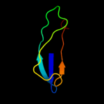
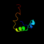
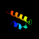
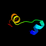
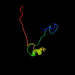
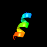
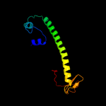
Top model
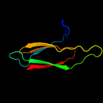
Image coloured by rainbow N → C terminus
Model (left) based on template d2ccva1
Top template information
Fold:HPA-like
Superfamily:Agglutinin HPA-like
Family:Agglutinin HPA-like
Confidence and coverage
Confidence: 82.1% Coverage: 42%
72 residues ( 42% of your sequence) have been modelled with 82.1% confidence by the single highest scoring template.
You may wish to submit your sequence to Phyrealarm. This will automatically scan your sequence every week for new potential templates as they appear in the Phyre2 library.
Please note: You must be registered and logged in to use Phyrealarm.
3D viewing
Interactive 3D view in Jmol

Sequence analysis 

Secondary structure and disorder prediction 

   1........10.........20.........30.........40.........50.........60
Sequence  MLQEELCSVVEASGASLEKGRHDQLLTALRALLLSRKNPFGDIKLDGTVQKALENLGLGE
Secondary structure 


SS confidence 


Disorder  ???????


?????
???


??????????????


?????
??????
?
Disorder confidence 


   .........70.........80.........90.........100.........110.........120
Sequence  AAKRNVGTGANQIPDMGSFMLSASVPGYQKLPSGLIIQWGPIDVPLTSQDTVTYFPIAFP
Secondary structure 


SS confidence 


Disorder  ?????


?


???????
??


???????


Disorder confidence 


   .........130.........140.........150.........160.........170..
Sequence  NRCLRVFATQDYTPGSANVGYIACAGYNQDPVKFISRAGVPGIGASFFALGC
Secondary structure 


SS confidence 


Disorder 


??
?????
?


?

?


??


?
Disorder confidence 


Confidence Key
High(9)                    Low (0)
?Disordered
Alpha helix
Beta strand

Domain analysis 

Hover over an aligned region to see model and summary info

Please note, only up to the top 20 hits are modelled to reduce computer load

RankAligned region

PDB 2ccv chain A domain 1

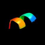
3D model

Region: 99 - 170
Aligned: 72
Modelled: 72
Confidence: 82.1%
Identity: 10%
Fold: HPA-like
Superfamily: Agglutinin HPA-like
Family: Agglutinin HPA-like

Phyre2

PDB 2vme chain C

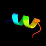
3D model

Region: 105 - 170
Aligned: 66
Modelled: 66
Confidence: 37.0%
Identity: 11%
PDB header:cell adhesion
Chain: C: PDB Molecule:discoidin-2;
PDBTitle: structure of the wild-type discoidin ii from dictyostelium2 discoideum

Phyre2

PDB 2w95 chain B

3D model

Region: 111 - 171
Aligned: 61
Modelled: 61
Confidence: 26.8%
Identity: 16%
PDB header:cell adhesion
Chain: B: PDB Molecule:discoidin-1 subunit a;
PDBTitle: structure of the discoidin i from dictyostelium discoideum2 in complex with galnac at 1.75 angstrom resolution

Phyre2

PDB 3a1y chain F

3D model

Region: 4 - 29
Aligned: 26
Modelled: 26
Confidence: 25.3%
Identity: 38%
PDB header:ribosomal protein
Chain: F: PDB Molecule:50s ribosomal protein p1 (l12p);
PDBTitle: the structure of protein complex

Phyre2

PDB 3hrz chain A

3D model

Region: 88 - 133
Aligned: 46
Modelled: 46
Confidence: 13.7%
Identity: 15%
PDB header:immune system
Chain: A: PDB Molecule:cobra venom factor;
PDBTitle: cobra venom factor (cvf) in complex with human factor b

Phyre2

PDB 2w1o chain A

3D model

Region: 3 - 47
Aligned: 45
Modelled: 45
Confidence: 12.1%
Identity: 9%
PDB header:translation
Chain: A: PDB Molecule:60s acidic ribosomal protein p2;
PDBTitle: nmr structure of dimerization domain of human ribosomal2 protein p2

Phyre2

PDB 3p45 chain F

3D model

Region: 4 - 38
Aligned: 32
Modelled: 35
Confidence: 12.1%
Identity: 22%
PDB header:hydrolase
Chain: F: PDB Molecule:caspase-6;
PDBTitle: crystal structure of apo-caspase-6 at physiological ph

Phyre2

PDB 3trk chain A

3D model

Region: 6 - 30
Aligned: 25
Modelled: 25
Confidence: 11.6%
Identity: 24%
PDB header:hydrolase
Chain: A: PDB Molecule:nonstructural polyprotein;
PDBTitle: structure of the chikungunya virus nsp2 protease

Phyre2

PDB 1jqp chain A domain 2

3D model

Region: 76 - 120
Aligned: 36
Modelled: 45
Confidence: 10.4%
Identity: 17%
Fold: Cysteine proteinases
Superfamily: Cysteine proteinases
Family: Papain-like

Phyre2

PDB 1ev0 chain A

3D model

Region: 1 - 12
Aligned: 12
Modelled: 12
Confidence: 9.3%
Identity: 17%
Fold: Cell division protein MinE topological specificity domain
Superfamily: Cell division protein MinE topological specificity domain
Family: Cell division protein MinE topological specificity domain

Phyre2

PDB 2jsn chain A

3D model

Region: 92 - 119
Aligned: 27
Modelled: 28
Confidence: 9.2%
Identity: 7%
PDB header:protein transport
Chain: A: PDB Molecule:trafficking protein particle complex subunit 4;
PDBTitle: solution structure of the atypical pdz-like domain of2 synbindin

Phyre2

PDB 1bqf chain A

3D model

Region: 85 - 94
Aligned: 10
Modelled: 10
Confidence: 9.0%
Identity: 50%
PDB header:hormone/growth factor
Chain: A: PDB Molecule:protein (growth-blocking peptide);
PDBTitle: growth-blocking peptide (gbp) from pseudaletia separata

Phyre2

PDB 1aoy chain A

3D model

Region: 3 - 15
Aligned: 13
Modelled: 13
Confidence: 8.2%
Identity: 23%
Fold: DNA/RNA-binding 3-helical bundle
Superfamily: "Winged helix" DNA-binding domain
Family: Arginine repressor (ArgR), N-terminal DNA-binding domain

Phyre2

PDB 2ct8 chain A

3D model

Region: 111 - 126
Aligned: 16
Modelled: 16
Confidence: 7.8%
Identity: 19%
PDB header:ligase/rna
Chain: A: PDB Molecule:methionyl-trna synthetase;
PDBTitle: crystal structure of aquifex aeolicus methionyl-trna2 synthetase complexed with trna(met) and methionyl-adenylate3 anologue

Phyre2

PDB 2p5k chain A domain 1

3D model

Region: 3 - 16
Aligned: 14
Modelled: 14
Confidence: 7.8%
Identity: 29%
Fold: DNA/RNA-binding 3-helical bundle
Superfamily: "Winged helix" DNA-binding domain
Family: Arginine repressor (ArgR), N-terminal DNA-binding domain

Phyre2

PDB 1f9n chain A domain 1

3D model

Region: 3 - 16
Aligned: 14
Modelled: 14
Confidence: 7.4%
Identity: 29%
Fold: DNA/RNA-binding 3-helical bundle
Superfamily: "Winged helix" DNA-binding domain
Family: Arginine repressor (ArgR), N-terminal DNA-binding domain

Phyre2

PDB 1fd9 chain A

3D model

Region: 5 - 121
Aligned: 114
Modelled: 117
Confidence: 7.2%
Identity: 15%
Fold: FKBP-like
Superfamily: FKBP-like
Family: FKBP immunophilin/proline isomerase

Phyre2

PDB 2kxw chain B

3D model

Region: 3 - 10
Aligned: 8
Modelled: 8
Confidence: 7.1%
Identity: 50%
PDB header:calcium-binding protein/metal transport
Chain: B: PDB Molecule:sodium channel protein type 2 subunit alpha;
PDBTitle: structure of the c-domain fragment of apo calmodulin bound to the iq2 motif of nav1.2

Phyre2

PDB 1b4a chain A domain 1

3D model

Region: 3 - 16
Aligned: 14
Modelled: 14
Confidence: 6.3%
Identity: 29%
Fold: DNA/RNA-binding 3-helical bundle
Superfamily: "Winged helix" DNA-binding domain
Family: Arginine repressor (ArgR), N-terminal DNA-binding domain

Phyre2

PDB 1b4a chain A

3D model

Region: 3 - 16
Aligned: 14
Modelled: 14
Confidence: 6.0%
Identity: 29%
PDB header:repressor
Chain: A: PDB Molecule:arginine repressor;
PDBTitle: structure of the arginine repressor from bacillus stearothermophilus

Phyre2
1

d2ccva1
2

c2vmeC_
3

c2w95B_
4

c3a1yF_
5

c3hrzA_
6

c2w1oA_
7

c3p45F_
8

c3trkA_
9

d1jqpa2
10

d1ev0a_
11

c2jsnA_
12

c1bqfA_
13

d1aoya_
14

c2ct8A_
15

d2p5ka1
16

d1f9na1
17

d1fd9a_
18

c2kxwB_
19

d1b4aa1
20

c1b4aA_
21


22


23


24


25


Detailed template information 

#
Template Alignment Coverage3D Model Confidence
% i.d. Template Information
1d2ccva1


82.1 10 Fold:HPA-like
Superfamily:Agglutinin HPA-like
Family:Agglutinin HPA-like
2c2vmeC_


37.0 11 PDB header:cell adhesion
Chain: C: PDB Molecule:discoidin-2;
PDBTitle: structure of the wild-type discoidin ii from dictyostelium2 discoideum
3c2w95B_


26.8 16 PDB header:cell adhesion
Chain: B: PDB Molecule:discoidin-1 subunit a;
PDBTitle: structure of the discoidin i from dictyostelium discoideum2 in complex with galnac at 1.75 angstrom resolution
4c3a1yF_


25.3 38 PDB header:ribosomal protein
Chain: F: PDB Molecule:50s ribosomal protein p1 (l12p);
PDBTitle: the structure of protein complex
5c3hrzA_


13.7 15 PDB header:immune system
Chain: A: PDB Molecule:cobra venom factor;
PDBTitle: cobra venom factor (cvf) in complex with human factor b
6c2w1oA_


12.1 9 PDB header:translation
Chain: A: PDB Molecule:60s acidic ribosomal protein p2;
PDBTitle: nmr structure of dimerization domain of human ribosomal2 protein p2
7c3p45F_


12.1 22 PDB header:hydrolase
Chain: F: PDB Molecule:caspase-6;
PDBTitle: crystal structure of apo-caspase-6 at physiological ph
8c3trkA_


11.6 24 PDB header:hydrolase
Chain: A: PDB Molecule:nonstructural polyprotein;
PDBTitle: structure of the chikungunya virus nsp2 protease
9d1jqpa2


10.4 17 Fold:Cysteine proteinases
Superfamily:Cysteine proteinases
Family:Papain-like
10d1ev0a_


9.3 17 Fold:Cell division protein MinE topological specificity domain
Superfamily:Cell division protein MinE topological specificity domain
Family:Cell division protein MinE topological specificity domain
11c2jsnA_


9.2 7 PDB header:protein transport
Chain: A: PDB Molecule:trafficking protein particle complex subunit 4;
PDBTitle: solution structure of the atypical pdz-like domain of2 synbindin
12c1bqfA_


9.0 50 PDB header:hormone/growth factor
Chain: A: PDB Molecule:protein (growth-blocking peptide);
PDBTitle: growth-blocking peptide (gbp) from pseudaletia separata
13d1aoya_


8.2 23 Fold:DNA/RNA-binding 3-helical bundle
Superfamily:"Winged helix" DNA-binding domain
Family:Arginine repressor (ArgR), N-terminal DNA-binding domain
14c2ct8A_


7.8 19 PDB header:ligase/rna
Chain: A: PDB Molecule:methionyl-trna synthetase;
PDBTitle: crystal structure of aquifex aeolicus methionyl-trna2 synthetase complexed with trna(met) and methionyl-adenylate3 anologue
15d2p5ka1


7.8 29 Fold:DNA/RNA-binding 3-helical bundle
Superfamily:"Winged helix" DNA-binding domain
Family:Arginine repressor (ArgR), N-terminal DNA-binding domain
16d1f9na1


7.4 29 Fold:DNA/RNA-binding 3-helical bundle
Superfamily:"Winged helix" DNA-binding domain
Family:Arginine repressor (ArgR), N-terminal DNA-binding domain
17d1fd9a_


7.2 15 Fold:FKBP-like
Superfamily:FKBP-like
Family:FKBP immunophilin/proline isomerase
18c2kxwB_


7.1 50 PDB header:calcium-binding protein/metal transport
Chain: B: PDB Molecule:sodium channel protein type 2 subunit alpha;
PDBTitle: structure of the c-domain fragment of apo calmodulin bound to the iq2 motif of nav1.2
19d1b4aa1


6.3 29 Fold:DNA/RNA-binding 3-helical bundle
Superfamily:"Winged helix" DNA-binding domain
Family:Arginine repressor (ArgR), N-terminal DNA-binding domain
20c1b4aA_


6.0 29 PDB header:repressor
Chain: A: PDB Molecule:arginine repressor;
PDBTitle: structure of the arginine repressor from bacillus stearothermophilus
21c1dfwA_


not modelled 6.0 67 PDB header:immune system
Chain: A: PDB Molecule:lung surfactant protein b;
PDBTitle: conformational mapping of the n-terminal segment of2 surfactant protein b in lipid using 13c-enhanced fourier3 transform infrared spectroscopy (ftir)
22d2sici_


not modelled 5.7 19 Fold:Subtilisin inhibitor
Superfamily:Subtilisin inhibitor
Family:Subtilisin inhibitor
23d2hgsa4


not modelled 5.5 40 Fold:ATP-grasp
Superfamily:Glutathione synthetase ATP-binding domain-like
Family:Eukaryotic glutathione synthetase ATP-binding domain
24c2xvcA_


not modelled 5.4 10 PDB header:cell cycle
Chain: A: PDB Molecule:escrt-iii;
PDBTitle: molecular and structural basis of escrt-iii recruitment to2 membranes during archaeal cell division
25d1m0wa2


not modelled 5.4 30 Fold:ATP-grasp
Superfamily:Glutathione synthetase ATP-binding domain-like
Family:Eukaryotic glutathione synthetase ATP-binding domain

Binding site prediction 

Due to computational demand, binding site predictions are not run for batch jobs

If you want to predict binding sites, please manually submit your model of choice to 3DLigandSite


Phyre is for academic use only

Please cite: Protein structure prediction on the web: a case study using the Phyre server
Kelley LA and Sternberg MJE. Nature Protocols 4, 363 - 371 (2009) [pdf] [Import into BibTeX]
 
If you use the binding site predictions from 3DLigandSite, please also cite:
3DLigandSite: predicting ligand-binding sites using similar structures.
Wass MN, Kelley LA and Sternberg MJ Nucleic Acids Research 38, W469-73 (2010) [PubMed]
 
© Structural Bioinformatics Group
Imperial College London
Lawrence Kelley, Benjamin Jefferys
Disclaimer
Terms and Conditions
Component software
Template detection: HHpred 1.51
Secondary structure prediction: Psi-pred 2.5
Disorder prediction: Disopred 2.4
Transmembrane prediction: Memsat_SVM
Multi-template modelling and ab initio: Poing 1.0