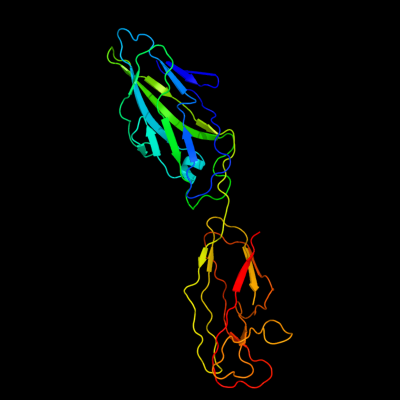
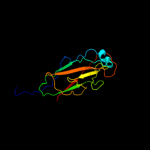
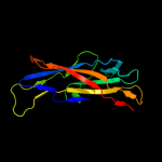
1 c1klfP_
99.9
14
PDB header: chaperone/adhesin complexChain: P: PDB Molecule: fimh protein;PDBTitle: fimh adhesin-fimc chaperone complex with d-mannose
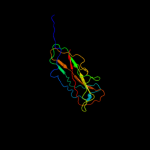
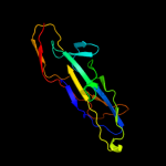
2 c2w07B_
99.9
17
PDB header: cell adhesionChain: B: PDB Molecule: minor pilin subunit papf;PDBTitle: structural determinants of polymerization reactivity of the2 p pilus adaptor subunit papf
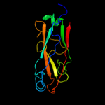
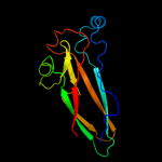
3 c3bfwA_
99.8
14
PDB header: structural protein/structural proteinChain: A: PDB Molecule: protein fimg;PDBTitle: crystal structure of truncated fimg (fimgt) in complex with the donor2 strand peptide of fimf (dsf)
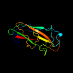
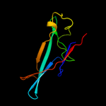
4 c3jwnK_
99.8
19
PDB header: protein binding/cell adhesionChain: K: PDB Molecule: protein fimf;PDBTitle: complex of fimc, fimf, fimg and fimh
5 c3jwnL_
99.8
19
PDB header: protein binding/cell adhesionChain: L: PDB Molecule: protein fimf;PDBTitle: complex of fimc, fimf, fimg and fimh
6 c3jwnE_
99.8
19
PDB header: protein binding/cell adhesionChain: E: PDB Molecule: protein fimf;PDBTitle: complex of fimc, fimf, fimg and fimh
7 c2jmrA_
99.8
21
PDB header: cell adhesionChain: A: PDB Molecule: fimf;PDBTitle: nmr structure of the e. coli type 1 pilus subunit fimf
8 d2j2zb1
99.8
14
Fold: Common fold of diphtheria toxin/transcription factors/cytochrome fSuperfamily: Bacterial adhesinsFamily: Pilus subunits9 c3jwnF_
99.8
18
PDB header: protein binding/cell adhesionChain: F: PDB Molecule: protein fimf;PDBTitle: complex of fimc, fimf, fimg and fimh
10 d1pdkb_
99.8
17
Fold: Common fold of diphtheria toxin/transcription factors/cytochrome fSuperfamily: Bacterial adhesinsFamily: Pilus subunits11 c2jtyA_
99.7
21
PDB header: structural proteinChain: A: PDB Molecule: type-1 fimbrial protein, a chain;PDBTitle: self-complemented variant of fima, the main subunit of type 1 pilus
12 d2uy6b1
99.7
16
Fold: Common fold of diphtheria toxin/transcription factors/cytochrome fSuperfamily: Bacterial adhesinsFamily: Pilus subunits13 d1n12a_
99.7
21
Fold: Common fold of diphtheria toxin/transcription factors/cytochrome fSuperfamily: Bacterial adhesinsFamily: Pilus subunits14 d1ze3h1
99.7
20
Fold: Common fold of diphtheria toxin/transcription factors/cytochrome fSuperfamily: Bacterial adhesinsFamily: Pilus subunits15 c3bwuF_
99.6
18
PDB header: chaperone, structural, membrane proteinChain: F: PDB Molecule: protein fimf;PDBTitle: crystal structure of the ternary complex of fimd (n-terminal domain,2 fimdn) with fimc and the n-terminally truncated pilus subunit fimf3 (fimft)
16 d2bsca1
90.9
13
Fold: Common fold of diphtheria toxin/transcription factors/cytochrome fSuperfamily: Bacterial adhesinsFamily: F17c-type adhesin17 d1oioa_
88.2
10
Fold: Common fold of diphtheria toxin/transcription factors/cytochrome fSuperfamily: Bacterial adhesinsFamily: F17c-type adhesin18 c2wmpB_
72.0
16
PDB header: chaperoneChain: B: PDB Molecule: papg protein;PDBTitle: structure of the e. coli chaperone papd in complex with the pilin2 domain of the papgii adhesin
19 d1p5vb_
48.9
12
Fold: Common fold of diphtheria toxin/transcription factors/cytochrome fSuperfamily: Bacterial adhesinsFamily: Pilus subunits20 c2wd6B_
31.6
15
PDB header: cell adhesionChain: B: PDB Molecule: agglutinin receptor;PDBTitle: crystal structure of the variable domain of the2 streptococcus gordonii surface protein sspb
21 d1jmma_
not modelled
10.9
10
Fold: SupersandwichSuperfamily: V-region of surface antigen I/II (SA I/II, PAC)Family: V-region of surface antigen I/II (SA I/II, PAC)22 c3fblA_
not modelled
9.6
55
PDB header: structural proteinChain: A: PDB Molecule: putative uncharacterized protein;PDBTitle: crystal structure of orf132 of the archaeal virus acidianus2 filamentous virus 1 (afv1)
23 c3osvC_
not modelled
6.2
13
PDB header: structural proteinChain: C: PDB Molecule: flagellar basal-body rod modification protein flgd;PDBTitle: the crytsal structure of flgd from p. aeruginosa
24 c2voyG_
not modelled
5.4
32
PDB header: hydrolaseChain: G: PDB Molecule: sarcoplasmic/endoplasmic reticulum calciumPDBTitle: cryoem model of copa, the copper transporting atpase from2 archaeoglobus fulgidus