| 1 |
|
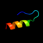
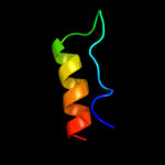
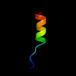
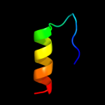
PDB 1eoq chain A
Region: 38 - 58
Aligned: 21
Modelled: 21
Confidence: 33.6%
Identity: 24%
Fold: Acyl carrier protein-like
Superfamily: Retrovirus capsid dimerization domain-like
Family: Retrovirus capsid protein C-terminal domain
Phyre2
| 2 |
|
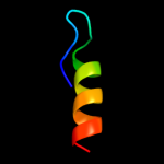
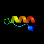
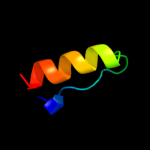
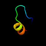
PDB 1a8o chain A
Region: 38 - 58
Aligned: 21
Modelled: 21
Confidence: 31.5%
Identity: 38%
Fold: Acyl carrier protein-like
Superfamily: Retrovirus capsid dimerization domain-like
Family: Retrovirus capsid protein C-terminal domain
Phyre2
| 3 |
|
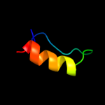
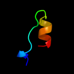
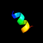
PDB 1qrj chain B domain 1
Region: 34 - 58
Aligned: 25
Modelled: 25
Confidence: 31.2%
Identity: 24%
Fold: Acyl carrier protein-like
Superfamily: Retrovirus capsid dimerization domain-like
Family: Retrovirus capsid protein C-terminal domain
Phyre2
| 4 |
|
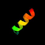
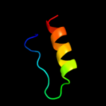
PDB 2eia chain A domain 1
Region: 34 - 59
Aligned: 26
Modelled: 26
Confidence: 27.4%
Identity: 31%
Fold: Acyl carrier protein-like
Superfamily: Retrovirus capsid dimerization domain-like
Family: Retrovirus capsid protein C-terminal domain
Phyre2
| 5 |
|
PDB 1baj chain A
Region: 34 - 58
Aligned: 25
Modelled: 25
Confidence: 23.6%
Identity: 36%
PDB header:viral protein
Chain: A: PDB Molecule:gag polyprotein;
PDBTitle: hiv-1 capsid protein c-terminal fragment plus gag p2 domain
Phyre2
| 6 |
|
PDB 2k1c chain A
Region: 34 - 53
Aligned: 20
Modelled: 20
Confidence: 21.7%
Identity: 45%
PDB header:hydrolase/hydrolase inhibitor
Chain: A: PDB Molecule:capsid protein p24;
PDBTitle: nmr structure of the c-terminal domain of hiv-1 capsid in complex with2 peptide inhibitor
Phyre2
| 7 |
|
PDB 3dik chain A
Region: 38 - 58
Aligned: 21
Modelled: 21
Confidence: 14.5%
Identity: 38%
PDB header:viral protein
Chain: A: PDB Molecule:capsid protein p24;
PDBTitle: pseudo-atomic model of the hiv-1 ca hexameric lattice
Phyre2
| 8 |
|
PDB 1bmx chain A
Region: 38 - 54
Aligned: 17
Modelled: 17
Confidence: 13.7%
Identity: 47%
PDB header:viral protein
Chain: A: PDB Molecule:human immunodeficiency virus type 1 capsid;
PDBTitle: hiv-1 capsid protein major homology region peptide analog,2 nmr, 8 structures
Phyre2
| 9 |
|
PDB 3gv2 chain E
Region: 34 - 58
Aligned: 25
Modelled: 25
Confidence: 13.7%
Identity: 36%
PDB header:viral protein
Chain: E: PDB Molecule:fusion protein consisting of capsid protein p24,
PDBTitle: x-ray structure of hexameric hiv-1 ca
Phyre2
| 10 |
|
PDB 1pzr chain A
Region: 30 - 41
Aligned: 12
Modelled: 12
Confidence: 12.6%
Identity: 58%
Fold: HLH-like
Superfamily: Docking domain B of the erythromycin polyketide synthase (DEBS)
Family: Docking domain B of the erythromycin polyketide synthase (DEBS)
Phyre2
| 11 |
|
PDB 1eia chain A
Region: 34 - 58
Aligned: 25
Modelled: 25
Confidence: 11.5%
Identity: 32%
PDB header:viral protein
Chain: A: PDB Molecule:eiav capsid protein p26;
PDBTitle: x-ray crystal structure of equine infectious anemia virus2 (eiav) capsid protein p26
Phyre2
| 12 |
|
PDB 1d1d chain A
Region: 38 - 58
Aligned: 21
Modelled: 21
Confidence: 9.6%
Identity: 24%
PDB header:viral protein
Chain: A: PDB Molecule:protein (capsid protein);
PDBTitle: nmr solution structure of the capsid protein from rous2 sarcoma virus
Phyre2
| 13 |
|
PDB 2x8q chain A
Region: 38 - 53
Aligned: 16
Modelled: 16
Confidence: 6.8%
Identity: 31%
PDB header:virus
Chain: A: PDB Molecule:capsid protein p27;
PDBTitle: cryo-em 3d model of the icosahedral particle2 composed of rous sarcoma virus capsid protein pentamers
Phyre2
| 14 |
|
PDB 3kev chain A
Region: 15 - 53
Aligned: 36
Modelled: 39
Confidence: 5.7%
Identity: 25%
PDB header:structural genomics, unknown function
Chain: A: PDB Molecule:galieria sulfuraria dcun1 domain-containing protein;
PDBTitle: x-ray crystal structure of a dcun1 domain-containing protein from2 galdieria sulfuraria
Phyre2