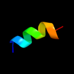
| 1 |
|
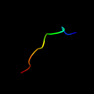
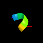
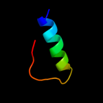
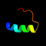
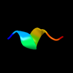
PDB 2pmf chain A
Region: 50 - 60
Aligned: 11
Modelled: 11
Confidence: 15.2%
Identity: 18%
PDB header:ligase
Chain: A: PDB Molecule:glycyl-trna synthetase;
PDBTitle: the crystal structure of a human glycyl-trna synthetase mutant
Phyre2
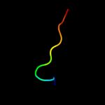
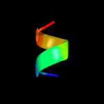
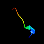
| 2 |
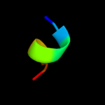
|
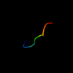
PDB 1qhd chain A domain 2
Region: 49 - 75
Aligned: 27
Modelled: 27
Confidence: 12.9%
Identity: 33%
Fold: Viral protein domain
Superfamily: Viral protein domain
Family: Top domain of virus capsid protein
Phyre2
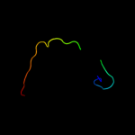
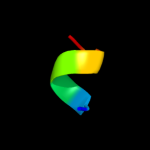
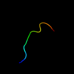
| 3 |
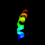
|
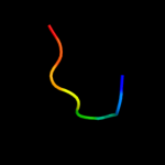
PDB 1m1c chain A
Region: 59 - 73
Aligned: 15
Modelled: 15
Confidence: 11.0%
Identity: 47%
Fold: L-A virus major coat protein
Superfamily: L-A virus major coat protein
Family: L-A virus major coat protein
Phyre2
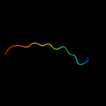
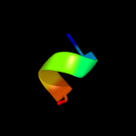
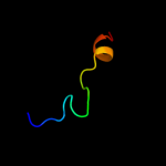
| 4 |
|
PDB 1m1c chain B
Region: 59 - 73
Aligned: 15
Modelled: 15
Confidence: 11.0%
Identity: 47%
PDB header:virus
Chain: B: PDB Molecule:major coat protein;
PDBTitle: structure of the l-a virus
Phyre2
| 5 |
|
PDB 1z8y chain N
Region: 21 - 27
Aligned: 7
Modelled: 7
Confidence: 9.7%
Identity: 43%
PDB header:virus
Chain: N: PDB Molecule:spike glycoprotein e2;
PDBTitle: mapping the e2 glycoprotein of alphaviruses
Phyre2
| 6 |
|
PDB 1z8y chain L
Region: 21 - 27
Aligned: 7
Modelled: 7
Confidence: 9.7%
Identity: 43%
PDB header:virus
Chain: L: PDB Molecule:spike glycoprotein e2;
PDBTitle: mapping the e2 glycoprotein of alphaviruses
Phyre2
| 7 |
|
PDB 1z8y chain J
Region: 21 - 27
Aligned: 7
Modelled: 7
Confidence: 9.7%
Identity: 43%
PDB header:virus
Chain: J: PDB Molecule:spike glycoprotein e2;
PDBTitle: mapping the e2 glycoprotein of alphaviruses
Phyre2
| 8 |
|
PDB 1z8y chain P
Region: 21 - 27
Aligned: 7
Modelled: 7
Confidence: 9.7%
Identity: 43%
PDB header:virus
Chain: P: PDB Molecule:spike glycoprotein e2;
PDBTitle: mapping the e2 glycoprotein of alphaviruses
Phyre2
| 9 |
|
PDB 3u2s chain C
Region: 35 - 58
Aligned: 24
Modelled: 24
Confidence: 9.4%
Identity: 25%
PDB header:immune system
Chain: C: PDB Molecule:envelope glycoprotein gp120;
PDBTitle: crystal structure of pg9 fab in complex with v1v2 region from hiv-12 strain zm109
Phyre2
| 10 |
|
PDB 2hv2 chain A domain 1
Region: 49 - 58
Aligned: 10
Modelled: 10
Confidence: 7.7%
Identity: 40%
Fold: SCP-like
Superfamily: SCP-like
Family: EF1021 C-terminal domain-like
Phyre2
| 11 |
|
PDB 1u5t chain B
Region: 53 - 65
Aligned: 13
Modelled: 13
Confidence: 7.2%
Identity: 15%
PDB header:transport protein
Chain: B: PDB Molecule:defective in vacuolar protein sorting; vps36p;
PDBTitle: structure of the escrt-ii endosomal trafficking complex
Phyre2
| 12 |
|
PDB 3m1c chain A
Region: 38 - 57
Aligned: 19
Modelled: 20
Confidence: 6.7%
Identity: 26%
PDB header:viral protein
Chain: A: PDB Molecule:envelope glycoprotein h;
PDBTitle: crystal structure of the conserved herpesvirus fusion regulator2 complex gh-gl
Phyre2
| 13 |
|
PDB 2jwu chain A
Region: 35 - 58
Aligned: 24
Modelled: 24
Confidence: 6.6%
Identity: 25%
PDB header:de novo protein
Chain: A: PDB Molecule:gb88;
PDBTitle: solution nmr structures of two designed proteins with 88%2 sequence identity but different fold and function
Phyre2
| 14 |
|
PDB 2j3m chain A
Region: 53 - 60
Aligned: 8
Modelled: 8
Confidence: 6.0%
Identity: 38%
PDB header:ligase
Chain: A: PDB Molecule:prolyl-trna synthetase;
PDBTitle: prolyl-trna synthetase from enterococcus faecalis complexed2 with atp, manganese and prolinol
Phyre2
| 15 |
|
PDB 1sqw chain A domain 1
Region: 49 - 56
Aligned: 8
Modelled: 8
Confidence: 5.9%
Identity: 25%
Fold: PUA domain-like
Superfamily: PUA domain-like
Family: PUA domain
Phyre2
| 16 |
|
PDB 1zs4 chain A domain 1
Region: 35 - 64
Aligned: 14
Modelled: 14
Confidence: 5.8%
Identity: 43%
Fold: lambda repressor-like DNA-binding domains
Superfamily: lambda repressor-like DNA-binding domains
Family: Bacteriophage CII protein
Phyre2
| 17 |
|
PDB 1s6c chain B
Region: 54 - 60
Aligned: 7
Modelled: 7
Confidence: 5.7%
Identity: 71%
PDB header:transport protein
Chain: B: PDB Molecule:potassium voltage-gated channel subfamily d member 2;
PDBTitle: crystal structure of the complex between kchip1 and kv4.2 n1-30
Phyre2
| 18 |
|
PDB 1ijd chain A
Region: 57 - 63
Aligned: 7
Modelled: 7
Confidence: 5.5%
Identity: 43%
Fold: Light-harvesting complex subunits
Superfamily: Light-harvesting complex subunits
Family: Light-harvesting complex subunits
Phyre2
| 19 |
|
PDB 2i00 chain A domain 1
Region: 49 - 57
Aligned: 9
Modelled: 9
Confidence: 5.5%
Identity: 33%
Fold: SCP-like
Superfamily: SCP-like
Family: EF1021 C-terminal domain-like
Phyre2
| 20 |
|
PDB 3a0h chain Y
Region: 1 - 18
Aligned: 18
Modelled: 18
Confidence: 5.2%
Identity: 39%
PDB header:electron transport
Chain: Y: PDB Molecule:photosystem ii reaction center protein ycf12;
PDBTitle: crystal structure of i-substituted photosystem ii complex
Phyre2
| 21 |
|
| 22 |
|