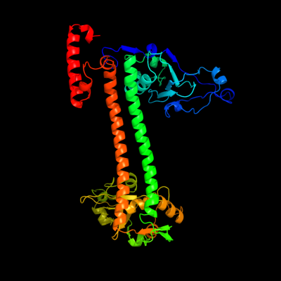
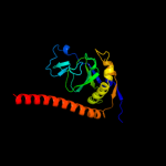
1 c2y7cA_
100.0
100
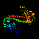
PDB header: transferaseChain: A: PDB Molecule: type-1 restriction enzyme ecoki specificity protein;PDBTitle: atomic model of the ocr-bound methylase complex from the2 type i restriction-modification enzyme ecoki (m2s1). based3 on fitting into em map 1534.
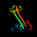
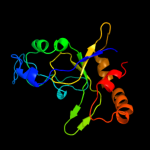
2 c1yf2A_
100.0
15
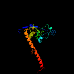
PDB header: hydrolase regulatorChain: A: PDB Molecule: type i restriction-modification enzyme, s subunit;PDBTitle: three-dimensional structure of dna sequence specificity (s) subunit of2 a type i restriction-modification enzyme and its functional3 implications
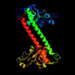
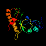
3 c3okgB_
100.0
20
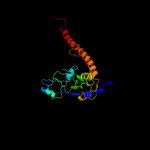
PDB header: dna binding proteinChain: B: PDB Molecule: restriction endonuclease s subunits;PDBTitle: crystal structure of hsds subunit from thermoanaerobacter2 tengcongensis
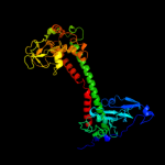
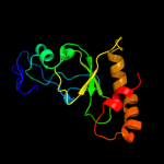
4 c1ydxA_
100.0
14
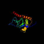
PDB header: dna binding proteinChain: A: PDB Molecule: type i restriction enzyme specificity protein mg438;PDBTitle: crystal structure of type-i restriction-modification system s subunit2 from m. genitalium
5 d1yf2a2
99.9
22
Fold: DNA methylase specificity domainSuperfamily: DNA methylase specificity domainFamily: Type I restriction modification DNA specificity domain6 d1yf2a1
99.9
21
Fold: DNA methylase specificity domainSuperfamily: DNA methylase specificity domainFamily: Type I restriction modification DNA specificity domain7 d1ydxa2
99.9
14
Fold: DNA methylase specificity domainSuperfamily: DNA methylase specificity domainFamily: Type I restriction modification DNA specificity domain8 d1ydxa1
99.8
18
Fold: DNA methylase specificity domainSuperfamily: DNA methylase specificity domainFamily: Type I restriction modification DNA specificity domain9 c1aqjB_
96.8
9
PDB header: methyltransferaseChain: B: PDB Molecule: adenine-n6-dna-methyltransferase taqi;PDBTitle: structure of adenine-n6-dna-methyltransferase taqi
10 c1g38A_
96.5
9
PDB header: transferase/dnaChain: A: PDB Molecule: modification methylase taqi;PDBTitle: adenine-specific methyltransferase m. taq i/dna complex
11 c3s1sA_
91.4
14
PDB header: hydrolase, transferaseChain: A: PDB Molecule: restriction endonuclease bpusi;PDBTitle: characterization and crystal structure of the type iig restriction2 endonuclease bpusi
12 d2ih2a2
78.3
10
Fold: DNA methylase specificity domainSuperfamily: DNA methylase specificity domainFamily: TaqI C-terminal domain-like13 c1yiuA_
61.7
38
PDB header: ligaseChain: A: PDB Molecule: itchy e3 ubiquitin protein ligase;PDBTitle: itch e3 ubiquitin ligase ww3 domain
14 c2kykA_
58.0
38
PDB header: ligaseChain: A: PDB Molecule: e3 ubiquitin-protein ligase itchy homolog;PDBTitle: the sandwich region between two lmp2a py motif regulates the2 interaction between aip4ww2domain and py motif
15 c2djyA_
51.1
46
PDB header: ligase/signaling proteinChain: A: PDB Molecule: smad ubiquitination regulatory factor 2;PDBTitle: solution structure of smurf2 ww3 domain-smad7 py peptide2 complex
16 c2jmfA_
49.5
46
PDB header: ligase/signaling proteinChain: A: PDB Molecule: e3 ubiquitin-protein ligase suppressor of deltex;PDBTitle: solution structure of the su(dx) ww4- notch py peptide2 complex
17 c2yscA_
48.8
43
PDB header: protein bindingChain: A: PDB Molecule: amyloid beta a4 precursor protein-binding familyPDBTitle: solution structure of the ww domain from the human amyloid2 beta a4 precursor protein-binding family b member 3, apbb3
18 d1tk7a2
46.7
46
Fold: WW domain-likeSuperfamily: WW domainFamily: WW domain19 c2lawA_
43.4
55
PDB header: signaling protein/transcriptionChain: A: PDB Molecule: yorkie homolog;PDBTitle: structure of the second ww domain from human yap in complex with a2 human smad1 derived peptide
20 d2jmfa1
42.4
55
Fold: WW domain-likeSuperfamily: WW domainFamily: WW domain21 d1tk7a1
not modelled
39.9
38
Fold: WW domain-likeSuperfamily: WW domainFamily: WW domain22 c2dmvA_
not modelled
39.3
31
PDB header: ligaseChain: A: PDB Molecule: itchy homolog e3 ubiquitin protein ligase;PDBTitle: solution structure of the second ww domain of itchy homolog2 e3 ubiquitin protein ligase (itch)
23 c2yshA_
not modelled
39.0
31
PDB header: protein bindingChain: A: PDB Molecule: growth-arrest-specific protein 7;PDBTitle: solution structure of the ww domain from the human growth-2 arrest-specific protein 7, gas-7
24 c2lb0A_
not modelled
38.2
40
PDB header: signaling protein/transcriptionChain: A: PDB Molecule: e3 ubiquitin-protein ligase smurf1;PDBTitle: structure of the first ww domain of human smurf1 in complex with a di-2 phosphorylated human smad1 derived peptide
25 c2lazA_
not modelled
38.2
40
PDB header: signaling protein/transcriptionChain: A: PDB Molecule: e3 ubiquitin-protein ligase smurf1;PDBTitle: structure of the first ww domain of human smurf1 in complex with a2 mono-phosphorylated human smad1 derived peptide
26 d1seda_
not modelled
36.0
20
Fold: Hypothetical protein YhaISuperfamily: Hypothetical protein YhaIFamily: Hypothetical protein YhaI27 d1gxha_
not modelled
35.9
35
Fold: Acyl carrier protein-likeSuperfamily: Colicin E immunity proteinsFamily: Colicin E immunity proteins28 d2vlqa1
not modelled
35.3
26
Fold: Acyl carrier protein-likeSuperfamily: Colicin E immunity proteinsFamily: Colicin E immunity proteins29 c1pqvS_
not modelled
35.2
20
PDB header: transferase/transcriptionChain: S: PDB Molecule: transcription elongation factor s-ii;PDBTitle: rna polymerase ii-tfiis complex
30 d1ayia_
not modelled
34.3
30
Fold: Acyl carrier protein-likeSuperfamily: Colicin E immunity proteinsFamily: Colicin E immunity proteins31 c1wmvA_
not modelled
34.1
43
PDB header: oxidoreductase, apoptosisChain: A: PDB Molecule: ww domain containing oxidoreductase;PDBTitle: solution structure of the second ww domain of wwox
32 c2ysgA_
not modelled
33.9
38
PDB header: protein bindingChain: A: PDB Molecule: syntaxin-binding protein 4;PDBTitle: solution structure of the ww domain from the human syntaxin-2 binding protein 4
33 c1wr7A_
not modelled
33.7
29
PDB header: ligaseChain: A: PDB Molecule: nedd4-2;PDBTitle: solution structure of the third ww domain of nedd4-2
34 d1pina1
not modelled
33.6
56
Fold: WW domain-likeSuperfamily: WW domainFamily: WW domain35 c2dwvB_
not modelled
32.9
29
PDB header: protein bindingChain: B: PDB Molecule: salvador homolog 1 protein;PDBTitle: solution structure of the second ww domain from mouse2 salvador homolog 1 protein (mww45)
36 c3l4hA_
not modelled
32.9
38
PDB header: protein bindingChain: A: PDB Molecule: e3 ubiquitin-protein ligase hecw1;PDBTitle: helical box domain and second ww domain of the human e3 ubiquitin-2 protein ligase hecw1
37 c2l4jA_
not modelled
32.9
70
PDB header: transcriptionChain: A: PDB Molecule: yes-associated protein 2 (yap2);PDBTitle: yap ww2
38 c1y1yS_
not modelled
32.5
20
PDB header: transferase/transcription/dna-rna hybridChain: S: PDB Molecule: transcription elongation factor s-ii;PDBTitle: rna polymerase ii-tfiis-dna/rna complex
39 c3m9hB_
not modelled
32.2
24
PDB header: chaperoneChain: B: PDB Molecule: proteasome-associated atpase;PDBTitle: crystal structure of the amino terminal coiled coil domain of the2 mycobacterium tuberculosis proteasomal atpase mpa
40 c2no8A_
not modelled
32.1
35
PDB header: immune systemChain: A: PDB Molecule: colicin-e2 immunity protein;PDBTitle: nmr structure analysis of the colicin immuntiy protein im2
41 d1i5hw_
not modelled
30.9
38
Fold: WW domain-likeSuperfamily: WW domainFamily: WW domain42 c2ysfA_
not modelled
30.7
38
PDB header: protein bindingChain: A: PDB Molecule: e3 ubiquitin-protein ligase itchy homolog;PDBTitle: solution structure of the fourth ww domain from the human2 e3 ubiquitin-protein ligase itchy homolog, itch
43 d2p0ta1
not modelled
30.7
16
Fold: PSPTO4464-likeSuperfamily: PSPTO4464-likeFamily: PSPTO4464-like44 c2p0tA_
not modelled
30.7
16
PDB header: structural genomics, unknown functionChain: A: PDB Molecule: upf0307 protein pspto_4464;PDBTitle: structural genomics, the crystal structure of a conserved putative2 protein from pseudomonas syringae pv. tomato str. dc3000
45 c2ysdA_
not modelled
30.0
50
PDB header: protein bindingChain: A: PDB Molecule: membrane-associated guanylate kinase, ww and pdzPDBTitle: solution structure of the first ww domain from the human2 membrane-associated guanylate kinase, ww and pdz domain-3 containing protein 1. magi-1
46 c3bc1F_
not modelled
29.9
17
PDB header: signaling protein/transport proteinChain: F: PDB Molecule: synaptotagmin-like protein 2;PDBTitle: crystal structure of the complex rab27a-slp2a
47 d1i8gb_
not modelled
29.2
56
Fold: WW domain-likeSuperfamily: WW domainFamily: WW domain48 c3p8cE_
not modelled
28.6
16
PDB header: protein bindingChain: E: PDB Molecule: probable protein brick1;PDBTitle: structure and control of the actin regulatory wave complex
49 d2crua1
not modelled
25.9
16
Fold: RuvA C-terminal domain-likeSuperfamily: Double-stranded DNA-binding domainFamily: Double-stranded DNA-binding domain50 c1yw5A_
not modelled
23.9
38
PDB header: isomeraseChain: A: PDB Molecule: peptidyl prolyl cis/trans isomerase;PDBTitle: peptidyl-prolyl isomerase ess1 from candida albicans
51 c2dmeA_
not modelled
23.4
23
PDB header: metal binding proteinChain: A: PDB Molecule: phd finger protein 3;PDBTitle: solution structure of the tfiis domain ii of human phd2 finger protein 3
52 c2gl2B_
not modelled
22.9
17
PDB header: cell adhesionChain: B: PDB Molecule: adhesion a;PDBTitle: crystal structure of the tetra muntant (t66g,r67g,f68g,2 y69g) of bacterial adhesin fada
53 c3le4A_
not modelled
22.3
44
PDB header: nuclear proteinChain: A: PDB Molecule: microprocessor complex subunit dgcr8;PDBTitle: crystal structure of the dgcr8 dimerization domain
54 c1tk7A_
not modelled
21.8
38
PDB header: signaling proteinChain: A: PDB Molecule: cg4244-pb;PDBTitle: nmr structure of ww domains (ww3-4) from suppressor of2 deltex
55 c1e0mA_
not modelled
21.7
44
PDB header: de novo proteinChain: A: PDB Molecule: wwprototype;PDBTitle: prototype ww domain
56 c1ymzA_
not modelled
21.4
40
PDB header: unknown functionChain: A: PDB Molecule: cc45;PDBTitle: cc45, an artificial ww domain designed using statistical2 coupling analysis
57 d2f21a1
not modelled
20.6
44
Fold: WW domain-likeSuperfamily: WW domainFamily: WW domain58 d2itka1
not modelled
20.5
63
Fold: WW domain-likeSuperfamily: WW domainFamily: WW domain59 c2ysbA_
not modelled
20.4
36
PDB header: protein bindingChain: A: PDB Molecule: salvador homolog 1 protein;PDBTitle: solution structure of the first ww domain from the mouse2 salvador homolog 1 protein (sav1)
60 d1k9ra_
not modelled
20.1
38
Fold: WW domain-likeSuperfamily: WW domainFamily: WW domain61 c2zajA_
not modelled
19.6
40
PDB header: protein bindingChain: A: PDB Molecule: membrane-associated guanylate kinase, ww and pdzPDBTitle: solution structure of the short-isoform of the second ww2 domain from the human membrane-associated guanylate kinase,3 ww and pdz domain-containing protein 1 (magi-1)
62 c1wr4A_
not modelled
18.5
44
PDB header: ligaseChain: A: PDB Molecule: ubiquitin-protein ligase nedd4-2;PDBTitle: solution structure of the second ww domain of nedd4-2
63 c2kq0A_
not modelled
18.4
44
PDB header: ligaseChain: A: PDB Molecule: e3 ubiquitin-protein ligase nedd4;PDBTitle: human nedd4 3rd ww domain complex with ebola zaire virus matrix2 protein vp40 derived peptide ilptappeymea
64 d3dlqi1
not modelled
18.0
27
Fold: 4-helical cytokinesSuperfamily: 4-helical cytokinesFamily: Interferons/interleukin-10 (IL-10)65 c2jv4A_
not modelled
18.0
38
PDB header: isomeraseChain: A: PDB Molecule: peptidyl-prolyl cis/trans isomerase;PDBTitle: structure characterisation of pina ww domain and comparison2 with other group iv ww domains, pin1 and ess1
66 c2ez5W_
not modelled
17.6
23
PDB header: signalling protein,ligaseChain: W: PDB Molecule: e3 ubiquitin-protein ligase nedd4;PDBTitle: solution structure of the dnedd4 ww3* domain- comm lpsy2 peptide complex
67 d1ig4a_
not modelled
17.2
17
Fold: DNA-binding domainSuperfamily: DNA-binding domainFamily: Methyl-CpG-binding domain, MBD68 d2ho2a1
not modelled
17.0
36
Fold: WW domain-likeSuperfamily: WW domainFamily: WW domain69 d1f8ab1
not modelled
16.8
36
Fold: WW domain-likeSuperfamily: WW domainFamily: WW domain70 c3olmA_
not modelled
15.6
63
PDB header: ligaseChain: A: PDB Molecule: e3 ubiquitin-protein ligase rsp5;PDBTitle: structure and function of a ubiquitin binding site within the2 catalytic domain of a hect ubiquitin ligase
71 d1dd5a_
not modelled
14.8
17
Fold: RRF/tRNA synthetase additional domain-likeSuperfamily: Ribosome recycling factor, RRFFamily: Ribosome recycling factor, RRF72 c2gleA_
not modelled
14.0
7
PDB header: protein bindingChain: A: PDB Molecule: neurabin-1;PDBTitle: solution structure of neurabin sam domain
73 c1bm4A_
not modelled
14.0
41
PDB header: viral proteinChain: A: PDB Molecule: protein (moloney murine leukemia virus capsid);PDBTitle: momlv capsid protein major homology region peptide analog
74 c2fh0A_
not modelled
14.0
13
PDB header: unknown functionChain: A: PDB Molecule: hypothetical 16.0 kda protein in abf2-chl12PDBTitle: nmr ensemble of the yeast saccharomyces cerevisiae protein2 ymr074cp core region
75 d1ek8a_
not modelled
13.3
20
Fold: RRF/tRNA synthetase additional domain-likeSuperfamily: Ribosome recycling factor, RRFFamily: Ribosome recycling factor, RRF76 d2ysca1
not modelled
13.1
44
Fold: WW domain-likeSuperfamily: WW domainFamily: WW domain77 c3uorB_
not modelled
12.6
18
PDB header: sugar binding proteinChain: B: PDB Molecule: abc transporter sugar binding protein;PDBTitle: the structure of the sugar-binding protein male from the phytopathogen2 xanthomonas citri
78 c3kxeD_
not modelled
12.1
13
PDB header: protein bindingChain: D: PDB Molecule: antitoxin protein pard-1;PDBTitle: a conserved mode of protein recognition and binding in a2 pard-pare toxin-antitoxin complex
79 d1wqga1
not modelled
12.0
19
Fold: RRF/tRNA synthetase additional domain-likeSuperfamily: Ribosome recycling factor, RRFFamily: Ribosome recycling factor, RRF80 c3cqxD_
not modelled
12.0
30
PDB header: chaperoneChain: D: PDB Molecule: bag family molecular chaperone regulator 2;PDBTitle: chaperone complex
81 d1nmva1
not modelled
11.6
46
Fold: WW domain-likeSuperfamily: WW domainFamily: WW domain82 d1zkea1
not modelled
11.4
12
Fold: ROP-likeSuperfamily: HP1531-likeFamily: HP1531-like83 d1ge9a_
not modelled
11.3
29
Fold: RRF/tRNA synthetase additional domain-likeSuperfamily: Ribosome recycling factor, RRFFamily: Ribosome recycling factor, RRF84 c2ky8A_
not modelled
11.3
19
PDB header: transcription/dnaChain: A: PDB Molecule: methyl-cpg-binding domain protein 2;PDBTitle: solution structure and dynamic analysis of chicken mbd2 methyl binding2 domain bound to a target methylated dna sequence
85 c2ysiA_
not modelled
11.2
23
PDB header: protein bindingChain: A: PDB Molecule: transcription elongation regulator 1;PDBTitle: solution structure of the first ww domain from the mouse2 transcription elongation regulator 1, transcription factor3 ca150
86 c2ejxA_
not modelled
10.8
36
PDB header: structural genomics, unknown functionChain: A: PDB Molecule: st0812;PDBTitle: crystal structure of the hypothetical protein st0812 from2 sulfolobus tokodaii
87 d1eh1a_
not modelled
10.8
14
Fold: RRF/tRNA synthetase additional domain-likeSuperfamily: Ribosome recycling factor, RRFFamily: Ribosome recycling factor, RRF88 c1f8aB_
not modelled
10.6
42
PDB header: isomeraseChain: B: PDB Molecule: peptidyl-prolyl cis-trans isomerase nima-PDBTitle: structural basis for the phosphoserine-proline recognition2 by group iv ww domains
89 d1is1a_
not modelled
10.4
17
Fold: RRF/tRNA synthetase additional domain-likeSuperfamily: Ribosome recycling factor, RRFFamily: Ribosome recycling factor, RRF90 d2gsva1
not modelled
10.0
21
Fold: Open three-helical up-and-down bundleSuperfamily: YvfG-likeFamily: YvfG-like91 c1lypA_
not modelled
9.4
17
PDB header: lipopolysaccharide-binding proteinChain: A: PDB Molecule: cap18;PDBTitle: the solution structure of the active domain of cap18: a2 lipopolysaccharide binding protein from rabbit leukocytes
92 d2clyb1
not modelled
9.3
12
Fold: ATP synthase D chain-likeSuperfamily: ATP synthase D chain-likeFamily: ATP synthase D chain-like93 c3npfB_
not modelled
9.2
13
PDB header: hydrolaseChain: B: PDB Molecule: putative dipeptidyl-peptidase vi;PDBTitle: crystal structure of a putative dipeptidyl-peptidase vi (bacova_00612)2 from bacteroides ovatus at 1.72 a resolution
94 d2evra2
not modelled
9.2
13
Fold: Cysteine proteinasesSuperfamily: Cysteine proteinasesFamily: NlpC/P6095 d2nrka1
not modelled
9.2
11
Fold: NucleotidyltransferaseSuperfamily: NucleotidyltransferaseFamily: GrpB-like96 c3qufB_
not modelled
9.0
17
PDB header: transport proteinChain: B: PDB Molecule: extracellular solute-binding protein, family 1;PDBTitle: the structure of a family 1 extracellular solute-binding protein from2 bifidobacterium longum subsp. infantis
97 c2oarA_
not modelled
8.9
16
PDB header: membrane proteinChain: A: PDB Molecule: large-conductance mechanosensitive channel;PDBTitle: mechanosensitive channel of large conductance (mscl)
98 c3pmdA_
not modelled
8.9
29
PDB header: lipid binding proteinChain: A: PDB Molecule: conserved domain protein;PDBTitle: crystal structure of the sporulation inhibitor pxo1-118 from bacillus2 anthracis
99 c2xzmW_
not modelled
8.7
14
PDB header: ribosomeChain: W: PDB Molecule: 40s ribosomal protein s4;PDBTitle: crystal structure of the eukaryotic 40s ribosomal2 subunit in complex with initiation factor 1. this file3 contains the 40s subunit and initiation factor for4 molecule 1