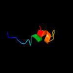
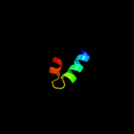
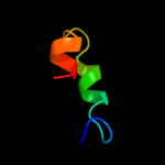
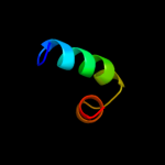
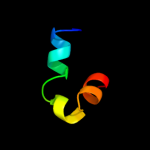
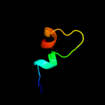
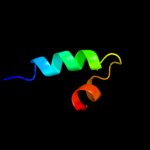
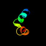
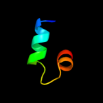
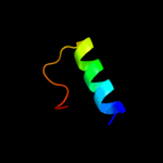
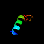
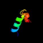
1 d1vhra_
86.9
20
Fold: (Phosphotyrosine protein) phosphatases IISuperfamily: (Phosphotyrosine protein) phosphatases IIFamily: Dual specificity phosphatase-like2 c2y96A_
86.7
20
PDB header: hydrolaseChain: A: PDB Molecule: dual specificity phosphatase dupd1;PDBTitle: structure of human dual-specificity phosphatase 27
3 c2r0bA_
84.0
28
PDB header: hydrolaseChain: A: PDB Molecule: serine/threonine/tyrosine-interacting protein;PDBTitle: crystal structure of human tyrosine phosphatase-like2 serine/threonine/tyrosine-interacting protein
4 c2esbA_
83.5
28
PDB header: hydrolaseChain: A: PDB Molecule: dual specificity protein phosphatase 18;PDBTitle: crystal structure of human dusp18
5 c1wrmA_
83.3
20
PDB header: hydrolaseChain: A: PDB Molecule: dual specificity phosphatase 22;PDBTitle: crystal structure of jsp-1
6 c2imgA_
83.0
24
PDB header: hydrolaseChain: A: PDB Molecule: dual specificity protein phosphatase 23;PDBTitle: crystal structure of dual specificity protein phosphatase2 23 from homo sapiens in complex with ligand malate ion
7 d1xria_
81.2
22
Fold: (Phosphotyrosine protein) phosphatases IISuperfamily: (Phosphotyrosine protein) phosphatases IIFamily: Dual specificity phosphatase-like8 c3emuA_
81.1
16
PDB header: hydrolaseChain: A: PDB Molecule: leucine rich repeat and phosphatase domainPDBTitle: crystal structure of a leucine rich repeat and phosphatase2 domain containing protein from entamoeba histolytica
9 c2gwoC_
80.8
20
PDB header: hydrolaseChain: C: PDB Molecule: dual specificity protein phosphatase 13;PDBTitle: crystal structure of tmdp
10 c2g6zB_
80.5
20
PDB header: hydrolaseChain: B: PDB Molecule: dual specificity protein phosphatase 5;PDBTitle: crystal structure of human dusp5
11 d1m3ga_
80.2
28
Fold: (Phosphotyrosine protein) phosphatases IISuperfamily: (Phosphotyrosine protein) phosphatases IIFamily: Dual specificity phosphatase-like12 c1zzwA_
79.9
24
PDB header: hydrolaseChain: A: PDB Molecule: dual specificity protein phosphatase 10;PDBTitle: crystal structure of catalytic domain of human map kinase2 phosphatase 5
13 c3rgqA_
79.8
28
PDB header: hydrolaseChain: A: PDB Molecule: protein-tyrosine phosphatase mitochondrial 1;PDBTitle: crystal structure of ptpmt1 in complex with pi(5)p
14 c3nmeA_
79.4
20
PDB header: hydrolaseChain: A: PDB Molecule: sex4 glucan phosphatase;PDBTitle: structure of a plant phosphatase
15 c2oudA_
78.5
24
PDB header: hydrolaseChain: A: PDB Molecule: dual specificity protein phosphatase 10;PDBTitle: crystal structure of the catalytic domain of human mkp5
16 c2nt2C_
78.2
28
PDB header: hydrolaseChain: C: PDB Molecule: protein phosphatase slingshot homolog 2;PDBTitle: crystal structure of slingshot phosphatase 2
17 c1fpzF_
77.9
24
PDB header: hydrolaseChain: F: PDB Molecule: cyclin-dependent kinase inhibitor 3;PDBTitle: crystal structure analysis of kinase associated phosphatase2 (kap) with a substitution of the catalytic site cysteine3 (cys140) to a serine
18 c1yz4A_
77.4
24
PDB header: hydrolaseChain: A: PDB Molecule: dual specificity phosphatase-like 15 isoform a;PDBTitle: crystal structure of dusp15
19 c2e0tA_
77.2
20
PDB header: hydrolaseChain: A: PDB Molecule: dual specificity phosphatase 26;PDBTitle: crystal structure of catalytic domain of dual specificity phosphatase2 26, ms0830 from homo sapiens
20 d1mkpa_
77.0
24
Fold: (Phosphotyrosine protein) phosphatases IISuperfamily: (Phosphotyrosine protein) phosphatases IIFamily: Dual specificity phosphatase-like21 c2wgpA_
not modelled
76.4
24
PDB header: hydrolaseChain: A: PDB Molecule: dual specificity protein phosphatase 14;PDBTitle: crystal structure of human dual specificity phosphatase 14
22 d1i9sa_
not modelled
75.5
36
Fold: (Phosphotyrosine protein) phosphatases IISuperfamily: (Phosphotyrosine protein) phosphatases IIFamily: Dual specificity phosphatase-like23 d1ohea2
not modelled
75.4
16
Fold: (Phosphotyrosine protein) phosphatases IISuperfamily: (Phosphotyrosine protein) phosphatases IIFamily: Dual specificity phosphatase-like24 c2hcmA_
not modelled
75.3
32
PDB header: hydrolaseChain: A: PDB Molecule: dual specificity protein phosphatase;PDBTitle: crystal structure of mouse putative dual specificity phosphatase2 complexed with zinc tungstate, new york structural genomics3 consortium
25 c1yn9B_
not modelled
74.9
20
PDB header: hydrolaseChain: B: PDB Molecule: polynucleotide 5'-phosphatase;PDBTitle: crystal structure of baculovirus rna 5'-phosphatase2 complexed with phosphate
26 c2j17A_
not modelled
72.4
24
PDB header: hydrolaseChain: A: PDB Molecule: tyrosine-protein phosphatase yil113w;PDBTitle: ptyr bound form of sdp-1
27 d1rxda_
not modelled
69.8
20
Fold: (Phosphotyrosine protein) phosphatases IISuperfamily: (Phosphotyrosine protein) phosphatases IIFamily: Dual specificity phosphatase-like28 d1v3aa_
not modelled
68.6
20
Fold: (Phosphotyrosine protein) phosphatases IISuperfamily: (Phosphotyrosine protein) phosphatases IIFamily: Dual specificity phosphatase-like29 c2c46B_
not modelled
68.3
36
PDB header: transferaseChain: B: PDB Molecule: mrna capping enzyme;PDBTitle: crystal structure of the human rna guanylyltransferase and2 5'-phosphatase
30 c2i6oA_
not modelled
67.9
24
PDB header: hydrolaseChain: A: PDB Molecule: sulfolobus solfataricus protein tyrosinePDBTitle: crystal structure of the complex of the archaeal sulfolobus2 ptp-fold phosphatase with phosphopeptides n-g-(p)y-k-n
31 d1fpza_
not modelled
65.4
28
Fold: (Phosphotyrosine protein) phosphatases IISuperfamily: (Phosphotyrosine protein) phosphatases IIFamily: Dual specificity phosphatase-like32 c3rz2B_
not modelled
65.1
20
PDB header: hydrolaseChain: B: PDB Molecule: protein tyrosine phosphatase type iva 1;PDBTitle: crystal of prl-1 complexed with peptide
33 c3s4oB_
not modelled
64.5
20
PDB header: structural genomics, unknown functionChain: B: PDB Molecule: protein tyrosine phosphatase-like protein;PDBTitle: protein tyrosine phosphatase (putative) from leishmania major
34 d1d5ra2
not modelled
46.1
32
Fold: (Phosphotyrosine protein) phosphatases IISuperfamily: (Phosphotyrosine protein) phosphatases IIFamily: Dual specificity phosphatase-like35 c2p4dA_
not modelled
45.6
32
PDB header: hydrolaseChain: A: PDB Molecule: dual specificity protein phosphatase;PDBTitle: structure-assisted discovery of variola major h12 phosphatase inhibitors
36 d1vdda_
not modelled
30.2
18
Fold: Recombination protein RecRSuperfamily: Recombination protein RecRFamily: Recombination protein RecR37 d1texa_
not modelled
24.8
36
Fold: P-loop containing nucleoside triphosphate hydrolasesSuperfamily: P-loop containing nucleoside triphosphate hydrolasesFamily: PAPS sulfotransferase38 d1kcfa1
not modelled
24.5
40
Fold: LEM/SAP HeH motifSuperfamily: SAP domainFamily: SAP domain39 c1vddC_
not modelled
20.1
17
PDB header: recombinationChain: C: PDB Molecule: recombination protein recr;PDBTitle: crystal structure of recombinational repair protein recr
40 c1oheA_
not modelled
19.5
16
PDB header: hydrolaseChain: A: PDB Molecule: cdc14b2 phosphatase;PDBTitle: structure of cdc14b phosphatase with a peptide ligand
41 c3ap3A_
not modelled
18.2
50
PDB header: transferaseChain: A: PDB Molecule: protein-tyrosine sulfotransferase 2;PDBTitle: crystal structure of human tyrosylprotein sulfotransferase-2 complexed2 with pap
42 c1b9qA_
not modelled
12.2
80
PDB header: collagen facit xivChain: A: PDB Molecule: protein (collagen alpha 1);PDBTitle: nmr structure of heparin binding site of non collagenous2 domain i (nc1) of collagen facit xiv
43 c1b9pA_
not modelled
12.2
80
PDB header: collagen facit xivChain: A: PDB Molecule: protein (collagen alpha 1);PDBTitle: nmr structure of heparin binding site of non collagenous2 domain i (nc1) of collagen facit xiv
44 c2zq5A_
not modelled
11.5
36
PDB header: transferaseChain: A: PDB Molecule: putative uncharacterized protein;PDBTitle: crystal structure of sulfotransferase stf1 from2 mycobacterium tuberculosis h37rv (type1 form)
45 c2ksdA_
not modelled
10.9
12
PDB header: transferaseChain: A: PDB Molecule: aerobic respiration control sensor protein arcb;PDBTitle: backbone structure of the membrane domain of e. coli2 histidine kinase receptor arcb, center for structures of3 membrane proteins (csmp) target 4310c
46 c3u4gA_
not modelled
10.4
50
PDB header: transferaseChain: A: PDB Molecule: namn:dmb phosphoribosyltransferase;PDBTitle: the structure of cobt from pyrococcus horikoshii
47 c2z6vA_
not modelled
9.1
36
PDB header: unknown functionChain: A: PDB Molecule: putative uncharacterized protein;PDBTitle: crystal structure of sulfotransferase stf9 from2 mycobacterium avium
48 c2jp3A_
not modelled
9.0
20
PDB header: transcriptionChain: A: PDB Molecule: fxyd domain-containing ion transport regulator 4;PDBTitle: solution structure of the human fxyd4 (chif) protein in sds2 micelles
49 c2f46A_
not modelled
7.6
13
PDB header: hydrolaseChain: A: PDB Molecule: hypothetical protein;PDBTitle: crystal structure of a putative phosphatase (nma1982) from neisseria2 meningitidis z2491 at 1.41 a resolution
50 c1vkjA_
not modelled
7.2
24
PDB header: transferaseChain: A: PDB Molecule: heparan sulfate (glucosamine) 3-o-PDBTitle: crystal structure of heparan sulfate 3-o-sulfotransferase2 isoform 1 in the presence of pap
51 d1vkja_
not modelled
7.2
24
Fold: P-loop containing nucleoside triphosphate hydrolasesSuperfamily: P-loop containing nucleoside triphosphate hydrolasesFamily: PAPS sulfotransferase52 c3awfC_
not modelled
6.8
32
PDB header: hydrolase, membrane proteinChain: C: PDB Molecule: voltage-sensor containing phosphatase;PDBTitle: crystal structure of pten-like domain of ci-vsp (236-576)
53 c3rnlA_
not modelled
6.2
21
PDB header: transferaseChain: A: PDB Molecule: sulfotransferase;PDBTitle: crystal structure of sulfotransferase from alicyclobacillus2 acidocaldarius
54 c3majA_
not modelled
6.1
22
PDB header: dna binding proteinChain: A: PDB Molecule: dna processing chain a;PDBTitle: crystal structure of putative dna processing protein dpra from2 rhodopseudomonas palustris cga009
55 d1cuka1
not modelled
5.7
18
Fold: RuvA C-terminal domain-likeSuperfamily: DNA helicase RuvA subunit, C-terminal domainFamily: DNA helicase RuvA subunit, C-terminal domain56 c2pqnB_
not modelled
5.6
25
PDB header: apoptosisChain: B: PDB Molecule: mitochondrial division protein 1;PDBTitle: crystal structure of yeast fis1 complexed with a fragment of yeast2 mdv1
57 d1jb0k_
not modelled
5.5
39
Fold: Photosystem I reaction center subunit X, PsaKSuperfamily: Photosystem I reaction center subunit X, PsaKFamily: Photosystem I reaction center subunit X, PsaK58 c1jb0K_
not modelled
5.5
39
PDB header: photosynthesisChain: K: PDB Molecule: photosystem 1 reaction centre subunit x;PDBTitle: crystal structure of photosystem i: a photosynthetic reaction center2 and core antenna system from cyanobacteria
59 d1sdia_
not modelled
5.5
22
Fold: YcfC-likeSuperfamily: YcfC-likeFamily: YcfC-like60 c3bd9A_
not modelled
5.3
24
PDB header: transferaseChain: A: PDB Molecule: heparan sulfate glucosamine 3-o-sulfotransferasePDBTitle: human 3-o-sulfotransferase isoform 5 with bound pap
61 c1q90G_
not modelled
5.2
31
PDB header: photosynthesisChain: G: PDB Molecule: cytochrome b6f complex subunit petg;PDBTitle: structure of the cytochrome b6f (plastohydroquinone : plastocyanin2 oxidoreductase) from chlamydomonas reinhardtii
62 d1q90g_
not modelled
5.2
31
Fold: Single transmembrane helixSuperfamily: PetG subunit of the cytochrome b6f complexFamily: PetG subunit of the cytochrome b6f complex


