| 1 |
|
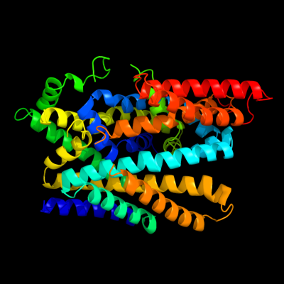
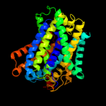
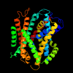
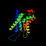
PDB 2xq2 chain A
Region: 2 - 483
Aligned: 459
Modelled: 459
Confidence: 100.0%
Identity: 18%
PDB header:transport protein
Chain: A: PDB Molecule:sodium/glucose cotransporter;
PDBTitle: structure of the k294a mutant of vsglt
Phyre2
| 2 |
|
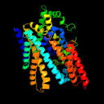
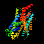
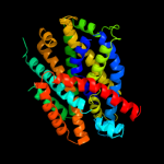
PDB 3dh4 chain A
Region: 24 - 483
Aligned: 455
Modelled: 440
Confidence: 100.0%
Identity: 16%
PDB header:transport protein
Chain: A: PDB Molecule:sodium/glucose cotransporter;
PDBTitle: crystal structure of sodium/sugar symporter with bound galactose from2 vibrio parahaemolyticus
Phyre2
| 3 |
|
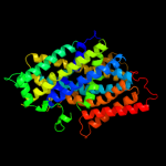
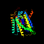
PDB 3gia chain A
Region: 40 - 474
Aligned: 419
Modelled: 419
Confidence: 99.8%
Identity: 12%
PDB header:transport protein
Chain: A: PDB Molecule:uncharacterized protein mj0609;
PDBTitle: crystal structure of apct transporter
Phyre2
| 4 |
|
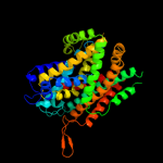
PDB 2jln chain A
Region: 26 - 482
Aligned: 435
Modelled: 435
Confidence: 99.7%
Identity: 13%
PDB header:membrane protein
Chain: A: PDB Molecule:mhp1;
PDBTitle: structure of mhp1, a nucleobase-cation-symport-1 family2 transporter
Phyre2
| 5 |
|
PDB 3lrc chain C
Region: 41 - 473
Aligned: 395
Modelled: 395
Confidence: 99.4%
Identity: 11%
PDB header:transport protein
Chain: C: PDB Molecule:arginine/agmatine antiporter;
PDBTitle: structure of e. coli adic (p1)
Phyre2
| 6 |
|
PDB 2w8a chain C
Region: 3 - 416
Aligned: 396
Modelled: 411
Confidence: 99.2%
Identity: 11%
PDB header:membrane protein
Chain: C: PDB Molecule:glycine betaine transporter betp;
PDBTitle: crystal structure of the sodium-coupled glycine betaine2 symporter betp from corynebacterium glutamicum with bound3 substrate
Phyre2
| 7 |
|
PDB 3hfx chain A
Region: 2 - 416
Aligned: 400
Modelled: 400
Confidence: 98.8%
Identity: 14%
PDB header:transport protein
Chain: A: PDB Molecule:l-carnitine/gamma-butyrobetaine antiporter;
PDBTitle: crystal structure of carnitine transporter
Phyre2
| 8 |
|
PDB 2a65 chain A domain 1
Region: 44 - 419
Aligned: 359
Modelled: 359
Confidence: 96.4%
Identity: 12%
Fold: SNF-like
Superfamily: SNF-like
Family: SNF-like
Phyre2
| 9 |
|
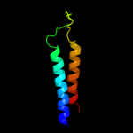
PDB 3mk7 chain F
Region: 3 - 66
Aligned: 64
Modelled: 64
Confidence: 15.3%
Identity: 8%
PDB header:oxidoreductase
Chain: F: PDB Molecule:cytochrome c oxidase, cbb3-type, subunit p;
PDBTitle: the structure of cbb3 cytochrome oxidase
Phyre2
| 10 |
|
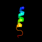
PDB 2k9y chain B
Region: 10 - 28
Aligned: 19
Modelled: 19
Confidence: 7.1%
Identity: 32%
PDB header:transferase
Chain: B: PDB Molecule:ephrin type-a receptor 2;
PDBTitle: epha2 dimeric structure in the lipidic bicelle at ph 5.0
Phyre2
| 11 |
|
PDB 2k9y chain A
Region: 10 - 28
Aligned: 19
Modelled: 19
Confidence: 7.1%
Identity: 32%
PDB header:transferase
Chain: A: PDB Molecule:ephrin type-a receptor 2;
PDBTitle: epha2 dimeric structure in the lipidic bicelle at ph 5.0
Phyre2
| 12 |
|
PDB 1eys chain H domain 2
Region: 7 - 36
Aligned: 26
Modelled: 30
Confidence: 6.8%
Identity: 23%
Fold: Single transmembrane helix
Superfamily: Photosystem II reaction centre subunit H, transmembrane region
Family: Photosystem II reaction centre subunit H, transmembrane region
Phyre2
| 13 |
|
PDB 1j4n chain A
Region: 270 - 482
Aligned: 207
Modelled: 213
Confidence: 5.8%
Identity: 12%
Fold: Aquaporin-like
Superfamily: Aquaporin-like
Family: Aquaporin-like
Phyre2