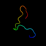
PDB header:hydrolase/hydrolase inhibitor Chain: H: PDB Molecule:fibrinogen beta chain; PDBTitle: crystal structure of the complex between thrombin and the central "e"2 region of fibrin
Confidence and coverage
Confidence:
28.2%
Coverage:
15%
13 residues ( 15% of your sequence) have been modelled with 28.2% confidence by the single highest scoring template.
You may wish to submit your sequence to Phyrealarm. This will automatically scan your sequence every week for new potential templates as they appear in the Phyre2 library.
Please note: You must be registered and logged in to use Phyrealarm.
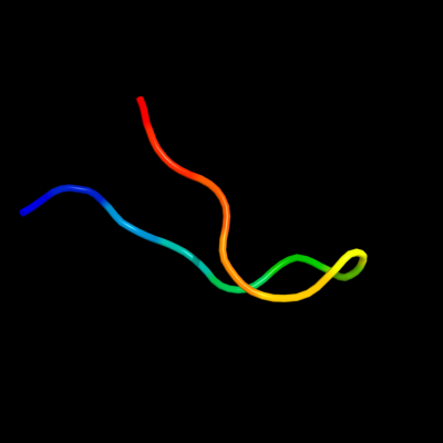
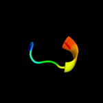
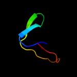
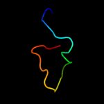
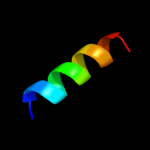
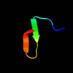
Region: 46 - 58 Aligned: 13 Modelled: 13 Confidence: 28.2% Identity: 62% PDB header:hydrolase/hydrolase inhibitor Chain: H: PDB Molecule:fibrinogen beta chain; PDBTitle: crystal structure of the complex between thrombin and the central "e"2 region of fibrin
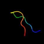
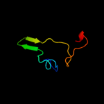
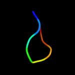
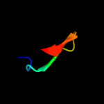
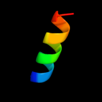
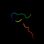
Region: 44 - 65 Aligned: 22 Modelled: 21 Confidence: 7.0% Identity: 36% PDB header:transport protein Chain: C: PDB Molecule:p2x purinoceptor; PDBTitle: crystal structure of the atp-gated p2x4 ion channel in the closed, apo2 state at 3.5 angstroms (r3)
Phyre2
21
22
23
24
25
26
Detailed template information
Binding site prediction
Due to computational demand, binding site predictions are not run for batch jobs
If you want to predict binding sites, please manually submit your model of choice to 3DLigandSite
Phyre is for academic use only
Please cite: Protein structure prediction on
the web: a case study using the Phyre server
Kelley LA and Sternberg MJE. Nature Protocols
4, 363 - 371 (2009) [pdf] [Import into BibTeX]
If you use the binding site
predictions from 3DLigandSite, please also cite:
3DLigandSite: predicting ligand-binding sites using similar structures.
Wass MN, Kelley LA and Sternberg
MJ Nucleic Acids Research 38, W469-73 (2010) [PubMed]