Phyre Home Retrieve Phyre Job Id
Emaill.a.kelley@imperial.ac.uk
DescriptionP0ACW6
DateThu Jan 5 11:19:17 GMT 2012
Unique Job ID1dc402a5b0f4f22e

Summary 

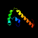
Top model
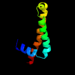
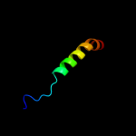
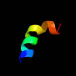
Image coloured by rainbow N → C terminus
Model (left) based on template c1zhcA_
Top template information
PDB header:unknown function
Chain: A: PDB Molecule:hypothetical protein hp1242;
PDBTitle: solution structure of hp1242 from helicobacter pylori
Confidence and coverage
Confidence:100.0% Coverage: 97%
72 residues ( 97% of your sequence) have been modelled with 100.0% confidence by the single highest scoring template.
3D viewing
Interactive 3D view in Jmol

Sequence analysis 

Secondary structure and disorder prediction 

   1........10.........20.........30.........40.........50.........60
Sequence  MFPEYRDLISRLKNENPRFMSLFDKHNKLDHEIARKEGSDGRGYNAEVVRMKKQKLQLKD
Secondary structure 


SS confidence 


Disorder  ????


????????


Disorder confidence 


   .........70....
Sequence  EMLKILQQESVKEV
Secondary structure 

SS confidence 


Disorder 


?

???
Disorder confidence 


Confidence Key
High(9)                    Low (0)
?Disordered
Alpha helix
Beta strand

Domain analysis 

Hover over an aligned region to see model and summary info

Please note, only up to the top 20 hits are modelled to reduce computer load

RankAligned region

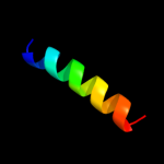
PDB 1zhc chain A

3D model

Region: 1 - 74
Aligned: 72
Modelled: 74
Confidence: 100.0%
Identity: 47%
PDB header:unknown function
Chain: A: PDB Molecule:hypothetical protein hp1242;
PDBTitle: solution structure of hp1242 from helicobacter pylori

Phyre2

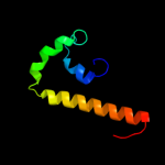
PDB 2a3d chain A

3D model

Region: 6 - 65
Aligned: 56
Modelled: 60
Confidence: 59.9%
Identity: 14%
PDB header:three-helix bundle
Chain: A: PDB Molecule:protein (de novo three-helix bundle);
PDBTitle: solution structure of a de novo designed single chain three-2 helix bundle (a3d)

Phyre2

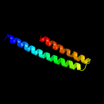
PDB 1yyb chain A

3D model

Region: 43 - 57
Aligned: 15
Modelled: 15
Confidence: 42.1%
Identity: 20%
PDB header:apoptosis
Chain: A: PDB Molecule:programmed cell death protein 5;
PDBTitle: solution structure of 1-26 fragment of human programmed2 cell death 5 protein

Phyre2

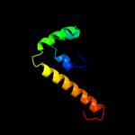
PDB 3dds chain B

3D model

Region: 12 - 74
Aligned: 63
Modelled: 63
Confidence: 42.0%
Identity: 14%
PDB header:transferase
Chain: B: PDB Molecule:glycogen phosphorylase, liver form;
PDBTitle: crystal structure of glycogen phosphorylase complexed with an2 anthranilimide based inhibitor gsk261

Phyre2

PDB 3dd7 chain D

3D model

Region: 16 - 33
Aligned: 18
Modelled: 17
Confidence: 41.7%
Identity: 33%
PDB header:ribosome inhibitor
Chain: D: PDB Molecule:prevent host death protein;
PDBTitle: structure of doch66y in complex with the c-terminal domain of phd

Phyre2

PDB 2xus chain A

3D model

Region: 46 - 64
Aligned: 19
Modelled: 19
Confidence: 38.9%
Identity: 26%
PDB header:protein binding
Chain: A: PDB Molecule:breast cancer metastasis-suppressor 1;
PDBTitle: crystal structure of the brms1 n-terminal region

Phyre2

PDB 2c4m chain A

3D model

Region: 12 - 74
Aligned: 63
Modelled: 63
Confidence: 35.9%
Identity: 13%
PDB header:transferase
Chain: A: PDB Molecule:glycogen phosphorylase;
PDBTitle: starch phosphorylase: structural studies explain oxyanion-2 dependent kinetic stability and regulatory control.

Phyre2

PDB 1hx1 chain B

3D model

Region: 3 - 66
Aligned: 64
Modelled: 64
Confidence: 35.8%
Identity: 14%
Fold: Spectrin repeat-like
Superfamily: BAG domain
Family: BAG domain

Phyre2

PDB 2ati chain A domain 1

3D model

Region: 12 - 74
Aligned: 63
Modelled: 63
Confidence: 34.0%
Identity: 14%
Fold: UDP-Glycosyltransferase/glycogen phosphorylase
Superfamily: UDP-Glycosyltransferase/glycogen phosphorylase
Family: Oligosaccharide phosphorylase

Phyre2

PDB 2gj4 chain A domain 1

3D model

Region: 12 - 74
Aligned: 63
Modelled: 63
Confidence: 30.2%
Identity: 13%
Fold: UDP-Glycosyltransferase/glycogen phosphorylase
Superfamily: UDP-Glycosyltransferase/glycogen phosphorylase
Family: Oligosaccharide phosphorylase

Phyre2

PDB 1i6z chain A

3D model

Region: 3 - 66
Aligned: 64
Modelled: 64
Confidence: 22.3%
Identity: 14%
Fold: Spectrin repeat-like
Superfamily: BAG domain
Family: BAG domain

Phyre2

PDB 1l5w chain A

3D model

Region: 12 - 74
Aligned: 63
Modelled: 63
Confidence: 20.0%
Identity: 16%
Fold: UDP-Glycosyltransferase/glycogen phosphorylase
Superfamily: UDP-Glycosyltransferase/glycogen phosphorylase
Family: Oligosaccharide phosphorylase

Phyre2

PDB 3he5 chain A

3D model

Region: 28 - 63
Aligned: 28
Modelled: 36
Confidence: 19.4%
Identity: 39%
PDB header:de novo protein
Chain: A: PDB Molecule:synzip1;
PDBTitle: heterospecific coiled-coil pair synzip2:synzip1

Phyre2

PDB 3swf chain A

3D model

Region: 18 - 48
Aligned: 31
Modelled: 31
Confidence: 17.2%
Identity: 19%
PDB header:transport protein
Chain: A: PDB Molecule:cgmp-gated cation channel alpha-1;
PDBTitle: cnga1 621-690 containing clz domain

Phyre2

PDB 3bbp chain F

3D model

Region: 12 - 39
Aligned: 28
Modelled: 28
Confidence: 14.6%
Identity: 25%
PDB header:protein transport/splicing
Chain: F: PDB Molecule:grip and coiled-coil domain-containing protein 2;
PDBTitle: rab6-gtp:gcc185 rab binding domain complex

Phyre2

PDB 3pxp chain A

3D model

Region: 3 - 27
Aligned: 24
Modelled: 25
Confidence: 14.2%
Identity: 13%
PDB header:transcription regulator
Chain: A: PDB Molecule:helix-turn-helix domain protein;
PDBTitle: crystal structure of a pas and dna binding domain containing protein2 (caur_2278) from chloroflexus aurantiacus j-10-fl at 2.30 a3 resolution

Phyre2

PDB 3swy chain B

3D model

Region: 10 - 35
Aligned: 26
Modelled: 26
Confidence: 13.0%
Identity: 23%
PDB header:transport protein
Chain: B: PDB Molecule:cyclic nucleotide-gated cation channel alpha-3;
PDBTitle: cnga3 626-672 containing clz domain

Phyre2

PDB 3dd7 chain B

3D model

Region: 17 - 33
Aligned: 17
Modelled: 16
Confidence: 12.7%
Identity: 35%
PDB header:ribosome inhibitor
Chain: B: PDB Molecule:prevent host death protein;
PDBTitle: structure of doch66y in complex with the c-terminal domain of phd

Phyre2

PDB 3nmd chain A

3D model

Region: 5 - 36
Aligned: 32
Modelled: 30
Confidence: 10.0%
Identity: 16%
PDB header:transferase
Chain: A: PDB Molecule:cgmp dependent protein kinase;
PDBTitle: crystal structure of the leucine zipper domain of cgmp dependent2 protein kinase i beta

Phyre2

PDB 1xbr chain A

3D model

Region: 7 - 25
Aligned: 19
Modelled: 19
Confidence: 9.2%
Identity: 26%
Fold: Common fold of diphtheria toxin/transcription factors/cytochrome f
Superfamily: p53-like transcription factors
Family: T-box

Phyre2
1

c1zhcA_
2

c2a3dA_
3

c1yybA_
4

c3ddsB_
5

c3dd7D_
6

c2xusA_
7

c2c4mA_
8

d1hx1b_
9

d2atia1
10

d2gj4a1
11

d1i6za_
12

d1l5wa_
13

c3he5A_
14

c3swfA_
15

c3bbpF_
16

c3pxpA_
17

c3swyB_
18

c3dd7B_
19

c3nmdA_
20

d1xbra_
21


22


23


24


25


26


27


28


29


Detailed template information 

#
Template Alignment Coverage3D Model Confidence
% i.d. Template Information
1c1zhcA_


100.0 47 PDB header:unknown function
Chain: A: PDB Molecule:hypothetical protein hp1242;
PDBTitle: solution structure of hp1242 from helicobacter pylori
2c2a3dA_


59.9 14 PDB header:three-helix bundle
Chain: A: PDB Molecule:protein (de novo three-helix bundle);
PDBTitle: solution structure of a de novo designed single chain three-2 helix bundle (a3d)
3c1yybA_


42.1 20 PDB header:apoptosis
Chain: A: PDB Molecule:programmed cell death protein 5;
PDBTitle: solution structure of 1-26 fragment of human programmed2 cell death 5 protein
4c3ddsB_


42.0 14 PDB header:transferase
Chain: B: PDB Molecule:glycogen phosphorylase, liver form;
PDBTitle: crystal structure of glycogen phosphorylase complexed with an2 anthranilimide based inhibitor gsk261
5c3dd7D_


41.7 33 PDB header:ribosome inhibitor
Chain: D: PDB Molecule:prevent host death protein;
PDBTitle: structure of doch66y in complex with the c-terminal domain of phd
6c2xusA_


38.9 26 PDB header:protein binding
Chain: A: PDB Molecule:breast cancer metastasis-suppressor 1;
PDBTitle: crystal structure of the brms1 n-terminal region
7c2c4mA_


35.9 13 PDB header:transferase
Chain: A: PDB Molecule:glycogen phosphorylase;
PDBTitle: starch phosphorylase: structural studies explain oxyanion-2 dependent kinetic stability and regulatory control.
8d1hx1b_


35.8 14 Fold:Spectrin repeat-like
Superfamily:BAG domain
Family:BAG domain
9d2atia1


34.0 14 Fold:UDP-Glycosyltransferase/glycogen phosphorylase
Superfamily:UDP-Glycosyltransferase/glycogen phosphorylase
Family:Oligosaccharide phosphorylase
10d2gj4a1


30.2 13 Fold:UDP-Glycosyltransferase/glycogen phosphorylase
Superfamily:UDP-Glycosyltransferase/glycogen phosphorylase
Family:Oligosaccharide phosphorylase
11d1i6za_


22.3 14 Fold:Spectrin repeat-like
Superfamily:BAG domain
Family:BAG domain
12d1l5wa_


20.0 16 Fold:UDP-Glycosyltransferase/glycogen phosphorylase
Superfamily:UDP-Glycosyltransferase/glycogen phosphorylase
Family:Oligosaccharide phosphorylase
13c3he5A_


19.4 39 PDB header:de novo protein
Chain: A: PDB Molecule:synzip1;
PDBTitle: heterospecific coiled-coil pair synzip2:synzip1
14c3swfA_


17.2 19 PDB header:transport protein
Chain: A: PDB Molecule:cgmp-gated cation channel alpha-1;
PDBTitle: cnga1 621-690 containing clz domain
15c3bbpF_


14.6 25 PDB header:protein transport/splicing
Chain: F: PDB Molecule:grip and coiled-coil domain-containing protein 2;
PDBTitle: rab6-gtp:gcc185 rab binding domain complex
16c3pxpA_


14.2 13 PDB header:transcription regulator
Chain: A: PDB Molecule:helix-turn-helix domain protein;
PDBTitle: crystal structure of a pas and dna binding domain containing protein2 (caur_2278) from chloroflexus aurantiacus j-10-fl at 2.30 a3 resolution
17c3swyB_


13.0 23 PDB header:transport protein
Chain: B: PDB Molecule:cyclic nucleotide-gated cation channel alpha-3;
PDBTitle: cnga3 626-672 containing clz domain
18c3dd7B_


12.7 35 PDB header:ribosome inhibitor
Chain: B: PDB Molecule:prevent host death protein;
PDBTitle: structure of doch66y in complex with the c-terminal domain of phd
19c3nmdA_


10.0 16 PDB header:transferase
Chain: A: PDB Molecule:cgmp dependent protein kinase;
PDBTitle: crystal structure of the leucine zipper domain of cgmp dependent2 protein kinase i beta
20d1xbra_


9.2 26 Fold:Common fold of diphtheria toxin/transcription factors/cytochrome f
Superfamily:p53-like transcription factors
Family:T-box
21c2w6aB_


not modelled 9.1 26 PDB header:signaling protein
Chain: B: PDB Molecule:arf gtpase-activating protein git1;
PDBTitle: x-ray structure of the dimeric git1 coiled-coil domain
22d1or7a2


not modelled 8.1 17 Fold:Sigma2 domain of RNA polymerase sigma factors
Superfamily:Sigma2 domain of RNA polymerase sigma factors
Family:Sigma2 domain of RNA polymerase sigma factors
23c2kq6A_


not modelled 6.7 19 PDB header:transport protein
Chain: A: PDB Molecule:polycystin-2;
PDBTitle: the structure of the ef-hand domain of polycystin-2 suggests a2 mechanism for ca2+-dependent regulation of polycystin-2 channel3 activity
24c3d7iB_


not modelled 5.8 13 PDB header:lyase
Chain: B: PDB Molecule:carboxymuconolactone decarboxylase family protein;
PDBTitle: crystal structure of carboxymuconolactone decarboxylase family protein2 possibly involved in oxygen detoxification (1591455) from3 methanococcus jannaschii at 1.75 a resolution
25c1ny9A_


not modelled 5.7 17 PDB header:transcription
Chain: A: PDB Molecule:transcriptional activator tipa-s;
PDBTitle: antibiotic binding domain of a tipa-class multidrug2 resistance transcriptional regulator
26d1ny9a_


not modelled 5.7 17 Fold:Antibiotic binding domain of TipA-like multidrug resistance regulators
Superfamily:Antibiotic binding domain of TipA-like multidrug resistance regulators
Family:Antibiotic binding domain of TipA-like multidrug resistance regulators
27c2f9jP_


not modelled 5.5 11 PDB header:rna binding protein
Chain: P: PDB Molecule:splicing factor 3b subunit 1;
PDBTitle: 3.0 angstrom resolution structure of a y22m mutant of the spliceosomal2 protein p14 bound to a region of sf3b155
28d1or7b2


not modelled 5.3 15 Fold:Sigma2 domain of RNA polymerase sigma factors
Superfamily:Sigma2 domain of RNA polymerase sigma factors
Family:Sigma2 domain of RNA polymerase sigma factors
29d1h6fa_


not modelled 5.2 41 Fold:Common fold of diphtheria toxin/transcription factors/cytochrome f
Superfamily:p53-like transcription factors
Family:T-box

Binding site prediction 

Due to computational demand, binding site predictions are not run for batch jobs

If you want to predict binding sites, please manually submit your model of choice to 3DLigandSite


Phyre is for academic use only

Please cite: Protein structure prediction on the web: a case study using the Phyre server
Kelley LA and Sternberg MJE. Nature Protocols 4, 363 - 371 (2009) [pdf] [Import into BibTeX]
 
If you use the binding site predictions from 3DLigandSite, please also cite:
3DLigandSite: predicting ligand-binding sites using similar structures.
Wass MN, Kelley LA and Sternberg MJ Nucleic Acids Research 38, W469-73 (2010) [PubMed]
 
© Structural Bioinformatics Group
Imperial College London
Lawrence Kelley, Benjamin Jefferys
Disclaimer
Terms and Conditions
Component software
Template detection: HHpred 1.51
Secondary structure prediction: Psi-pred 2.5
Disorder prediction: Disopred 2.4
Transmembrane prediction: Memsat_SVM
Multi-template modelling and ab initio: Poing 1.0