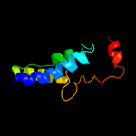
| Secondary structure and disorder prediction | |
| | |
1 | . | . | . | . | . | . | . | . | 10 | . | . | . | . | . | . | . | . | . | 20 | . | . | . | . | . | . | . | . | . | 30 | . | . | . | . | . | . | . | . | . | 40 | . | . | . | . | . | . | . | . | . | 50 | . | . | . | . | . | . | . | . | . | 60 |
| Sequence | |
M | T | A | Y | W | L | A | Q | G | V | G | V | I | A | F | L | I | G | I | T | T | F | F | N | R | D | E | R | R | F | K | K | Q | L | S | V | Y | S | A | V | I | G | V | H | F | F | L | L | G | T | Y | P | A | G | A | S | A | I | L | N |
| Secondary structure | |
|
|  | | | | | | | | | | | | | | | | | | | | | |
|
| | | | | | | | | | | | | | | | | | | | | |
|
|
| | | | | | | | | | |
| SS confidence | |
|
|
|
|
|
|
|
|
|
|
|
|
|
|
|
|
|
|
|
|
|
|
|
|
|
|
|
|
|
|
|
|
|
|
|
|
|
|
|
|
|
|
|
|
|
|
|
|
|
|
|
|
|
|
|
|
|
|
|
|
| Disorder | |
? | ? | ? |
|
|
|
|
|
|
|
|
|
|
|
|
|
|
|
|
|
|
|
|
|
|
|
|
|
|
|
|
|
|
|
|
|
|
|
|
|
|
|
|
|
|
|
|
|
|
|
|
|
|
|
|
|
|
|
|
|
| Disorder confidence | |
|
|
|
|
|
|
|
|
|
|
|
|
|
|
|
|
|
|
|
|
|
|
|
|
|
|
|
|
|
|
|
|
|
|
|
|
|
|
|
|
|
|
|
|
|
|
|
|
|
|
|
|
|
|
|
|
|
|
|
|
| |
| | |
. | . | . | . | . | . | . | . | . | 70 | . | . | . | . | . | . | . | . | . | 80 | . | . | . | . | . | . | . | . | . | 90 | . | . | . | . | . | . | . | . | . | 100 | . | . | . | . | . | . | . | . | . | 110 | . | . | . | . | . | . | . | . | . | 120 |
| Sequence | |
A | I | R | T | L | I | T | L | R | T | R | S | L | W | V | M | A | I | F | I | V | L | T | G | G | I | G | L | A | K | F | H | H | P | V | E | L | L | P | V | I | G | T | I | V | S | T | W | A | L | F | C | C | K | G | L | T | M | R | C |
| Secondary structure | |
| | | | | | | | | |
|
| | | | | | | | | | | | | | | | | | | | | | | | | | | | | | | | | | | | | | | | | |
|
| | | | | |
| SS confidence | |
|
|
|
|
|
|
|
|
|
|
|
|
|
|
|
|
|
|
|
|
|
|
|
|
|
|
|
|
|
|
|
|
|
|
|
|
|
|
|
|
|
|
|
|
|
|
|
|
|
|
|
|
|
|
|
|
|
|
|
|
| Disorder | |
|
|
|
|
|
|
|
|
|
|
|
|
|
|
|
|
|
|
|
|
|
|
|
|
|
|
|
|
|
|
|
|
|
|
|
|
|
|
|
|
|
|
|
|
|
|
|
|
|
|
|
| ? |
|
|
|
|
|
|
|
| Disorder confidence | |
|
|
|
|
|
|
|
|
|
|
|
|
|
|
|
|
|
|
|
|
|
|
|
|
|
|
|
|
|
|
|
|
|
|
|
|
|
|
|
|
|
|
|
|
|
|
|
|
|
|
|
|
|
|
|
|
|
|
|
|
| |
| | |
. | . | . | . | . | . | . | . | . | 130 | . | . | . | . | . | . | . | . | . | 140 | . | . | . | . | . | . | . | . | . | 150 | . | . | . | . | . | . | . | . | . | 160 | . | . | . | . | . | . | . | . | . | 170 | . | . | . | . | . | . | . | . | . | 180 |
| Sequence | |
V | M | W | F | S | T | C | C | W | V | I | H | N | F | W | A | G | S | I | G | G | T | M | I | E | G | S | F | L | L | M | N | G | L | N | I | I | R | F | W | R | M | Q | K | R | G | I | D | P | F | K | V | E | K | T | P | S | A | V | D |
| Secondary structure | |
| | | | | | | | | | | | | | | |
|
| | | | | | | | | | | | | | | | | | | | | | | | | | | | |
|
|
|
|
|
|
|
|
|
|
| | | |
| SS confidence | |
|
|
|
|
|
|
|
|
|
|
|
|
|
|
|
|
|
|
|
|
|
|
|
|
|
|
|
|
|
|
|
|
|
|
|
|
|
|
|
|
|
|
|
|
|
|
|
|
|
|
|
|
|
|
|
|
|
|
|
|
| Disorder | |
|
|
|
|
|
|
|
|
|
|
|
|
|
|
|
|
|
|
|
|
|
|
|
|
|
|
|
|
|
|
|
|
|
|
|
|
|
|
|
|
|
|
|
| ? | ? | ? | ? | ? | ? | ? | ? | ? | ? | ? | ? | ? | ? | ? | ? |
| Disorder confidence | |
|
|
|
|
|
|
|
|
|
|
|
|
|
|
|
|
|
|
|
|
|
|
|
|
|
|
|
|
|
|
|
|
|
|
|
|
|
|
|
|
|
|
|
|
|
|
|
|
|
|
|
|
|
|
|
|
|
|
|
|
| |
| | |
. | . | . |
| Sequence | |
E | R | G |
| Secondary structure | |
|
|
|
| SS confidence | |
|
|
|
| Disorder | |
? | ? | ? |
| Disorder confidence | |
|
|
|
| |
| Confidence Key |
| High(9) | |
|
|
|
|
|
|
|
|
|
Low (0) |
| ? | Disordered |
| Alpha helix |
  | Beta strand |
Hover over an aligned region to see model and summary info
Please note, only up to the top 20 hits are modelled to reduce computer load
|
| 1 |
|
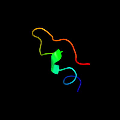
PDB 2dch chain X
Region: 158 - 178
Aligned: 21
Modelled: 21
Confidence: 14.0%
Identity: 38%
PDB header:hydrolase
Chain: X: PDB Molecule:putative homing endonuclease;
PDBTitle: crystal structure of archaeal intron-encoded homing endonuclease i-2 tsp061i
Phyre2
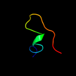
| 2 |
|
PDB 1sfu chain A
Region: 159 - 177
Aligned: 19
Modelled: 19
Confidence: 10.0%
Identity: 16%
Fold: DNA/RNA-binding 3-helical bundle
Superfamily: "Winged helix" DNA-binding domain
Family: Z-DNA binding domain
Phyre2
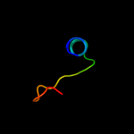
| 3 |
|
PDB 3mku chain A
Region: 94 - 175
Aligned: 76
Modelled: 82
Confidence: 7.7%
Identity: 21%
PDB header:transport protein
Chain: A: PDB Molecule:multi antimicrobial extrusion protein (na(+)/drug
PDBTitle: structure of a cation-bound multidrug and toxin compound extrusion2 (mate) transporter
Phyre2
| 4 |
|
PDB 3lj4 chain I
Region: 172 - 183
Aligned: 12
Modelled: 12
Confidence: 7.1%
Identity: 33%
PDB header:viral protein
Chain: I: PDB Molecule:portal protein;
PDBTitle: bacteriophage p22 portal protein bound to middle tail factor gp4. this2 file contain the first biological assembly
Phyre2
| 5 |
|
PDB 1xrd chain A domain 1
Region: 25 - 49
Aligned: 25
Modelled: 25
Confidence: 6.1%
Identity: 16%
Fold: Light-harvesting complex subunits
Superfamily: Light-harvesting complex subunits
Family: Light-harvesting complex subunits
Phyre2
| 6 |
|
PDB 1xmk chain A domain 1
Region: 159 - 176
Aligned: 18
Modelled: 18
Confidence: 5.4%
Identity: 17%
Fold: DNA/RNA-binding 3-helical bundle
Superfamily: "Winged helix" DNA-binding domain
Family: Z-DNA binding domain
Phyre2
| 7 |
|
PDB 1v54 chain F
Region: 162 - 182
Aligned: 21
Modelled: 21
Confidence: 5.3%
Identity: 33%
Fold: Rubredoxin-like
Superfamily: Rubredoxin-like
Family: Cytochrome c oxidase Subunit F
Phyre2
| 8 |
|
PDB 3be3 chain A
Region: 160 - 181
Aligned: 22
Modelled: 22
Confidence: 5.3%
Identity: 18%
PDB header:structural genomics, unknown function
Chain: A: PDB Molecule:uncharacterized protein;
PDBTitle: crystal structure of a protein belonging to pfam duf16532 from bordetella bronchiseptica
Phyre2
|
| Detailed template information | |
Due to computational demand, binding site predictions are not run for batch jobs
If you want to predict binding sites, please manually submit your model of choice to 3DLigandSite
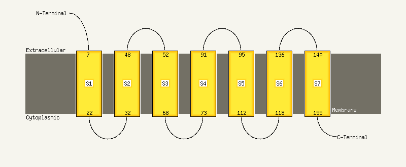
| Transmembrane helix prediction | |
Transmembrane helices have been predicted in your sequence to adopt the topology shown below
Phyre is for academic use only
| Please cite: Protein structure prediction on
the web: a case study using the Phyre server |
| Kelley LA and Sternberg MJE. Nature Protocols
4, 363 - 371 (2009) [pdf] [Import into BibTeX] |
| |
| If you use the binding site
predictions from 3DLigandSite, please also cite: |
| 3DLigandSite: predicting ligand-binding sites using similar structures. |
| Wass MN, Kelley LA and Sternberg
MJ Nucleic Acids Research 38, W469-73 (2010) [PubMed] |
| |
|
|
|
|