Phyre Home Retrieve Phyre Job Id
Emaill.a.kelley@imperial.ac.uk
DescriptionP0AB49
DateThu Jan 5 11:14:38 GMT 2012
Unique Job ID1d1f16c28ca30004

Summary 

Top model
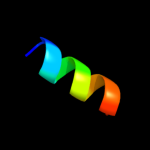
Image coloured by rainbow N → C terminus
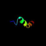
Model (left) based on template c3hefB_
Top template information
PDB header:viral protein
Chain: B: PDB Molecule:gene 1 protein;
PDBTitle: crystal structure of the bacteriophage sf6 terminase small2 subunit
Confidence and coverage
Confidence: 39.5% Coverage: 29%
27 residues ( 29% of your sequence) have been modelled with 39.5% confidence by the single highest scoring template.
You may wish to submit your sequence to Phyrealarm. This will automatically scan your sequence every week for new potential templates as they appear in the Phyre2 library.
Please note: You must be registered and logged in to use Phyrealarm.
3D viewing
Interactive 3D view in Jmol

Sequence analysis 

Secondary structure and disorder prediction 

   1........10.........20.........30.........40.........50.........60
Sequence  MKRKNASLLGNVLMGLGLVVMVVGVGYSILNQLPQFNMPQYFAHGAVLSIFVGAILWLAG
Secondary structure 


SS confidence 


Disorder  ???????


?


Disorder confidence 


   .........70.........80.........90..
Sequence  ARVGGHEQVCDRYWWVRHYDKRCRRSDNRRHS
Secondary structure 


SS confidence 


Disorder 
?
????


??????????
Disorder confidence 


Confidence Key
High(9)                    Low (0)
?Disordered
Alpha helix
Beta strand

Domain analysis 

Hover over an aligned region to see model and summary info

Please note, only up to the top 20 hits are modelled to reduce computer load

RankAligned region

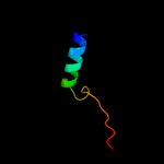
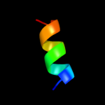
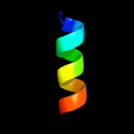
PDB 3hef chain B

3D model

Region: 66 - 92
Aligned: 27
Modelled: 27
Confidence: 39.5%
Identity: 11%
PDB header:viral protein
Chain: B: PDB Molecule:gene 1 protein;
PDBTitle: crystal structure of the bacteriophage sf6 terminase small2 subunit

Phyre2

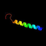
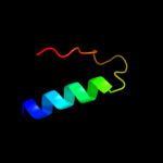
PDB 1v54 chain J

3D model

Region: 8 - 39
Aligned: 31
Modelled: 32
Confidence: 25.2%
Identity: 23%
Fold: Single transmembrane helix
Superfamily: Mitochondrial cytochrome c oxidase subunit VIIa
Family: Mitochondrial cytochrome c oxidase subunit VIIa

Phyre2

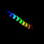
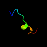
PDB 2y69 chain W

3D model

Region: 8 - 40
Aligned: 32
Modelled: 33
Confidence: 21.7%
Identity: 22%
PDB header:electron transport
Chain: W: PDB Molecule:cytochrome c oxidase polypeptide 7a1;
PDBTitle: bovine heart cytochrome c oxidase re-refined with molecular2 oxygen

Phyre2

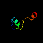
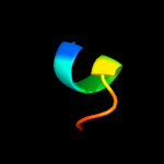
PDB 1ulv chain A domain 1

3D model

Region: 39 - 74
Aligned: 33
Modelled: 36
Confidence: 17.4%
Identity: 30%
Fold: alpha/alpha toroid
Superfamily: Six-hairpin glycosidases
Family: Bacterial glucoamylase C-terminal domain-like

Phyre2

PDB 2qts chain A

3D model

Region: 32 - 54
Aligned: 23
Modelled: 23
Confidence: 16.6%
Identity: 22%
PDB header:membrane protein
Chain: A: PDB Molecule:acid-sensing ion channel;
PDBTitle: structure of an acid-sensing ion channel 1 at 1.9 a resolution and low2 ph

Phyre2

PDB 1ijd chain A

3D model

Region: 34 - 56
Aligned: 23
Modelled: 23
Confidence: 15.2%
Identity: 26%
Fold: Light-harvesting complex subunits
Superfamily: Light-harvesting complex subunits
Family: Light-harvesting complex subunits

Phyre2

PDB 1nkz chain A

3D model

Region: 34 - 56
Aligned: 23
Modelled: 23
Confidence: 12.8%
Identity: 22%
Fold: Light-harvesting complex subunits
Superfamily: Light-harvesting complex subunits
Family: Light-harvesting complex subunits

Phyre2

PDB 1h2s chain B

3D model

Region: 50 - 61
Aligned: 12
Modelled: 12
Confidence: 12.6%
Identity: 67%
Fold: Transmembrane helix hairpin
Superfamily: Htr2 transmembrane domain-like
Family: Htr2 transmembrane domain-like

Phyre2

PDB 1h2s chain B

3D model

Region: 50 - 61
Aligned: 12
Modelled: 12
Confidence: 12.6%
Identity: 67%
PDB header:membrane protein
Chain: B: PDB Molecule:sensory rhodopsin ii transducer;
PDBTitle: molecular basis of transmenbrane signalling by sensory2 rhodopsin ii-transducer complex

Phyre2

PDB 1s21 chain A

3D model

Region: 61 - 77
Aligned: 17
Modelled: 17
Confidence: 11.9%
Identity: 18%
Fold: ADP-ribosylation
Superfamily: ADP-ribosylation
Family: AvrPphF ORF2, a type III effector

Phyre2

PDB 1s21 chain A

3D model

Region: 61 - 77
Aligned: 17
Modelled: 17
Confidence: 11.9%
Identity: 18%
PDB header:chaperone
Chain: A: PDB Molecule:orf2;
PDBTitle: crystal structure of avrpphf orf2, a type iii effector from2 p. syringae

Phyre2

PDB 2j5d chain A

3D model

Region: 33 - 53
Aligned: 19
Modelled: 21
Confidence: 11.5%
Identity: 37%
PDB header:membrane protein
Chain: A: PDB Molecule:bcl2/adenovirus e1b 19 kda protein-interacting
PDBTitle: nmr structure of bnip3 transmembrane domain in lipid2 bicelles

Phyre2

PDB 2f95 chain B

3D model

Region: 50 - 61
Aligned: 12
Modelled: 12
Confidence: 11.4%
Identity: 67%
PDB header:membrane protein
Chain: B: PDB Molecule:sensory rhodopsin ii transducer;
PDBTitle: m intermediate structure of sensory rhodopsin ii/transducer complex in2 combination with the ground state structure

Phyre2

PDB 3ij4 chain A

3D model

Region: 32 - 54
Aligned: 23
Modelled: 23
Confidence: 10.7%
Identity: 22%
PDB header:transport protein
Chain: A: PDB Molecule:amiloride-sensitive cation channel 2, neuronal;
PDBTitle: cesium sites in the crystal structure of a functional acid2 sensing ion channel in the desensitized state

Phyre2

PDB 1q90 chain L

3D model

Region: 41 - 60
Aligned: 20
Modelled: 20
Confidence: 10.4%
Identity: 15%
PDB header:photosynthesis
Chain: L: PDB Molecule:cytochrome b6f complex subunit petl;
PDBTitle: structure of the cytochrome b6f (plastohydroquinone : plastocyanin2 oxidoreductase) from chlamydomonas reinhardtii

Phyre2

PDB 1q90 chain L

3D model

Region: 41 - 60
Aligned: 20
Modelled: 20
Confidence: 10.4%
Identity: 15%
Fold: Single transmembrane helix
Superfamily: PetL subunit of the cytochrome b6f complex
Family: PetL subunit of the cytochrome b6f complex

Phyre2

PDB 3dl8 chain D

3D model

Region: 47 - 59
Aligned: 13
Modelled: 13
Confidence: 8.9%
Identity: 31%
PDB header:protein transport
Chain: D: PDB Molecule:sece;
PDBTitle: structure of the complex of aquifex aeolicus secyeg and2 bacillus subtilis seca

Phyre2

PDB 3eps chain B

3D model

Region: 65 - 92
Aligned: 28
Modelled: 28
Confidence: 8.9%
Identity: 21%
PDB header:transferase, hydrolase
Chain: B: PDB Molecule:isocitrate dehydrogenase kinase/phosphatase;
PDBTitle: the crystal structure of isocitrate dehydrogenase kinase/phosphatase2 from e. coli

Phyre2

PDB 1t3t chain A domain 5

3D model

Region: 66 - 81
Aligned: 16
Modelled: 16
Confidence: 7.5%
Identity: 19%
Fold: Bacillus chorismate mutase-like
Superfamily: PurM N-terminal domain-like
Family: PurM N-terminal domain-like

Phyre2

PDB 1cf7 chain B

3D model

Region: 11 - 19
Aligned: 9
Modelled: 9
Confidence: 6.6%
Identity: 44%
Fold: DNA/RNA-binding 3-helical bundle
Superfamily: "Winged helix" DNA-binding domain
Family: Cell cycle transcription factor e2f-dp

Phyre2
1

c3hefB_
2

d1v54j_
3

c2y69W_
4

d1ulva1
5

c2qtsA_
6

d1ijda_
7

d1nkza_
8

d1h2sb_
9

c1h2sB_
10

d1s21a_
11

c1s21A_
12

c2j5dA_
13

c2f95B_
14

c3ij4A_
15

c1q90L_
16

d1q90l_
17

c3dl8D_
18

c3epsB_
19

d1t3ta5
20

d1cf7b_
21


22


23


24


Detailed template information 

#
Template Alignment Coverage3D Model Confidence
% i.d. Template Information
1c3hefB_


39.5 11 PDB header:viral protein
Chain: B: PDB Molecule:gene 1 protein;
PDBTitle: crystal structure of the bacteriophage sf6 terminase small2 subunit
2d1v54j_


25.2 23 Fold:Single transmembrane helix
Superfamily:Mitochondrial cytochrome c oxidase subunit VIIa
Family:Mitochondrial cytochrome c oxidase subunit VIIa
3c2y69W_


21.7 22 PDB header:electron transport
Chain: W: PDB Molecule:cytochrome c oxidase polypeptide 7a1;
PDBTitle: bovine heart cytochrome c oxidase re-refined with molecular2 oxygen
4d1ulva1


17.4 30 Fold:alpha/alpha toroid
Superfamily:Six-hairpin glycosidases
Family:Bacterial glucoamylase C-terminal domain-like
5c2qtsA_


16.6 22 PDB header:membrane protein
Chain: A: PDB Molecule:acid-sensing ion channel;
PDBTitle: structure of an acid-sensing ion channel 1 at 1.9 a resolution and low2 ph
6d1ijda_


15.2 26 Fold:Light-harvesting complex subunits
Superfamily:Light-harvesting complex subunits
Family:Light-harvesting complex subunits
7d1nkza_


12.8 22 Fold:Light-harvesting complex subunits
Superfamily:Light-harvesting complex subunits
Family:Light-harvesting complex subunits
8d1h2sb_


12.6 67 Fold:Transmembrane helix hairpin
Superfamily:Htr2 transmembrane domain-like
Family:Htr2 transmembrane domain-like
9c1h2sB_


12.6 67 PDB header:membrane protein
Chain: B: PDB Molecule:sensory rhodopsin ii transducer;
PDBTitle: molecular basis of transmenbrane signalling by sensory2 rhodopsin ii-transducer complex
10d1s21a_


11.9 18 Fold:ADP-ribosylation
Superfamily:ADP-ribosylation
Family:AvrPphF ORF2, a type III effector
11c1s21A_


11.9 18 PDB header:chaperone
Chain: A: PDB Molecule:orf2;
PDBTitle: crystal structure of avrpphf orf2, a type iii effector from2 p. syringae
12c2j5dA_


11.5 37 PDB header:membrane protein
Chain: A: PDB Molecule:bcl2/adenovirus e1b 19 kda protein-interacting
PDBTitle: nmr structure of bnip3 transmembrane domain in lipid2 bicelles
13c2f95B_


11.4 67 PDB header:membrane protein
Chain: B: PDB Molecule:sensory rhodopsin ii transducer;
PDBTitle: m intermediate structure of sensory rhodopsin ii/transducer complex in2 combination with the ground state structure
14c3ij4A_


10.7 22 PDB header:transport protein
Chain: A: PDB Molecule:amiloride-sensitive cation channel 2, neuronal;
PDBTitle: cesium sites in the crystal structure of a functional acid2 sensing ion channel in the desensitized state
15c1q90L_


10.4 15 PDB header:photosynthesis
Chain: L: PDB Molecule:cytochrome b6f complex subunit petl;
PDBTitle: structure of the cytochrome b6f (plastohydroquinone : plastocyanin2 oxidoreductase) from chlamydomonas reinhardtii
16d1q90l_


10.4 15 Fold:Single transmembrane helix
Superfamily:PetL subunit of the cytochrome b6f complex
Family:PetL subunit of the cytochrome b6f complex
17c3dl8D_


8.9 31 PDB header:protein transport
Chain: D: PDB Molecule:sece;
PDBTitle: structure of the complex of aquifex aeolicus secyeg and2 bacillus subtilis seca
18c3epsB_


8.9 21 PDB header:transferase, hydrolase
Chain: B: PDB Molecule:isocitrate dehydrogenase kinase/phosphatase;
PDBTitle: the crystal structure of isocitrate dehydrogenase kinase/phosphatase2 from e. coli
19d1t3ta5


7.5 19 Fold:Bacillus chorismate mutase-like
Superfamily:PurM N-terminal domain-like
Family:PurM N-terminal domain-like
20d1cf7b_


6.6 44 Fold:DNA/RNA-binding 3-helical bundle
Superfamily:"Winged helix" DNA-binding domain
Family:Cell cycle transcription factor e2f-dp
21c2jh9A_


not modelled 6.3 44 PDB header:viral protein
Chain: A: PDB Molecule:vp4 core protein;
PDBTitle: the structure of bluetongue virus vp4 reveals a2 multifunctional rna-capping production-line
22c2ve7A_


not modelled 5.9 23 PDB header:cell cycle
Chain: A: PDB Molecule:kinetochore protein hec1, kinetochore protein spc25;
PDBTitle: crystal structure of a bonsai version of the human ndc802 complex
23c1spfA_


not modelled 5.8 50 PDB header:lipoprotein(surface film)
Chain: A: PDB Molecule:pulmonary surfactant-associated polypeptide c;
PDBTitle: the nmr structure of the pulmonary surfactant-associated2 polypeptide sp-c in an apolar solvent contains a valyl-3 rich alpha-helix
24c2v0xB_


not modelled 5.7 29 PDB header:cell cycle
Chain: B: PDB Molecule:lamina-associated polypeptide 2 isoforms
PDBTitle: the dimerization domain of lap2alpha

Binding site prediction 

Due to computational demand, binding site predictions are not run for batch jobs

If you want to predict binding sites, please manually submit your model of choice to 3DLigandSite


Phyre is for academic use only

Please cite: Protein structure prediction on the web: a case study using the Phyre server
Kelley LA and Sternberg MJE. Nature Protocols 4, 363 - 371 (2009) [pdf] [Import into BibTeX]
 
If you use the binding site predictions from 3DLigandSite, please also cite:
3DLigandSite: predicting ligand-binding sites using similar structures.
Wass MN, Kelley LA and Sternberg MJ Nucleic Acids Research 38, W469-73 (2010) [PubMed]
 
© Structural Bioinformatics Group
Imperial College London
Lawrence Kelley, Benjamin Jefferys
Disclaimer
Terms and Conditions
Component software
Template detection: HHpred 1.51
Secondary structure prediction: Psi-pred 2.5
Disorder prediction: Disopred 2.4
Transmembrane prediction: Memsat_SVM
Multi-template modelling and ab initio: Poing 1.0