1 d3d85d1
45.0
50
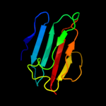
Fold: Immunoglobulin-like beta-sandwichSuperfamily: ImmunoglobulinFamily: I set domains2 c2voyG_
34.7
42
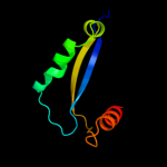
PDB header: hydrolaseChain: G: PDB Molecule: sarcoplasmic/endoplasmic reticulum calciumPDBTitle: cryoem model of copa, the copper transporting atpase from2 archaeoglobus fulgidus
3 d1r46a1
32.3
18
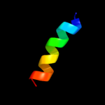
Fold: Glycosyl hydrolase domainSuperfamily: Glycosyl hydrolase domainFamily: alpha-Amylases, C-terminal beta-sheet domain4 c3p24C_
25.6
19
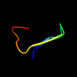
PDB header: hydrolaseChain: C: PDB Molecule: bft-3;PDBTitle: structure of profragilysin-3 from bacteroides fragilis
5 c2kncB_
22.8
35
PDB header: cell adhesionChain: B: PDB Molecule: integrin beta-3;PDBTitle: platelet integrin alfaiib-beta3 transmembrane-cytoplasmic2 heterocomplex
6 c2kvgA_
20.5
35
PDB header: transcriptionChain: A: PDB Molecule: zinc finger and btb domain-containing protein 32;PDBTitle: structure of the three-cys2his2 domain of mouse testis zinc2 finger protein
7 c2jgsA_
20.3
43
PDB header: biotin-binding proteinChain: A: PDB Molecule: circular permutant of avidin;PDBTitle: circular permutant of avidin
8 c3fkhB_
18.8
35
PDB header: oxidoreductaseChain: B: PDB Molecule: putative pyridoxamine 5'-phosphate oxidase;PDBTitle: crystal structure of putative pyridoxamine 5'-phosphate oxidase2 (np_601736.1) from corynebacterium glutamicum atcc 13032 kitasato at3 2.51 a resolution
9 d1htwa_
17.4
30
Fold: P-loop containing nucleoside triphosphate hydrolasesSuperfamily: P-loop containing nucleoside triphosphate hydrolasesFamily: YjeE-like10 c3idwA_
15.3
46
PDB header: endocytosisChain: A: PDB Molecule: actin cytoskeleton-regulatory complex protein sla1;PDBTitle: crystal structure of sla1 homology domain 2
11 c2g9tT_
13.8
30
PDB header: viral proteinChain: T: PDB Molecule: PDBTitle: crystal structure of the sars coronavirus nsp10 at 2.1a
12 c2a45L_
13.3
30
PDB header: hydrolase/hydrolase inhibitorChain: L: PDB Molecule: fibrinogen gamma chain;PDBTitle: crystal structure of the complex between thrombin and the central "e"2 region of fibrin
13 d2okqa1
11.6
25
Fold: Ferredoxin-likeSuperfamily: Dimeric alpha+beta barrelFamily: YbaA-like14 c3nicA_
11.5
28
PDB header: hydrolase/dnaChain: A: PDB Molecule: eco29kir;PDBTitle: dna binding and cleavage by the giy-yig endonuclease r.eco29ki2 inactive variant y49f
15 d2ea9a1
11.1
47
Fold: Profilin-likeSuperfamily: YeeU-likeFamily: YagB/YeeU/YfjZ-like16 d1lwub1
10.3
17
Fold: Fibrinogen C-terminal domain-likeSuperfamily: Fibrinogen C-terminal domain-likeFamily: Fibrinogen C-terminal domain-like17 c2okqB_
10.0
29
PDB header: structural genomics, unknown functionChain: B: PDB Molecule: hypothetical protein ybaa;PDBTitle: crystal structure of unknown conserved ybaa protein from2 shigella flexneri
18 c2ph7B_
9.6
36
PDB header: structural genomics, unknown functionChain: B: PDB Molecule: uncharacterized protein af_2093;PDBTitle: crystal structure of af2093 from archaeoglobus fulgidus
19 c1ei3E_
9.2
12
PDB header: PDB COMPND: 20 c2gbxF_
9.2
40
PDB header: oxidoreductaseChain: F: PDB Molecule: biphenyl 2,3-dioxygenase beta subunit;PDBTitle: crystal structure of biphenyl 2,3-dioxygenase from sphingomonas2 yanoikuyae b1 bound to biphenyl
21 c3izxE_
not modelled
9.2
11
PDB header: virusChain: E: PDB Molecule: viral structural protein 5;PDBTitle: 3.1 angstrom cryoem structure of cytoplasmic polyhedrosis virus
22 d1u5tb1
not modelled
8.7
38
Fold: DNA/RNA-binding 3-helical bundleSuperfamily: "Winged helix" DNA-binding domainFamily: Vacuolar sorting protein domain23 d2h28a1
not modelled
8.1
35
Fold: Profilin-likeSuperfamily: YeeU-likeFamily: YagB/YeeU/YfjZ-like24 c1tolA_
not modelled
8.1
18
PDB header: viral proteinChain: A: PDB Molecule: protein (fusion protein consisting of minor coatPDBTitle: fusion of n-terminal domain of the minor coat protein from2 gene iii in phage m13, and c-terminal domain of e. coli3 protein-tola
25 c3qa8A_
not modelled
8.0
25
PDB header: immune system, signaling proteinChain: A: PDB Molecule: mgc80376 protein;PDBTitle: crystal structure of inhibitor of kappa b kinase beta
26 c1jdmA_
not modelled
8.0
44
PDB header: membrane proteinChain: A: PDB Molecule: sarcolipin;PDBTitle: nmr structure of sarcolipin
27 c2vbeA_
not modelled
7.8
71
PDB header: viral proteinChain: A: PDB Molecule: tailspike-protein;PDBTitle: tailspike protein of bacteriophage sf6
28 c1deqO_
not modelled
7.6
20
PDB header: PDB COMPND: 29 d1f60a2
not modelled
7.6
22
Fold: Elongation factor/aminomethyltransferase common domainSuperfamily: EF-Tu/eEF-1alpha/eIF2-gamma C-terminal domainFamily: EF-Tu/eEF-1alpha/eIF2-gamma C-terminal domain30 d2inwa1
not modelled
7.5
35
Fold: Profilin-likeSuperfamily: YeeU-likeFamily: YagB/YeeU/YfjZ-like31 c2kbcA_
not modelled
7.4
63
PDB header: hormoneChain: A: PDB Molecule: insl5_a-chain;PDBTitle: solution structure of human insulin-like peptide 5 (insl5)
32 c2k1vA_
not modelled
7.4
63
PDB header: hormoneChain: A: PDB Molecule: insulin-like peptide insl5;PDBTitle: r3/i5 relaxin chimera
33 c2jmbA_
not modelled
7.3
50
PDB header: structural genomics, unknown functionChain: A: PDB Molecule: hypothetical protein atu4866;PDBTitle: solution structure of the protein atu4866 from agrobacterium2 tumefaciens
34 c2kzvA_
not modelled
7.1
28
PDB header: structural genomics, unknown functionChain: A: PDB Molecule: uncharacterized protein;PDBTitle: solution nmr structure of cv_0373(175-257) protein from2 chromobacterium violaceum, northeast structural genomics consortium3 target cvr118a
35 c3rykB_
not modelled
6.8
30
PDB header: isomeraseChain: B: PDB Molecule: dtdp-4-dehydrorhamnose 3,5-epimerase;PDBTitle: 1.63 angstrom resolution crystal structure of dtdp-4-dehydrorhamnose2 3,5-epimerase (rfbc) from bacillus anthracis str. ames with tdp and3 ppi bound
36 c2wryA_
not modelled
6.6
14
PDB header: immune systemChain: A: PDB Molecule: interleukin-1beta;PDBTitle: crystal structure of chicken cytokine interleukin 1 beta
37 d1zunb2
not modelled
6.4
28
Fold: Elongation factor/aminomethyltransferase common domainSuperfamily: EF-Tu/eEF-1alpha/eIF2-gamma C-terminal domainFamily: EF-Tu/eEF-1alpha/eIF2-gamma C-terminal domain38 d2bo9b2
not modelled
6.0
20
Fold: Cystatin-likeSuperfamily: Cystatin/monellinFamily: Latexin-like39 d1l8na1
not modelled
5.8
18
Fold: TIM beta/alpha-barrelSuperfamily: (Trans)glycosidasesFamily: alpha-D-glucuronidase/Hyaluronidase catalytic domain40 d2f0ca2
not modelled
5.7
53
Fold: Triple-stranded beta-helixSuperfamily: Phage fibre proteinsFamily: Lactophage receptor-binding protein domain41 c1s8kA_
not modelled
5.6
45
PDB header: toxinChain: A: PDB Molecule: toxin bmkk4;PDBTitle: solution structure of bmkk4, a novel potassium channel2 blocker from scorpion buthus martensii karsch, 253 structures
42 d1jnya2
not modelled
5.4
33
Fold: Elongation factor/aminomethyltransferase common domainSuperfamily: EF-Tu/eEF-1alpha/eIF2-gamma C-terminal domainFamily: EF-Tu/eEF-1alpha/eIF2-gamma C-terminal domain43 c2zx3B_
not modelled
5.3
30
PDB header: immune system, sugar binding proteinChain: B: PDB Molecule: csl3;PDBTitle: rhamnose-binding lectin csl3
44 d1cmwa1
not modelled
5.3
40
Fold: SAM domain-likeSuperfamily: 5' to 3' exonuclease, C-terminal subdomainFamily: 5' to 3' exonuclease, C-terminal subdomain45 c1fqjC_
not modelled
5.2
30
PDB header: signaling proteinChain: C: PDB Molecule: retinal rod rhodopsin-sensitive cgmp 3',5'-PDBTitle: crystal structure of the heterotrimeric complex of the rgs2 domain of rgs9, the gamma subunit of phosphodiesterase and3 the gt/i1 chimera alpha subunit [(rgs9)-(pdegamma)-4 (gt/i1alpha)-(gdp)-(alf4-)-(mg2+)]