Phyre Home Retrieve Phyre Job Id
Emaill.a.kelley@imperial.ac.uk
DescriptionP38394
DateThu Jan 5 11:58:08 GMT 2012
Unique Job ID05a29e5ba3fd1def

Summary 

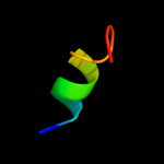
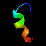
Top model
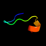
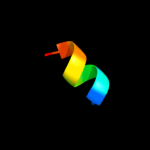
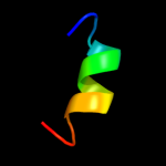
Image coloured by rainbow N → C terminus
Model (left) based on template d1jjda_
Top template information
Fold:Metallothionein
Superfamily:Metallothionein
Family:Metallothionein
Confidence and coverage
Confidence: 33.3% Coverage: 27%
15 residues ( 27% of your sequence) have been modelled with 33.3% confidence by the single highest scoring template.
You may wish to submit your sequence to Phyrealarm. This will automatically scan your sequence every week for new potential templates as they appear in the Phyre2 library.
Please note: You must be registered and logged in to use Phyrealarm.
3D viewing
Interactive 3D view in Jmol

Sequence analysis 

Secondary structure and disorder prediction 

   1........10.........20.........30.........40.........50......
Sequence  MTKKIKCAYHLCKKDVEESKAIERMLHFMHGILSKDEPRKYCSEACAEKDQMAHEL
Secondary structure 


SS confidence 


Disorder  ???????


??


????


???
Disorder confidence 


Confidence Key
High(9)                    Low (0)
?Disordered
Alpha helix
Beta strand

Domain analysis 

Hover over an aligned region to see model and summary info

Please note, only up to the top 20 hits are modelled to reduce computer load

RankAligned region

PDB 1jjd chain A

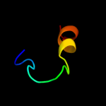
3D model

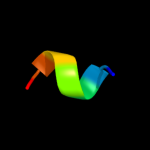
Region: 34 - 48
Aligned: 15
Modelled: 15
Confidence: 33.3%
Identity: 47%
Fold: Metallothionein
Superfamily: Metallothionein
Family: Metallothionein

Phyre2

PDB 2jz3 chain A

3D model

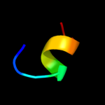
Region: 10 - 16
Aligned: 7
Modelled: 7
Confidence: 29.1%
Identity: 71%
PDB header:transcription inhibitor/transcription
Chain: A: PDB Molecule:suppressor of cytokine signaling 3;
PDBTitle: socs box elonginbc ternary complex

Phyre2

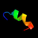
PDB 1lv3 chain A

3D model

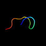
Region: 41 - 50
Aligned: 10
Modelled: 10
Confidence: 27.5%
Identity: 40%
Fold: Glucocorticoid receptor-like (DNA-binding domain)
Superfamily: Glucocorticoid receptor-like (DNA-binding domain)
Family: Hypothetical zinc finger protein YacG

Phyre2

PDB 1u5s chain B domain 1

3D model

Region: 7 - 17
Aligned: 9
Modelled: 11
Confidence: 19.9%
Identity: 33%
Fold: Glucocorticoid receptor-like (DNA-binding domain)
Superfamily: Glucocorticoid receptor-like (DNA-binding domain)
Family: LIM domain

Phyre2

PDB 1c94 chain B

3D model

Region: 20 - 28
Aligned: 9
Modelled: 9
Confidence: 11.4%
Identity: 44%
PDB header:gene regulation
Chain: B: PDB Molecule:retro-gcn4 leucine zipper;
PDBTitle: reversing the sequence of the gcn4 leucine zipper does not2 affect its fold.

Phyre2

PDB 2w0t chain A

3D model

Region: 36 - 47
Aligned: 12
Modelled: 12
Confidence: 9.6%
Identity: 25%
PDB header:transcription
Chain: A: PDB Molecule:lethal(3)malignant brain tumor-like 2 protein;
PDBTitle: solution structure of the fcs zinc finger domain of human2 lmbl2

Phyre2

PDB 2odd chain A

3D model

Region: 41 - 54
Aligned: 14
Modelled: 14
Confidence: 8.5%
Identity: 43%
PDB header:metal binding protein
Chain: A: PDB Molecule:protein cbfa2t1;
PDBTitle: solution structure of the mynd domain from aml1-eto2 complexed with smrt, a corepressor

Phyre2

PDB 1qcr chain D

3D model

Region: 47 - 56
Aligned: 10
Modelled: 10
Confidence: 8.3%
Identity: 30%
PDB header:
PDB COMPND:

Phyre2

PDB 2dj8 chain A domain 1

3D model

Region: 41 - 54
Aligned: 14
Modelled: 14
Confidence: 8.2%
Identity: 43%
Fold: HIT/MYND zinc finger-like
Superfamily: HIT/MYND zinc finger-like
Family: MYND zinc finger

Phyre2

PDB 1pg5 chain B domain 2

3D model

Region: 4 - 26
Aligned: 21
Modelled: 23
Confidence: 7.8%
Identity: 29%
Fold: Rubredoxin-like
Superfamily: Aspartate carbamoyltransferase, Regulatory-chain, C-terminal domain
Family: Aspartate carbamoyltransferase, Regulatory-chain, C-terminal domain

Phyre2

PDB 2dj8 chain A

3D model

Region: 41 - 54
Aligned: 14
Modelled: 14
Confidence: 7.5%
Identity: 43%
PDB header:metal binding protein
Chain: A: PDB Molecule:protein cbfa2t1;
PDBTitle: solution structure of zf-mynd domain of protein cbfa2ti2 (protein mtg8)

Phyre2

PDB 3qwv chain A

3D model

Region: 41 - 56
Aligned: 16
Modelled: 16
Confidence: 6.7%
Identity: 38%
PDB header:transferase
Chain: A: PDB Molecule:set and mynd domain-containing protein 2;
PDBTitle: crystal structure of histone lysine methyltransferase smyd2 in complex2 with the cofactor product adohcy

Phyre2

PDB 2jw6 chain A

3D model

Region: 41 - 54
Aligned: 14
Modelled: 14
Confidence: 6.7%
Identity: 50%
PDB header:transcription
Chain: A: PDB Molecule:deformed epidermal autoregulatory factor 1 homolog;
PDBTitle: solution structure of the deaf1 mynd domain

Phyre2

PDB 2d8q chain A domain 1

3D model

Region: 41 - 54
Aligned: 14
Modelled: 14
Confidence: 6.6%
Identity: 36%
Fold: HIT/MYND zinc finger-like
Superfamily: HIT/MYND zinc finger-like
Family: MYND zinc finger

Phyre2

PDB 1u6p chain A

3D model

Region: 6 - 14
Aligned: 7
Modelled: 9
Confidence: 6.4%
Identity: 71%
Fold: Retrovirus zinc finger-like domains
Superfamily: Retrovirus zinc finger-like domains
Family: Retrovirus zinc finger-like domains

Phyre2

PDB 1mv4 chain B

3D model

Region: 12 - 18
Aligned: 7
Modelled: 7
Confidence: 6.4%
Identity: 29%
PDB header:de novo protein
Chain: B: PDB Molecule:tropomyosin 1 alpha chain;
PDBTitle: tm9a251-284: a peptide model of the c-terminus of a rat2 striated alpha tropomyosin

Phyre2

PDB 1ppj chain D domain 1

3D model

Region: 47 - 56
Aligned: 10
Modelled: 10
Confidence: 6.2%
Identity: 30%
Fold: Cytochrome c
Superfamily: Cytochrome c
Family: Cytochrome bc1 domain

Phyre2

PDB 2jw6 chain A domain 1

3D model

Region: 41 - 54
Aligned: 14
Modelled: 14
Confidence: 6.1%
Identity: 50%
Fold: HIT/MYND zinc finger-like
Superfamily: HIT/MYND zinc finger-like
Family: MYND zinc finger

Phyre2

PDB 1a6s chain A

3D model

Region: 9 - 29
Aligned: 19
Modelled: 21
Confidence: 5.9%
Identity: 37%
Fold: Retroviral matrix proteins
Superfamily: Retroviral matrix proteins
Family: GAG polyprotein M-domain

Phyre2

PDB 2dan chain A

3D model

Region: 41 - 54
Aligned: 14
Modelled: 14
Confidence: 5.6%
Identity: 36%
PDB header:metal binding protein
Chain: A: PDB Molecule:zinc finger mynd domain containing protein 10;
PDBTitle: the solution structure of the mynd domain (leu384-cys430)2 of human zinc finger mynd domain containing protein 10

Phyre2
1

d1jjda_
2

c2jz3A_
3

d1lv3a_
4

d1u5sb1
5

c1c94B_
6

c2w0tA_
7

c2oddA_
8

c1qcrD_
9

d2dj8a1
10

d1pg5b2
11

c2dj8A_
12

c3qwvA_
13

c2jw6A_
14

d2d8qa1
15

d1u6pa_
16

c1mv4B_
17

d1ppjd1
18

d2jw6a1
19

d1a6sa_
20

c2danA_
21


22


23


24


25


Detailed template information 

#
Template Alignment Coverage3D Model Confidence
% i.d. Template Information
1d1jjda_


33.3 47 Fold:Metallothionein
Superfamily:Metallothionein
Family:Metallothionein
2c2jz3A_


29.1 71 PDB header:transcription inhibitor/transcription
Chain: A: PDB Molecule:suppressor of cytokine signaling 3;
PDBTitle: socs box elonginbc ternary complex
3d1lv3a_


27.5 40 Fold:Glucocorticoid receptor-like (DNA-binding domain)
Superfamily:Glucocorticoid receptor-like (DNA-binding domain)
Family:Hypothetical zinc finger protein YacG
4d1u5sb1


19.9 33 Fold:Glucocorticoid receptor-like (DNA-binding domain)
Superfamily:Glucocorticoid receptor-like (DNA-binding domain)
Family:LIM domain
5c1c94B_


11.4 44 PDB header:gene regulation
Chain: B: PDB Molecule:retro-gcn4 leucine zipper;
PDBTitle: reversing the sequence of the gcn4 leucine zipper does not2 affect its fold.
6c2w0tA_


9.6 25 PDB header:transcription
Chain: A: PDB Molecule:lethal(3)malignant brain tumor-like 2 protein;
PDBTitle: solution structure of the fcs zinc finger domain of human2 lmbl2
7c2oddA_


8.5 43 PDB header:metal binding protein
Chain: A: PDB Molecule:protein cbfa2t1;
PDBTitle: solution structure of the mynd domain from aml1-eto2 complexed with smrt, a corepressor
8c1qcrD_


8.3 30 PDB header:
PDB COMPND:
9d2dj8a1


8.2 43 Fold:HIT/MYND zinc finger-like
Superfamily:HIT/MYND zinc finger-like
Family:MYND zinc finger
10d1pg5b2


7.8 29 Fold:Rubredoxin-like
Superfamily:Aspartate carbamoyltransferase, Regulatory-chain, C-terminal domain
Family:Aspartate carbamoyltransferase, Regulatory-chain, C-terminal domain
11c2dj8A_


7.5 43 PDB header:metal binding protein
Chain: A: PDB Molecule:protein cbfa2t1;
PDBTitle: solution structure of zf-mynd domain of protein cbfa2ti2 (protein mtg8)
12c3qwvA_


6.7 38 PDB header:transferase
Chain: A: PDB Molecule:set and mynd domain-containing protein 2;
PDBTitle: crystal structure of histone lysine methyltransferase smyd2 in complex2 with the cofactor product adohcy
13c2jw6A_


6.7 50 PDB header:transcription
Chain: A: PDB Molecule:deformed epidermal autoregulatory factor 1 homolog;
PDBTitle: solution structure of the deaf1 mynd domain
14d2d8qa1


6.6 36 Fold:HIT/MYND zinc finger-like
Superfamily:HIT/MYND zinc finger-like
Family:MYND zinc finger
15d1u6pa_


6.4 71 Fold:Retrovirus zinc finger-like domains
Superfamily:Retrovirus zinc finger-like domains
Family:Retrovirus zinc finger-like domains
16c1mv4B_


6.4 29 PDB header:de novo protein
Chain: B: PDB Molecule:tropomyosin 1 alpha chain;
PDBTitle: tm9a251-284: a peptide model of the c-terminus of a rat2 striated alpha tropomyosin
17d1ppjd1


6.2 30 Fold:Cytochrome c
Superfamily:Cytochrome c
Family:Cytochrome bc1 domain
18d2jw6a1


6.1 50 Fold:HIT/MYND zinc finger-like
Superfamily:HIT/MYND zinc finger-like
Family:MYND zinc finger
19d1a6sa_


5.9 37 Fold:Retroviral matrix proteins
Superfamily:Retroviral matrix proteins
Family:GAG polyprotein M-domain
20c2danA_


5.6 36 PDB header:metal binding protein
Chain: A: PDB Molecule:zinc finger mynd domain containing protein 10;
PDBTitle: the solution structure of the mynd domain (leu384-cys430)2 of human zinc finger mynd domain containing protein 10
21c3iz5w_


not modelled 5.6 21 PDB header:ribosome
Chain: W: PDB Molecule:60s ribosomal protein l22 (l22e);
PDBTitle: localization of the large subunit ribosomal proteins into a 5.5 a2 cryo-em map of triticum aestivum translating 80s ribosome
22c2yiuE_


not modelled 5.6 50 PDB header:oxidoreductase
Chain: E: PDB Molecule:cytochrome c1, heme protein;
PDBTitle: x-ray structure of the dimeric cytochrome bc1 complex from2 the soil bacterium paracoccus denitrificans at 2.73 angstrom resolution
23d1ubdc2


not modelled 5.5 44 Fold:beta-beta-alpha zinc fingers
Superfamily:beta-beta-alpha zinc fingers
Family:Classic zinc finger, C2H2
24d2dana1


not modelled 5.5 36 Fold:HIT/MYND zinc finger-like
Superfamily:HIT/MYND zinc finger-like
Family:MYND zinc finger
25d3cx5d1


not modelled 5.4 40 Fold:Cytochrome c
Superfamily:Cytochrome c
Family:Cytochrome bc1 domain

Binding site prediction 

Due to computational demand, binding site predictions are not run for batch jobs

If you want to predict binding sites, please manually submit your model of choice to 3DLigandSite


Phyre is for academic use only

Please cite: Protein structure prediction on the web: a case study using the Phyre server
Kelley LA and Sternberg MJE. Nature Protocols 4, 363 - 371 (2009) [pdf] [Import into BibTeX]
 
If you use the binding site predictions from 3DLigandSite, please also cite:
3DLigandSite: predicting ligand-binding sites using similar structures.
Wass MN, Kelley LA and Sternberg MJ Nucleic Acids Research 38, W469-73 (2010) [PubMed]
 
© Structural Bioinformatics Group
Imperial College London
Lawrence Kelley, Benjamin Jefferys
Disclaimer
Terms and Conditions
Component software
Template detection: HHpred 1.51
Secondary structure prediction: Psi-pred 2.5
Disorder prediction: Disopred 2.4
Transmembrane prediction: Memsat_SVM
Multi-template modelling and ab initio: Poing 1.0