Phyre Home Retrieve Phyre Job Id
Emaill.a.kelley@imperial.ac.uk
DescriptionP15081
DateThu Jan 5 11:34:37 GMT 2012
Unique Job ID01e70705cf6c2b9c

Summary 

Top model
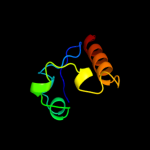
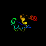
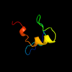
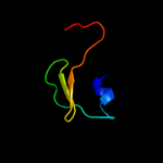
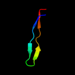
Image coloured by rainbow N → C terminus
Model (left) based on template c3fc7B_
Top template information
PDB header:transferase
Chain: B: PDB Molecule:htr-like protein;
PDBTitle: the crystal structure of a domain of htr-like protein from haloarcula2 marismortui atcc 43049
Confidence and coverage
Confidence: 48.8% Coverage: 49%
58 residues ( 49% of your sequence) have been modelled with 48.8% confidence by the single highest scoring template.
You may wish to submit your sequence to Phyrealarm. This will automatically scan your sequence every week for new potential templates as they appear in the Phyre2 library.
Please note: You must be registered and logged in to use Phyrealarm.
3D viewing
Interactive 3D view in Jmol

Sequence analysis 

Secondary structure and disorder prediction 

   1........10.........20.........30.........40.........50.........60
Sequence  MVSALITVAVIAWCAQLALGGWQISRFNRAFDTLCQQGRVGVGRSSGRFKPRVVVAIALD
Secondary structure 


SS confidence 


Disorder  ?


??

??????


Disorder confidence 


   .........70.........80.........90.........100.........110.........
Sequence  DQQRIVDTLFMKGLTVFARPQKIPAITGMHAGDLQPDVIFPHDPLSQNALSLALKLKRG
Secondary structure 


SS confidence 


Disorder 


?


???


???
Disorder confidence 


Confidence Key
High(9)                    Low (0)
?Disordered
Alpha helix
Beta strand

Domain analysis 

Hover over an aligned region to see model and summary info

Please note, only up to the top 20 hits are modelled to reduce computer load

RankAligned region

PDB 3fc7 chain B

3D model

Region: 55 - 118
Aligned: 58
Modelled: 64
Confidence: 48.8%
Identity: 9%
PDB header:transferase
Chain: B: PDB Molecule:htr-like protein;
PDBTitle: the crystal structure of a domain of htr-like protein from haloarcula2 marismortui atcc 43049

Phyre2

PDB 3fg8 chain B

3D model

Region: 49 - 118
Aligned: 60
Modelled: 70
Confidence: 37.6%
Identity: 18%
PDB header:structural genomics, unknown function
Chain: B: PDB Molecule:uncharacterized protein rha05790;
PDBTitle: crystal structure of pas domain of rha05790

Phyre2

PDB 1cq3 chain A

3D model

Region: 68 - 90
Aligned: 23
Modelled: 23
Confidence: 32.0%
Identity: 26%
Fold: Soluble secreted chemokine inhibitor, VCCI
Superfamily: Soluble secreted chemokine inhibitor, VCCI
Family: Soluble secreted chemokine inhibitor, VCCI

Phyre2

PDB 1xod chain A domain 1

3D model

Region: 54 - 112
Aligned: 57
Modelled: 59
Confidence: 29.4%
Identity: 18%
Fold: PH domain-like barrel
Superfamily: PH domain-like
Family: Enabled/VASP homology 1 domain (EVH1 domain)

Phyre2

PDB 1v9y chain A

3D model

Region: 55 - 115
Aligned: 55
Modelled: 61
Confidence: 21.6%
Identity: 13%
PDB header:signaling protein
Chain: A: PDB Molecule:heme pas sensor protein;
PDBTitle: crystal structure of the heme pas sensor domain of ec dos (ferric2 form)

Phyre2

PDB 1v9y chain A

3D model

Region: 55 - 115
Aligned: 55
Modelled: 61
Confidence: 21.6%
Identity: 13%
Fold: Profilin-like
Superfamily: PYP-like sensor domain (PAS domain)
Family: Heme-binding PAS domain

Phyre2

PDB 3ewk chain A

3D model

Region: 55 - 119
Aligned: 55
Modelled: 65
Confidence: 21.1%
Identity: 11%
PDB header:flavoprotein
Chain: A: PDB Molecule:sensor protein;
PDBTitle: structure of the redox sensor domain of methylococcus capsulatus2 (bath) mmos

Phyre2

PDB 3ajv chain A

3D model

Region: 31 - 65
Aligned: 35
Modelled: 35
Confidence: 21.0%
Identity: 26%
PDB header:hydrolase
Chain: A: PDB Molecule:putative uncharacterized protein;
PDBTitle: splicing endonuclease from aeropyrum pernix

Phyre2

PDB 1wa9 chain A

3D model

Region: 55 - 98
Aligned: 37
Modelled: 44
Confidence: 16.2%
Identity: 16%
PDB header:circadian rhythm
Chain: A: PDB Molecule:period circadian protein;
PDBTitle: crystal structure of the pas repeat region of the2 drosophila clock protein period

Phyre2

PDB 2grg chain A domain 1

3D model

Region: 51 - 68
Aligned: 18
Modelled: 18
Confidence: 13.8%
Identity: 33%
Fold: Profilin-like
Superfamily: YNR034W-A-like
Family: YNR034W-A-like

Phyre2

PDB 3flj chain A

3D model

Region: 54 - 71
Aligned: 18
Modelled: 18
Confidence: 13.8%
Identity: 28%
PDB header:structural genomics, unknown function
Chain: A: PDB Molecule:uncharacterized protein conserved in bacteria with a
PDBTitle: crystal structure of uncharacterized protein conserved in bacteria2 with a cystatin-like fold (yp_168589.1) from silicibacter pomeroyi3 dss-3 at 2.00 a resolution

Phyre2

PDB 2k9k chain A

3D model

Region: 53 - 73
Aligned: 21
Modelled: 21
Confidence: 13.7%
Identity: 19%
PDB header:metal transport
Chain: A: PDB Molecule:tonb2;
PDBTitle: molecular characterization of the tonb2 protein from vibrio2 anguillarum

Phyre2

PDB 2zyz chain A

3D model

Region: 29 - 65
Aligned: 37
Modelled: 37
Confidence: 13.6%
Identity: 14%
PDB header:splicing
Chain: A: PDB Molecule:putative uncharacterized protein pae0789;
PDBTitle: pyrobaculum aerophilum splicing endonuclease

Phyre2

PDB 2gsk chain B domain 1

3D model

Region: 53 - 73
Aligned: 21
Modelled: 21
Confidence: 13.4%
Identity: 14%
Fold: TolA/TonB C-terminal domain
Superfamily: TolA/TonB C-terminal domain
Family: TonB

Phyre2

PDB 1ujk chain A

3D model

Region: 10 - 40
Aligned: 31
Modelled: 31
Confidence: 13.3%
Identity: 19%
Fold: alpha-alpha superhelix
Superfamily: ENTH/VHS domain
Family: VHS domain

Phyre2

PDB 3mjq chain B

3D model

Region: 55 - 115
Aligned: 55
Modelled: 60
Confidence: 13.3%
Identity: 5%
PDB header:structural genomics, unknown function
Chain: B: PDB Molecule:uncharacterized protein;
PDBTitle: crystal structure of the pas domain of q24qt8_deshy protein from2 desulfitobacterium hafniense. northeast structural genomics3 consortium target dhr85c.

Phyre2

PDB 1xx3 chain A

3D model

Region: 53 - 73
Aligned: 21
Modelled: 21
Confidence: 11.5%
Identity: 14%
PDB header:transport protein
Chain: A: PDB Molecule:tonb protein;
PDBTitle: solution structure of escherichia coli tonb-ctd

Phyre2

PDB 1u07 chain A

3D model

Region: 53 - 73
Aligned: 21
Modelled: 21
Confidence: 10.4%
Identity: 14%
Fold: TolA/TonB C-terminal domain
Superfamily: TolA/TonB C-terminal domain
Family: TonB

Phyre2

PDB 3bzk chain A domain 5

3D model

Region: 37 - 71
Aligned: 33
Modelled: 35
Confidence: 10.2%
Identity: 27%
Fold: Ribonuclease H-like motif
Superfamily: Ribonuclease H-like
Family: Tex RuvX-like domain-like

Phyre2

PDB 3f8x chain D

3D model

Region: 54 - 71
Aligned: 18
Modelled: 17
Confidence: 9.6%
Identity: 28%
PDB header:isomerase
Chain: D: PDB Molecule:putative delta-5-3-ketosteroid isomerase;
PDBTitle: crystal structure of a putative delta-5-3-ketosteroid isomerase2 (eca2236) from pectobacterium atrosepticum scri1043 at 1.55 a3 resolution

Phyre2
1

c3fc7B_
2

c3fg8B_
3

d1cq3a_
4

d1xoda1
5

c1v9yA_
6

d1v9ya_
7

c3ewkA_
8

c3ajvA_
9

c1wa9A_
10

d2grga1
11

c3fljA_
12

c2k9kA_
13

c2zyzA_
14

d2gskb1
15

d1ujka_
16

c3mjqB_
17

c1xx3A_
18

d1u07a_
19

d3bzka5
20

c3f8xD_
21


22


23


24


25


26


27


28


29


30


31


32


33


34


35


36


37


38


39


Detailed template information 

#
Template Alignment Coverage3D Model Confidence
% i.d. Template Information
1c3fc7B_


48.8 9 PDB header:transferase
Chain: B: PDB Molecule:htr-like protein;
PDBTitle: the crystal structure of a domain of htr-like protein from haloarcula2 marismortui atcc 43049
2c3fg8B_


37.6 18 PDB header:structural genomics, unknown function
Chain: B: PDB Molecule:uncharacterized protein rha05790;
PDBTitle: crystal structure of pas domain of rha05790
3d1cq3a_


32.0 26 Fold:Soluble secreted chemokine inhibitor, VCCI
Superfamily:Soluble secreted chemokine inhibitor, VCCI
Family:Soluble secreted chemokine inhibitor, VCCI
4d1xoda1


29.4 18 Fold:PH domain-like barrel
Superfamily:PH domain-like
Family:Enabled/VASP homology 1 domain (EVH1 domain)
5c1v9yA_


21.6 13 PDB header:signaling protein
Chain: A: PDB Molecule:heme pas sensor protein;
PDBTitle: crystal structure of the heme pas sensor domain of ec dos (ferric2 form)
6d1v9ya_


21.6 13 Fold:Profilin-like
Superfamily:PYP-like sensor domain (PAS domain)
Family:Heme-binding PAS domain
7c3ewkA_


21.1 11 PDB header:flavoprotein
Chain: A: PDB Molecule:sensor protein;
PDBTitle: structure of the redox sensor domain of methylococcus capsulatus2 (bath) mmos
8c3ajvA_


21.0 26 PDB header:hydrolase
Chain: A: PDB Molecule:putative uncharacterized protein;
PDBTitle: splicing endonuclease from aeropyrum pernix
9c1wa9A_


16.2 16 PDB header:circadian rhythm
Chain: A: PDB Molecule:period circadian protein;
PDBTitle: crystal structure of the pas repeat region of the2 drosophila clock protein period
10d2grga1


13.8 33 Fold:Profilin-like
Superfamily:YNR034W-A-like
Family:YNR034W-A-like
11c3fljA_


13.8 28 PDB header:structural genomics, unknown function
Chain: A: PDB Molecule:uncharacterized protein conserved in bacteria with a
PDBTitle: crystal structure of uncharacterized protein conserved in bacteria2 with a cystatin-like fold (yp_168589.1) from silicibacter pomeroyi3 dss-3 at 2.00 a resolution
12c2k9kA_


13.7 19 PDB header:metal transport
Chain: A: PDB Molecule:tonb2;
PDBTitle: molecular characterization of the tonb2 protein from vibrio2 anguillarum
13c2zyzA_


13.6 14 PDB header:splicing
Chain: A: PDB Molecule:putative uncharacterized protein pae0789;
PDBTitle: pyrobaculum aerophilum splicing endonuclease
14d2gskb1


13.4 14 Fold:TolA/TonB C-terminal domain
Superfamily:TolA/TonB C-terminal domain
Family:TonB
15d1ujka_


13.3 19 Fold:alpha-alpha superhelix
Superfamily:ENTH/VHS domain
Family:VHS domain
16c3mjqB_


13.3 5 PDB header:structural genomics, unknown function
Chain: B: PDB Molecule:uncharacterized protein;
PDBTitle: crystal structure of the pas domain of q24qt8_deshy protein from2 desulfitobacterium hafniense. northeast structural genomics3 consortium target dhr85c.
17c1xx3A_


11.5 14 PDB header:transport protein
Chain: A: PDB Molecule:tonb protein;
PDBTitle: solution structure of escherichia coli tonb-ctd
18d1u07a_


10.4 14 Fold:TolA/TonB C-terminal domain
Superfamily:TolA/TonB C-terminal domain
Family:TonB
19d3bzka5


10.2 27 Fold:Ribonuclease H-like motif
Superfamily:Ribonuclease H-like
Family:Tex RuvX-like domain-like
20c3f8xD_


9.6 28 PDB header:isomerase
Chain: D: PDB Molecule:putative delta-5-3-ketosteroid isomerase;
PDBTitle: crystal structure of a putative delta-5-3-ketosteroid isomerase2 (eca2236) from pectobacterium atrosepticum scri1043 at 1.55 a3 resolution
21c3mxqC_


not modelled 8.8 11 PDB header:transferase
Chain: C: PDB Molecule:sensor protein;
PDBTitle: crystal structure of sensory box sensor histidine kinase from vibrio2 cholerae
22c3rheA_


not modelled 8.6 4 PDB header:oxidoreductase
Chain: A: PDB Molecule:nad-dependent benzaldehyde dehydrogenase;
PDBTitle: the crystal structure of nad-dependent benzaldehyde dehydrogenase from2 legionella pneumophila
23d1ul5a_


not modelled 7.8 8 Fold:SBT domain
Superfamily:SBT domain
Family:SBT domain
24d1ul4a_


not modelled 7.4 8 Fold:SBT domain
Superfamily:SBT domain
Family:SBT domain
25d1mqea_


not modelled 7.2 29 Fold:Nudix
Superfamily:Nudix
Family:MutT-like
26c2w4eA_


not modelled 6.7 22 PDB header:hydrolase
Chain: A: PDB Molecule:mutt/nudix family protein;
PDBTitle: structure of an n-terminally truncated nudix hydrolase2 dr2204 from deinococcus radiodurans
27c2l5fA_


not modelled 6.3 13 PDB header:protein binding
Chain: A: PDB Molecule:pre-mrna-processing factor 40 homolog a;
PDBTitle: solution structure of the tandem ww domains from hypa/fbp11
28c2ic6B_


not modelled 5.9 32 PDB header:viral protein
Chain: B: PDB Molecule:nucleocapsid protein;
PDBTitle: the coiled-coil domain (residues 1-75) structure of the sin2 nombre virus nucleocapsid protein
29c3rtyA_


not modelled 5.9 11 PDB header:circadian clock protein
Chain: A: PDB Molecule:period circadian protein;
PDBTitle: structure of an enclosed dimer formed by the drosophila period protein
30c4a1qB_


not modelled 5.9 33 PDB header:viral protein
Chain: B: PDB Molecule:orf e73;
PDBTitle: solution structure of e73 protein from sulfolobus spindle-2 shaped virus ragged hills, a hyperthermophilic3 crenarchaeal virus from yellowstone national park
31c2kelB_


not modelled 5.6 18 PDB header:transcription repressor
Chain: B: PDB Molecule:uncharacterized protein 56b;
PDBTitle: structure of the transcription regulator svtr from the2 hyperthermophilic archaeal virus sirv1
32d1ltqa1


not modelled 5.5 26 Fold:HAD-like
Superfamily:HAD-like
Family:phosphatase domain of polynucleotide kinase
33c3kebB_


not modelled 5.5 10 PDB header:oxidoreductase
Chain: B: PDB Molecule:probable thiol peroxidase;
PDBTitle: thiol peroxidase from chromobacterium violaceum
34c2jt1A_


not modelled 5.4 16 PDB header:transcription
Chain: A: PDB Molecule:pefi protein;
PDBTitle: solution nmr structure of pefi (plasmid-encoded fimbriae regulatory)2 protein from salmonella typhimurium. northeast structural genomics3 target str82
35c3euhF_


not modelled 5.3 26 PDB header:cell cycle
Chain: F: PDB Molecule:muke;
PDBTitle: crystal structure of the muke-mukf complex
36c3ak5B_


not modelled 5.3 21 PDB header:hydrolase
Chain: B: PDB Molecule:hemoglobin-binding protease hbp;
PDBTitle: hemoglobin protease (hbp) passenger missing domain-2
37c3doaA_


not modelled 5.2 18 PDB header:protein binding
Chain: A: PDB Molecule:fibrinogen binding protein;
PDBTitle: the crystal structure of the fibrinogen binding protein from2 staphylococcus aureus
38c2qhkA_


not modelled 5.1 23 PDB header:signaling protein
Chain: A: PDB Molecule:methyl-accepting chemotaxis protein;
PDBTitle: crystal structure of methyl-accepting chemotaxis protein from vibrio2 parahaemolyticus rimd 2210633
39c3rj1P_


not modelled 5.1 0 PDB header:transcription
Chain: P: PDB Molecule:mediator of rna polymerase ii transcription subunit 17;
PDBTitle: architecture of the mediator head module

Binding site prediction 

Due to computational demand, binding site predictions are not run for batch jobs

If you want to predict binding sites, please manually submit your model of choice to 3DLigandSite

Transmembrane helix prediction 

Transmembrane helices have been predicted in your sequence to adopt the topology shown below


Phyre is for academic use only

Please cite: Protein structure prediction on the web: a case study using the Phyre server
Kelley LA and Sternberg MJE. Nature Protocols 4, 363 - 371 (2009) [pdf] [Import into BibTeX]
 
If you use the binding site predictions from 3DLigandSite, please also cite:
3DLigandSite: predicting ligand-binding sites using similar structures.
Wass MN, Kelley LA and Sternberg MJ Nucleic Acids Research 38, W469-73 (2010) [PubMed]
 
© Structural Bioinformatics Group
Imperial College London
Lawrence Kelley, Benjamin Jefferys
Disclaimer
Terms and Conditions
Component software
Template detection: HHpred 1.51
Secondary structure prediction: Psi-pred 2.5
Disorder prediction: Disopred 2.4
Transmembrane prediction: Memsat_SVM
Multi-template modelling and ab initio: Poing 1.0