1 c3nc7A_
20.7
13
PDB header: oxidoreductaseChain: A: PDB Molecule: cytochrome p450 cypx;PDBTitle: cyp134a1 2-phenylimidazole bound structure
2 d1wj2a_
13.8
67
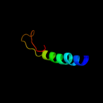
Fold: WRKY DNA-binding domainSuperfamily: WRKY DNA-binding domainFamily: WRKY DNA-binding domain3 c2ve3A_
10.3
13
PDB header: oxidoreductaseChain: A: PDB Molecule: putative cytochrome p450 120;PDBTitle: retinoic acid bound cyanobacterial cyp120a1
4 c2rnmC_
10.3
15
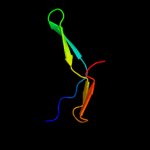
PDB header: protein fibrilChain: C: PDB Molecule: small s protein;PDBTitle: structure of the het-s(218-289) prion in its amyloid form2 obtained by solid-state nmr
5 c2aydA_
10.0
67
PDB header: transcriptionChain: A: PDB Molecule: wrky transcription factor 1;PDBTitle: crystal structure of the c-terminal wrky domainof atwrky1,2 an sa-induced and partially npr1-dependent transcription3 factor
6 d1m5ha1
9.7
18
Fold: Ferredoxin-likeSuperfamily: Formylmethanofuran:tetrahydromethanopterin formyltransferaseFamily: Formylmethanofuran:tetrahydromethanopterin formyltransferase7 d3czha1
9.5
16
Fold: Cytochrome P450Superfamily: Cytochrome P450Family: Cytochrome P4508 c2de0X_
9.2
39
PDB header: transferaseChain: X: PDB Molecule: alpha-(1,6)-fucosyltransferase;PDBTitle: crystal structure of human alpha 1,6-fucosyltransferase, fut8
9 d1qb5d_
9.1
27
Fold: OB-foldSuperfamily: Bacterial enterotoxinsFamily: Bacterial AB5 toxins, B-subunits10 c3iyuY_
8.8
24
PDB header: virusChain: Y: PDB Molecule: outer capsid protein vp4;PDBTitle: atomic model of an infectious rotavirus particle
11 c3g1qC_
8.4
11
PDB header: oxidoreductaseChain: C: PDB Molecule: sterol 14-alpha-demethylase;PDBTitle: crystal structure of sterol 14-alpha demethylase (cyp51)2 from trypanosoma brucei in ligand free state
12 c2fhjD_
8.4
13
PDB header: transferaseChain: D: PDB Molecule: formylmethanofuran--tetrahydromethanopterinPDBTitle: crystal structure of formylmethanofuran:2 tetrahydromethanopterin formyltransferase in complex with3 its coenzymes
13 d1tqna_
8.1
14
Fold: Cytochrome P450Superfamily: Cytochrome P450Family: Cytochrome P45014 d1po5a_
7.1
9
Fold: Cytochrome P450Superfamily: Cytochrome P450Family: Cytochrome P45015 c3k9vB_
6.8
12
PDB header: oxidoreductaseChain: B: PDB Molecule: 1,25-dihydroxyvitamin d(3) 24-hydroxylase,PDBTitle: crystal structure of rat mitochondrial p450 24a1 s57d in2 complex with chaps
16 c3p5nA_
6.8
14
PDB header: transport proteinChain: A: PDB Molecule: riboflavin uptake protein;PDBTitle: structure and mechanism of the s component of a bacterial ecf2 transporter
17 c2c0zA_
6.8
26
PDB header: isomeraseChain: A: PDB Molecule: novw;PDBTitle: the 1.6 a resolution crystal structure of novw: a 4-keto-6-2 deoxy sugar epimerase from the novobiocin biosynthetic3 gene cluster of streptomyces spheroides
18 c3na0B_
6.8
9
PDB header: oxidoreductase, electron transportChain: B: PDB Molecule: cholesterol side-chain cleavage enzyme, mitochondrial;PDBTitle: crystal structure of human cyp11a1 in complex with 20,22-2 dihydroxycholesterol
19 d1nr6a_
6.5
7
Fold: Cytochrome P450Superfamily: Cytochrome P450Family: Cytochrome P45020 d2nnja1
5.9
7
Fold: Cytochrome P450Superfamily: Cytochrome P450Family: Cytochrome P45021 d1bvp12
not modelled
5.7
26
Fold: Viral protein domainSuperfamily: Viral protein domainFamily: Top domain of virus capsid protein22 c3m3wA_
not modelled
5.6
25
PDB header: endocytosisChain: A: PDB Molecule: protein kinase c and casein kinase ii substrate protein 3;PDBTitle: crystal strcuture of mouse pacsin3 bar domain mutant
23 c1yewF_
not modelled
5.4
38
PDB header: oxidoreductase, membrane proteinChain: F: PDB Molecule: particulate methane monooxygenase, a subunit;PDBTitle: crystal structure of particulate methane monooxygenase
24 d1m5sa1
not modelled
5.2
13
Fold: Ferredoxin-likeSuperfamily: Formylmethanofuran:tetrahydromethanopterin formyltransferaseFamily: Formylmethanofuran:tetrahydromethanopterin formyltransferase25 d1h3ga1
not modelled
5.2
19
Fold: Immunoglobulin-like beta-sandwichSuperfamily: E set domainsFamily: E-set domains of sugar-utilizing enzymes26 c3e4eA_
not modelled
5.1
7
PDB header: oxidoreductaseChain: A: PDB Molecule: cytochrome p450 2e1;PDBTitle: human cytochrome p450 2e1 in complex with the inhibitor 4-2 methylpyrazole
27 c1g92A_
not modelled
5.1
35
PDB header: toxinChain: A: PDB Molecule: poneratoxin;PDBTitle: solution structure of poneratoxin
28 c3o0rC_
not modelled
5.0
15
PDB header: immune system/oxidoreductaseChain: C: PDB Molecule: nitric oxide reductase subunit c;PDBTitle: crystal structure of nitric oxide reductase from pseudomonas2 aeruginosa in complex with antibody fragment