Phyre Home Retrieve Phyre Job Id
Emaill.a.kelley@imperial.ac.uk
DescriptionP0ADY3
DateWed Jan 25 15:20:29 GMT 2012
Unique Job IDff02aebd34072e44

Summary 

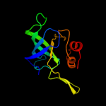
Top model
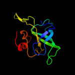
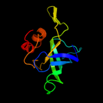
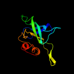
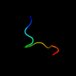
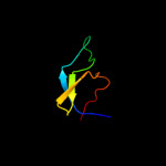
Image coloured by rainbow N → C terminus
Model (left) based on template d1whia_
Top template information
Fold:Ribosomal protein L14
Superfamily:Ribosomal protein L14
Family:Ribosomal protein L14
Confidence and coverage
Confidence:100.0% Coverage: 99%
122 residues ( 99% of your sequence) have been modelled with 100.0% confidence by the single highest scoring template.
3D viewing
Interactive 3D view in Jmol

Sequence analysis 

Secondary structure and disorder prediction 

   1........10.........20.........30.........40.........50.........60
Sequence  MIQEQTMLNVADNSGARRVMCIKVLGGSHRRYAGVGDIIKITIKEAIPRGKVKKGDVLKA
Secondary structure 


SS confidence 


Disorder  ??????


??
??


???????
?


Disorder confidence 


   .........70.........80.........90.........100.........110.........120
Sequence  VVVRTKKGVRRPDGSVIRFDGNACVLLNNNSEQPIGTRIFGPVTRELRSEKFMKIISLAP
Secondary structure 


SS confidence 


Disorder 


?

?


?


????
Disorder confidence 


   ...
Sequence  EVL
Secondary structure 


SS confidence 


Disorder  ???
Disorder confidence 


Confidence Key
High(9)                    Low (0)
?Disordered
Alpha helix
Beta strand

Domain analysis 

Hover over an aligned region to see model and summary info

Please note, only up to the top 20 hits are modelled to reduce computer load

RankAligned region

PDB 1whi chain A

3D model

Region: 1 - 123
Aligned: 122
Modelled: 123
Confidence: 100.0%
Identity: 69%
Fold: Ribosomal protein L14
Superfamily: Ribosomal protein L14
Family: Ribosomal protein L14

Phyre2

PDB 2j01 chain O domain 1

3D model

Region: 1 - 123
Aligned: 122
Modelled: 123
Confidence: 100.0%
Identity: 69%
Fold: Ribosomal protein L14
Superfamily: Ribosomal protein L14
Family: Ribosomal protein L14

Phyre2

PDB 2zjr chain H domain 1

3D model

Region: 1 - 123
Aligned: 122
Modelled: 123
Confidence: 100.0%
Identity: 66%
Fold: Ribosomal protein L14
Superfamily: Ribosomal protein L14
Family: Ribosomal protein L14

Phyre2

PDB 2gyc chain I domain 1

3D model

Region: 2 - 122
Aligned: 121
Modelled: 121
Confidence: 100.0%
Identity: 100%
Fold: Ribosomal protein L14
Superfamily: Ribosomal protein L14
Family: Ribosomal protein L14

Phyre2

PDB 3bbo chain M

3D model

Region: 1 - 123
Aligned: 121
Modelled: 123
Confidence: 100.0%
Identity: 59%
PDB header:ribosome
Chain: M: PDB Molecule:ribosomal protein l14;
PDBTitle: homology model for the spinach chloroplast 50s subunit2 fitted to 9.4a cryo-em map of the 70s chlororibosome

Phyre2

PDB 2zkr chain K

3D model

Region: 2 - 122
Aligned: 116
Modelled: 121
Confidence: 100.0%
Identity: 38%
PDB header:ribosomal protein/rna
Chain: K: PDB Molecule:rna expansion segment es19;
PDBTitle: structure of a mammalian ribosomal 60s subunit within an2 80s complex obtained by docking homology models of the rna3 and proteins into an 8.7 a cryo-em map

Phyre2

PDB 1vqo chain K domain 1

3D model

Region: 2 - 123
Aligned: 117
Modelled: 122
Confidence: 100.0%
Identity: 39%
Fold: Ribosomal protein L14
Superfamily: Ribosomal protein L14
Family: Ribosomal protein L14

Phyre2

PDB 1zv9 chain A domain 1

3D model

Region: 30 - 44
Aligned: 15
Modelled: 15
Confidence: 21.2%
Identity: 33%
Fold: Common fold of diphtheria toxin/transcription factors/cytochrome f
Superfamily: Carbohydrate-binding domain
Family: Cellulose-binding domain family III

Phyre2

PDB 1wyh chain A domain 1

3D model

Region: 92 - 102
Aligned: 11
Modelled: 11
Confidence: 20.3%
Identity: 64%
Fold: Glucocorticoid receptor-like (DNA-binding domain)
Superfamily: Glucocorticoid receptor-like (DNA-binding domain)
Family: LIM domain

Phyre2

PDB 1tyj chain A domain 1

3D model

Region: 30 - 44
Aligned: 15
Modelled: 15
Confidence: 19.0%
Identity: 33%
Fold: Common fold of diphtheria toxin/transcription factors/cytochrome f
Superfamily: Carbohydrate-binding domain
Family: Cellulose-binding domain family III

Phyre2

PDB 3ghp chain A

3D model

Region: 30 - 44
Aligned: 15
Modelled: 15
Confidence: 17.9%
Identity: 53%
PDB header:structural protein
Chain: A: PDB Molecule:cellulosomal scaffoldin adaptor protein b;
PDBTitle: structure of the second type ii cohesin module from the2 adaptor scaa scaffoldin of acetivibrio cellulolyticus3 (including long c-terminal linker)

Phyre2

PDB 2bm3 chain A domain 1

3D model

Region: 30 - 44
Aligned: 15
Modelled: 15
Confidence: 17.2%
Identity: 40%
Fold: Common fold of diphtheria toxin/transcription factors/cytochrome f
Superfamily: Carbohydrate-binding domain
Family: Cellulose-binding domain family III

Phyre2

PDB 2b59 chain A

3D model

Region: 30 - 44
Aligned: 15
Modelled: 15
Confidence: 17.1%
Identity: 40%
PDB header:hydrolase/structural protein
Chain: A: PDB Molecule:cog1196: chromosome segregation atpases;
PDBTitle: the type ii cohesin dockerin complex

Phyre2

PDB 1oj5 chain A

3D model

Region: 67 - 98
Aligned: 31
Modelled: 32
Confidence: 16.5%
Identity: 10%
Fold: Profilin-like
Superfamily: PYP-like sensor domain (PAS domain)
Family: PAS domain of steroid receptor coactivator 1A, NCo-A1

Phyre2

PDB 3fnk chain A

3D model

Region: 30 - 44
Aligned: 15
Modelled: 15
Confidence: 16.3%
Identity: 53%
PDB header:structural protein
Chain: A: PDB Molecule:cellulosomal scaffoldin adaptor protein b;
PDBTitle: crystal structure of the second type ii cohesin module from2 the cellulosomal adaptor scaa scaffoldin of acetivibrio3 cellulolyticus

Phyre2

PDB 1gd7 chain A

3D model

Region: 2 - 100
Aligned: 84
Modelled: 87
Confidence: 13.5%
Identity: 23%
Fold: OB-fold
Superfamily: Nucleic acid-binding proteins
Family: Myf domain

Phyre2

PDB 2e5q chain A

3D model

Region: 55 - 96
Aligned: 39
Modelled: 42
Confidence: 9.6%
Identity: 15%
PDB header:transcription
Chain: A: PDB Molecule:phd finger protein 19;
PDBTitle: solution structure of the tudor domain of phd finger2 protein 19, isoform b [homo sapiens]

Phyre2

PDB 2xk0 chain A

3D model

Region: 55 - 83
Aligned: 24
Modelled: 29
Confidence: 9.2%
Identity: 25%
PDB header:transcription
Chain: A: PDB Molecule:polycomb protein pcl;
PDBTitle: solution structure of the tudor domain from drosophila2 polycomblike (pcl)

Phyre2

PDB 3icy chain B

3D model

Region: 63 - 96
Aligned: 33
Modelled: 34
Confidence: 9.2%
Identity: 9%
PDB header:structural genomics, unknown function
Chain: B: PDB Molecule:sensor protein;
PDBTitle: the crystal structure of sensory box histidine2 kinase/response regulator domain from chlorobium tepidum3 tls

Phyre2

PDB 3ewk chain A

3D model

Region: 61 - 96
Aligned: 35
Modelled: 36
Confidence: 9.1%
Identity: 11%
PDB header:flavoprotein
Chain: A: PDB Molecule:sensor protein;
PDBTitle: structure of the redox sensor domain of methylococcus capsulatus2 (bath) mmos

Phyre2
1

d1whia_
2

d2j01o1
3

d2zjrh1
4

d2gyci1
5

c3bboM_
6

c2zkrk_
7

d1vqok1
8

d1zv9a1
9

d1wyha1
10

d1tyja1
11

c3ghpA_
12

d2bm3a1
13

c2b59A_
14

d1oj5a_
15

c3fnkA_
16

d1gd7a_
17

c2e5qA_
18

c2xk0A_
19

c3icyB_
20

c3ewkA_
21


22


23


24


25


26


27


28


29


30


31


32


Detailed template information 

#
Template Alignment Coverage3D Model Confidence
% i.d. Template Information
1d1whia_


100.0 69 Fold:Ribosomal protein L14
Superfamily:Ribosomal protein L14
Family:Ribosomal protein L14
2d2j01o1


100.0 69 Fold:Ribosomal protein L14
Superfamily:Ribosomal protein L14
Family:Ribosomal protein L14
3d2zjrh1


100.0 66 Fold:Ribosomal protein L14
Superfamily:Ribosomal protein L14
Family:Ribosomal protein L14
4d2gyci1


100.0 100 Fold:Ribosomal protein L14
Superfamily:Ribosomal protein L14
Family:Ribosomal protein L14
5c3bboM_


100.0 59 PDB header:ribosome
Chain: M: PDB Molecule:ribosomal protein l14;
PDBTitle: homology model for the spinach chloroplast 50s subunit2 fitted to 9.4a cryo-em map of the 70s chlororibosome
6c2zkrk_


100.0 38 PDB header:ribosomal protein/rna
Chain: K: PDB Molecule:rna expansion segment es19;
PDBTitle: structure of a mammalian ribosomal 60s subunit within an2 80s complex obtained by docking homology models of the rna3 and proteins into an 8.7 a cryo-em map
7d1vqok1


100.0 39 Fold:Ribosomal protein L14
Superfamily:Ribosomal protein L14
Family:Ribosomal protein L14
8d1zv9a1


21.2 33 Fold:Common fold of diphtheria toxin/transcription factors/cytochrome f
Superfamily:Carbohydrate-binding domain
Family:Cellulose-binding domain family III
9d1wyha1


20.3 64 Fold:Glucocorticoid receptor-like (DNA-binding domain)
Superfamily:Glucocorticoid receptor-like (DNA-binding domain)
Family:LIM domain
10d1tyja1


19.0 33 Fold:Common fold of diphtheria toxin/transcription factors/cytochrome f
Superfamily:Carbohydrate-binding domain
Family:Cellulose-binding domain family III
11c3ghpA_


17.9 53 PDB header:structural protein
Chain: A: PDB Molecule:cellulosomal scaffoldin adaptor protein b;
PDBTitle: structure of the second type ii cohesin module from the2 adaptor scaa scaffoldin of acetivibrio cellulolyticus3 (including long c-terminal linker)
12d2bm3a1


17.2 40 Fold:Common fold of diphtheria toxin/transcription factors/cytochrome f
Superfamily:Carbohydrate-binding domain
Family:Cellulose-binding domain family III
13c2b59A_


17.1 40 PDB header:hydrolase/structural protein
Chain: A: PDB Molecule:cog1196: chromosome segregation atpases;
PDBTitle: the type ii cohesin dockerin complex
14d1oj5a_


16.5 10 Fold:Profilin-like
Superfamily:PYP-like sensor domain (PAS domain)
Family:PAS domain of steroid receptor coactivator 1A, NCo-A1
15c3fnkA_


16.3 53 PDB header:structural protein
Chain: A: PDB Molecule:cellulosomal scaffoldin adaptor protein b;
PDBTitle: crystal structure of the second type ii cohesin module from2 the cellulosomal adaptor scaa scaffoldin of acetivibrio3 cellulolyticus
16d1gd7a_


13.5 23 Fold:OB-fold
Superfamily:Nucleic acid-binding proteins
Family:Myf domain
17c2e5qA_


9.6 15 PDB header:transcription
Chain: A: PDB Molecule:phd finger protein 19;
PDBTitle: solution structure of the tudor domain of phd finger2 protein 19, isoform b [homo sapiens]
18c2xk0A_


9.2 25 PDB header:transcription
Chain: A: PDB Molecule:polycomb protein pcl;
PDBTitle: solution structure of the tudor domain from drosophila2 polycomblike (pcl)
19c3icyB_


9.2 9 PDB header:structural genomics, unknown function
Chain: B: PDB Molecule:sensor protein;
PDBTitle: the crystal structure of sensory box histidine2 kinase/response regulator domain from chlorobium tepidum3 tls
20c3ewkA_


9.1 11 PDB header:flavoprotein
Chain: A: PDB Molecule:sensor protein;
PDBTitle: structure of the redox sensor domain of methylococcus capsulatus2 (bath) mmos
21d1b12a_


not modelled 9.0 13 Fold:LexA/Signal peptidase
Superfamily:LexA/Signal peptidase
Family:Type 1 signal peptidase
22c2q2iA_


not modelled 7.6 18 PDB header:chaperone
Chain: A: PDB Molecule:secretion chaperone;
PDBTitle: crystal structure of the protein secretion chaperone csaa from2 agrobacterium tumefaciens.
23d1ijdb_


not modelled 7.3 44 Fold:Light-harvesting complex subunits
Superfamily:Light-harvesting complex subunits
Family:Light-harvesting complex subunits
24c3s6xA_


not modelled 6.3 21 PDB header:viral protein
Chain: A: PDB Molecule:outer capsid protein sigma-1;
PDBTitle: structure of reovirus attachment protein sigma1 in complex with alpha-2 2,3-sialyllactose
25d2do3a1


not modelled 6.3 24 Fold:SH3-like barrel
Superfamily:Translation proteins SH3-like domain
Family:SPT5 KOW domain-like
26d1k8ke_


not modelled 6.1 25 Fold:Arp2/3 complex 21 kDa subunit ARPC3
Superfamily:Arp2/3 complex 21 kDa subunit ARPC3
Family:Arp2/3 complex 21 kDa subunit ARPC3
27d2zjrm1


not modelled 5.7 19 Fold:SH3-like barrel
Superfamily:Translation proteins SH3-like domain
Family:Ribosomal protein L19
28c2eqjA_


not modelled 5.7 13 PDB header:transcription
Chain: A: PDB Molecule:metal-response element-binding transcription
PDBTitle: solution structure of the tudor domain of metal-response2 element-binding transcription factor 2
29c2dyoB_


not modelled 5.1 100 PDB header:protein turnover/protein turnover
Chain: B: PDB Molecule:autophagy protein 16;
PDBTitle: the crystal structure of saccharomyces cerevisiae atg5-2 atg16(1-57) complex
30d1mb4a1


not modelled 5.1 13 Fold:NAD(P)-binding Rossmann-fold domains
Superfamily:NAD(P)-binding Rossmann-fold domains
Family:Glyceraldehyde-3-phosphate dehydrogenase-like, N-terminal domain
31d1x4ka1


not modelled 5.1 50 Fold:Glucocorticoid receptor-like (DNA-binding domain)
Superfamily:Glucocorticoid receptor-like (DNA-binding domain)
Family:LIM domain
32d1t3ta4


not modelled 5.1 33 Fold:Bacillus chorismate mutase-like
Superfamily:PurM N-terminal domain-like
Family:PurM N-terminal domain-like

Binding site prediction 

Due to computational demand, binding site predictions are not run for batch jobs

If you want to predict binding sites, please manually submit your model of choice to 3DLigandSite


Phyre is for academic use only

Please cite: Protein structure prediction on the web: a case study using the Phyre server
Kelley LA and Sternberg MJE. Nature Protocols 4, 363 - 371 (2009) [pdf] [Import into BibTeX]
 
If you use the binding site predictions from 3DLigandSite, please also cite:
3DLigandSite: predicting ligand-binding sites using similar structures.
Wass MN, Kelley LA and Sternberg MJ Nucleic Acids Research 38, W469-73 (2010) [PubMed]
 
© Structural Bioinformatics Group
Imperial College London
Lawrence Kelley, Benjamin Jefferys
Disclaimer
Terms and Conditions
Component software
Template detection: HHpred 1.51
Secondary structure prediction: Psi-pred 2.5
Disorder prediction: Disopred 2.4
Transmembrane prediction: Memsat_SVM
Multi-template modelling and ab initio: Poing 1.0