| Secondary structure and disorder prediction | |
| | |
1 | . | . | . | . | . | . | . | . | 10 | . | . | . | . | . | . | . | . | . | 20 | . | . | . | . | . | . | . | . | . | 30 | . | . | . | . | . | . | . | . | . | 40 | . | . | . | . | . | . | . | . | . | 50 | . | . | . | . | . | . | . |
| Sequence | |
M | A | L | F | S | K | I | L | I | F | Y | V | I | G | V | N | I | S | F | V | I | I | W | F | I | S | H | E | K | T | H | I | R | L | L | S | A | F | L | V | G | I | T | W | P | M | S | L | P | V | A | L | L | F | S | L | F |
| Secondary structure | |
|  | | | | | | | | | | | | | | | | | | | | | | | | | |
 | | |
|
| SS confidence | |
|
|
|
|
|
|
|
|
|
|
|
|
|
|
|
|
|
|
|
|
|
|
|
|
|
|
|
|
|
|
|
|
|
|
|
|
|
|
|
|
|
|
|
|
|
|
|
|
|
|
|
|
|
|
|
|
|
| Disorder | |
? | ? |
|
|
|
|
|
|
|
|
|
|
|
|
|
|
|
|
|
|
|
|
|
|
|
|
|
|
|
|
|
|
|
|
|
|
|
|
|
|
|
|
|
|
|
|
|
|
|
|
|
|
|
|
| ? | ? |
| Disorder confidence | |
|
|
|
|
|
|
|
|
|
|
|
|
|
|
|
|
|
|
|
|
|
|
|
|
|
|
|
|
|
|
|
|
|
|
|
|
|
|
|
|
|
|
|
|
|
|
|
|
|
|
|
|
|
|
|
|
|
| |
| Confidence Key |
| High(9) | |
|
|
|
|
|
|
|
|
|
Low (0) |
| ? | Disordered |
| Alpha helix |
  | Beta strand |
Hover over an aligned region to see model and summary info
Please note, only up to the top 20 hits are modelled to reduce computer load
|
| 1 |
|
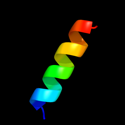
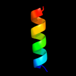
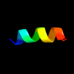
PDB 2p7t chain C domain 1
Region: 14 - 29
Aligned: 16
Modelled: 16
Confidence: 23.5%
Identity: 25%
Fold: Voltage-gated potassium channels
Superfamily: Voltage-gated potassium channels
Family: Voltage-gated potassium channels
Phyre2
| 2 |
|
PDB 1kzf chain A
Region: 43 - 50
Aligned: 8
Modelled: 8
Confidence: 14.6%
Identity: 75%
Fold: Acyl-CoA N-acyltransferases (Nat)
Superfamily: Acyl-CoA N-acyltransferases (Nat)
Family: Autoinducer synthetase
Phyre2
| 3 |
|
PDB 1ifl chain A
Region: 39 - 51
Aligned: 13
Modelled: 13
Confidence: 13.3%
Identity: 38%
PDB header:virus
Chain: A: PDB Molecule:inovirus;
PDBTitle: molecular models and structural comparisons of native and2 mutant class i filamentous bacteriophages ff (fd, f1, m13),3 if1 and ike
Phyre2
| 4 |
|
PDB 1v54 chain K
Region: 37 - 47
Aligned: 11
Modelled: 11
Confidence: 11.9%
Identity: 45%
Fold: Single transmembrane helix
Superfamily: Mitochondrial cytochrome c oxidase subunit VIIb
Family: Mitochondrial cytochrome c oxidase subunit VIIb
Phyre2
| 5 |
|
PDB 2y69 chain X
Region: 37 - 47
Aligned: 11
Modelled: 11
Confidence: 11.9%
Identity: 45%
PDB header:electron transport
Chain: X: PDB Molecule:cytochrome c oxidase polypeptide 7b;
PDBTitle: bovine heart cytochrome c oxidase re-refined with molecular2 oxygen
Phyre2
| 6 |
|
PDB 1tub chain B domain 1
Region: 33 - 49
Aligned: 17
Modelled: 17
Confidence: 6.7%
Identity: 35%
Fold: Tubulin nucleotide-binding domain-like
Superfamily: Tubulin nucleotide-binding domain-like
Family: Tubulin, GTPase domain
Phyre2
|
| Detailed template information | |
Due to computational demand, binding site predictions are not run for batch jobs
If you want to predict binding sites, please manually submit your model of choice to 3DLigandSite
Phyre is for academic use only
| Please cite: Protein structure prediction on
the web: a case study using the Phyre server |
| Kelley LA and Sternberg MJE. Nature Protocols
4, 363 - 371 (2009) [pdf] [Import into BibTeX] |
| |
| If you use the binding site
predictions from 3DLigandSite, please also cite: |
| 3DLigandSite: predicting ligand-binding sites using similar structures. |
| Wass MN, Kelley LA and Sternberg
MJ Nucleic Acids Research 38, W469-73 (2010) [PubMed] |
| |
|
|
|
|